Pelvic Floor – Structure, Function and Muscles
Walls Of Pelvis
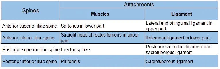
Attachments of iliac spines:
Table of Contents
Read And Learn More: General Histology Questions and Answers
Internal Iliac Artery
Internal Iliac Artery Introduction: This is the artery supplying the external genitalia, structures in the lateral pelvic wall, pelvic organs, and gluteal region.
1. Origin:
The internal iliac artery is one of the two terminal branches of the common iliac artery given at the sacroiliac joint. It supplies the pelvic walls, pelvic viscera, gluteal region, perineum and adductor compartment of the thigh.
2. Distribution:
It supplies
- Pelvic organs
- Perineum
- The gluteal region, and
- Iliac fossa.
3. Features:
1. It lies within the parietal pelvic fascia.
2. However, its branches pierce pelvic fascia to distribute the structures, except the obturator artery.
3. It is approximately 4 cm long.
4. The internal iliac artery in the foetus, however, is double the size of the external iliac artery.
5. Because it transmits blood to the placenta through the umbilical artery.
6. The umbilical artery with internal iliac artery then forms a direct continuation of the common iliac artery.
4. Course:
- It begins in front of the sacroiliac joint at the level of the intervertebral disc between L5 and the sacrum.
- It lies medial to the psoas major muscle.
- It runs downwards and backward and ends near the upper margin of the greater sciatic notch.
- It divides into anterior and posterior divisions.
5. Relations:
The artery is related
Anteriorly:
- In male ♂ and female ♀ to ureter.
- In females ♀ to the
- Ovary, and
- The lateral end of the uterine tube.
Posteriorly:
- Internal iliac vein
- Lumbosacral trunk, and
- Sacroiliac joint.
Laterally:
- External iliac vein, and
- Obturator nerve.
Medially:
- Peritoneum, and
- Tributaries of internal iliac vein.
6. Branches:
They are divided into
1. Anterior division:
In male ♂ :
1. Collateral branches
- Obliterated umbilical artery
- Obturator artery
- Superior vesical artery
- Inferior vesical artery
- Middle rectal artery
2. Terminal branches
- Inferior gluteal artery, and
- Internal pudendal artery.
In female ♀:
It is important to note that in females ♀ the inferior vesical artery is absent. It is replaced by two arteries, namely, the uterine and the vaginal.

2. Posterior division:
Breaks into three branches. They are all parietal branches. The keyword is.
- Superior gluteal (S)
- Iliolumbar and (I)
- Lateral sacral arteries (L).
Mnemonics:
Note: The branches of anterior division of internal iliac artery can be remembered by the keyword
O, O, SIM, GENERAL PRACTITIONER
- The letter “O” represents an obliterated umbilical artery.
- The next letter “O” represents the obturator.
- The letter “S” represents superior vesical.
- The letter “I” represents inferior vesical.
- The letter “M” represents the middle rectal.
- The letter “G” represents the inferior gluteal.
- The letter “P” represents internal pudendal
Things to note:
Note: Obliterated umbilical artery is a continuation of the superior vesical, which is usually the 1st highest branch to arise from anterior division. It is to be noted that the internal pudendal and inferior gluteal vessels are considered terminal branches of the anterior division.
7. Applied anatomy:
1. Inferior mesenteric arteriogram is done by passing a catheter through internal iliac artery.
2. Occasionally, the internal iliac artery is ligated to control pelvic haemorrhage.
3. In the kidney, transplantation renal vessels are anastomosed end-to-end to the internal iliac vessels.
4. Blockage of the internal iliac artery leads to impotence in males ♂
Inferior Vesical Artery
Inferior Vesical Artery Introduction: The inferior vesical artery is also called the vesicular artery, although this adjective is derived from the vesicula or vesicle, meaning ‘a small bladder’. It is present only in the male ♂. Please remember that there is no inferior vesical artery in females ♀. Instead, it is represented by the uterine and the vaginal arteries.
1. Origin:
It is a branch of anterior division of the internal iliac artery.
2. Course And Relations:
The inferior vesical artery runs forwards and medially, in the lateral true ligaments of the bladder, with the vesical veins.
3. Distribution:
It supplies
- Trigone
- The lower part of the urinary bladder
- The lower end of the ureter
- Urethra
- Ducts deferens
- A seminal vesicle, and
- Prostate.
The main artery of the prostate:
Note: Please note, the inferior vesical artery is the main artery of the prostate.
4. Branches Of Vesical Artery:
- Urethral, and
- Capsular
- The urethral vessels enter at the prostatovesical junction principally posteriorly at the 5 and 7 ‘O’ clock positions but also, anteriorly at 1 and 11 ‘O’clock
- The capsular arteries run posterolaterally and inferiorly in the neurovascular bundle. It provides perpendicular perforating vessels to the prostate.
5. Vaginal Artery:
1. They are often 2 or 3 in number
2. It corresponds with the inferior vesical artery of the males ♂ it passes downwards and medially to the vagina.

3. The vaginal artery anastomoses in front as well as behind the vaginal walls.
It forms two longitudinal vessels
- Anterior, and
- Posterior azygous arteries of the vagina.
It also gives branches to the lower part of the bladder wall and the anterior rectal wall.
Inferior Gluteal Artery
Inferior Gluteal Artery Introduction: It is the axial artery of the lower limb.
1. Origin:
It is the largest branch of the anterior division of the internal iliac artery.
2. Course And Relations:
It passes posteriorly between the 1st and the 2nd sacral ventral rami and pyriformis.
It enters the gluteal region through the greater sciatic foramen.
It then descends deep into the gluteus maximus muscle, over the obturator internus and gemelli, the quadratus femoris and extends into the upper part of the thigh.
3. Branches:
In pelvis, it supplies
1. Muscular branches to all the muscles of pelvic walls.
2. Visceral branches to the base of the bladder.

3. In males ♂, it supplies the seminal vesicles and the prostate.
4. Its other branches are as follows
- It gives a fine branch to the sciatic nerve called ‘arteria nervi ischidia.
- It provides an anastomotic branch to the cruciate anastomosis.
- A coccygeal branch runs towards the coccyx.
- Some branches are also given to the hip joint and to the skin of the gluteal region.
4. Applied Anatomy:
1. Pus from pelvic cavity may enter the gluteal region along with inferior gluteal artery.
2. Inferior gluteal artery is likely to bleed profusely during amputation of the thigh.
3. One of the collateral pathways to the internal iliac artery is through inferior gluteal and deep artery of the thigh.
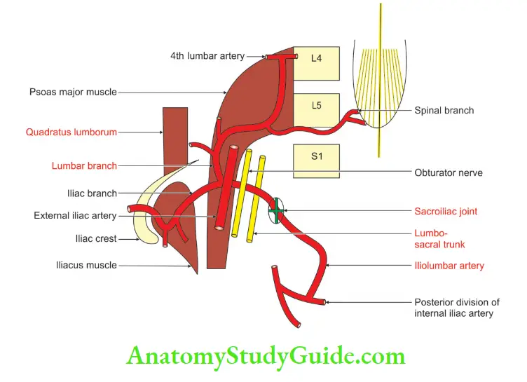
Iliolumbar Artery
1. Origin:
The iliolumbar artery is the 1st branch of the posterior trunk.
2. Course And Relations:
- It lies anterior to the sacroiliac joint and lumbosacral nerve trunk.
- It lies posterior to the obturator nerve and external iliac vessels and reaches the medial border of psoas major.
- It then ascends superolaterally in a recurrent fashion turning sharply backward to the iliac fossa.
3. Iliolumbar Artery Branches:
It divides behind psoas major into
1. Lumbar, and
2. Iliac branches.
3. The lumbar branch supplies psoas major and quadratus lumborum and anastomoses with the 4th lumbar artery.
4. It sends a small spinal branch through the intervertebral foramen.
5. It passes between the 5th lumbar and 1st sacral vertebrae, to supply the cauda equina.
6. The iliac branch supplies iliacus.
7. It anastomoses with the iliac branches of the obturator artery.
8. The site of anastomosis is between iliacus muscle and the ilium bone.
- A large nutrient branch enters an oblique canal in the ileum.
- Other branches run around the iliac crest to supply the gluteal and abdominal muscles.
- They anastomose with the
- Superior gluteal
- Circumflex iliac, and
- Lateral circumflex femoral arteries.
4. Applied Anatomy:
- In case of fracture dislocation of the sacroiliac joints, the iliolumbar artery, which forms an anterior relation of the joint, is torn and bleeds.
- The bleeding from the iliolumbar artery is in the retroperitoneal space, it can be alarming and may cause death.
Leave a Reply