Placenta Introduction
Placenta is a temporary membranous vascular organ that develops in females during pregnancy. It is expelled after childbirth. Placenta forms a link between the fetus and the mother. It is considered an anchor for the growing fetus. It is not only the physical attachment between the fetus and mother but also forms the physiological connection between the two.
Table of Contents
- Placenta is implanted in the wall of the uterus. It is formed from both embryonic and maternal tissues. So, it consists of two parts namely the fetal pad and the mother’s part. It is connected to the fetus by an umbilical cord which contains blood vessels and connective- tissue. The development of the placenta is explained.
Read And Learn More: Medical Physiology Notes
- The delivery of the fetus is followed by the expulsion of the placenta. After the expulsion of the placenta, the umbilical cord is cut. The site of the attachment of the placenta in the center of the anterior abdomen of a fetus is called the navel or umbilicus.
Functions Of Placenta
Nutritive Function
The various nutritive substances, electrolytes, and hormones necessary for the development of the fetus diffuse from the mother’s blood into the fetal blood through the placenta.
Excretory Function
The metabolic end products and other waste products from the fetal body are excreted into the mother’s blood through the placenta.
Respiratory Function
Fetal lungs are nonfunctioning and the placenta forms the respiratory organ for the fetus. Oxygen is necessary for the fetus is received by diffusion from the maternal blood and, carbon dioxide from the fetal blood diffuses into the U-a mother’s blood through the placenta.
Exchange of Respiratory Gases between Fete/ Blood and Maternal Blood
- The exchange of respiratory gases between fetal blood and maternal blood occurs mainly because of the pressure gradient. The partial pressure of oxygen in the maternal blood is 50 mm Hg. In the fetal blood, the partial pressure of oxygen is 30 mm Hg. This pressure gradient of 20 mm Hg causes the diffusion of oxygen into the fetal blood.
- This pressure gradient is very low, compared to the gradient existing between partial pressure of oxygen in arterial blood and alveoli in adults. Still, an adequate quantity of oxygen is available for the fetus.
It is because of two reasons:
- The hemoglobin in fetal blood has 20 times more affinity for oxygen than the adult hemoglobin
- The concentration of hemoglobin is about 50% more in fetal blood than in adult blood.
Bohr’s Effect and Double Bohr’s Effect
- Bohr’s effect is the decrease in the affinity of hemoglobin for oxygen due to increased carbon dioxide tension. When carbon dioxide tension decreases, the affinity of hemoglobin for oxygen is increased. All the metabolic end products including carbon dioxide are completely excreted from the fetus into the maternal blood.
- It develops low partial pressure of carbon dioxide in fetal blood. So, the affinity of fetal hemoglobin for oxygen increases resulting in the diffusion of more amount of oxygen from the mother’s blood into fetal blood.
- At the same time, because of the entrance of fetal carbon dioxide into maternal blood, the partial pressure of carbon dioxide is very high in the mother’s blood.
- It decreases the affinity of the mother’s hemoglobin for oxygen resulting in the diffusion of more amount of oxygen into the fetal blood. Double Bohr’s effect is the operation of Bohr’s effect in both fetal blood and maternal blood.
Endocrine Function
Hormones secreted by the placenta are:
- Human chorionic gonadotropin
- Estrogen
- Progesterone
- Human chorionic somatomammotropin
- Relaxin.
1. Human Chorionic Gonadotropin
Human chorionic gonadotropin (hCG) is a glycoprotein. Its chemical structure is similar to that of LH.
Actions of hCG
- On corpus luteum: hCG is responsible for the preservation and the secretory activity of corpus luteum. Progesterone and estrogen secreted by the corpus luteum are essential for the maintenance of pregnancy. Deficiency or absence of hCG during the first two months of pregnancy leads to termination of pregnancy (abortion), because of involution of corpus luteum.
- On fetal testes: The action of hCG on fetal testes is similar to that of LH in adults. It stimulates the interstitial cells of Leydig and causes the secretion of testosterone. Testosterone is necessary for the development of sex organs in the male fetus.
2. Estrogen
Placental estrogen is similar to ovarian estrogen in structure and function.
Actions of placental estrogen
- On uterus: Causes enlargement of the uterus so that, the growing fetus can be accommodated
- On breasts: Responsible for the enlargement of the breasts and growth of the duct system in the breasts
- On external genitalia: Causes enlargement of the female external genitalia
- On pelvis: Relaxes pelvic ligaments. It facilitates the passage of the fetus through the birth canal at the time of labor.
3. Progesterone
Placenta! Progesterone is similar to ovarian progesterone In structure and function.
Aotmnn of placenta! progesterone
- On endometrium of the uterus: Accelerates the prolate and development of decidual cells in the sr. momentum of the uterus. The decidual cells are responsible for the supply of nutrition to the embryo in the early stage
- On the movements of the uterus: Inhibits the contraction of muscles in the pregnant uterus. It is an important function of progesterone as it prevents the expulsion of the fetus during pregnancy
- On breasts: Causes enlargement of breasts and growth of duct system of the breasts.
Progesterone is responsible for the further development and preparation of mammary glands for lactation.
4. Human Chorionic Somatomammotropin (HCS)
- Human chorionic somatomammotropin (HCS) is a protein hormone secreted from the placenta. It is often called placental lactogen. It acts like prolactin and growth hormone secreted from the pituitary.
- So, it is believed to act on mammary glands and enhance the growth of the fetus by influencing metabolic activities. It increases the amount of glucose and lipids in the maternal blood which are transferred to the fetus.
Actions of HCS
- On breasts: In experimental animals, administration of HCS causes enlargement of mammary glands and induces lactation. That is why, it is named mammotropin. However, the action of this hormone on the breasts of pregnant women is not known
- On protein metabolism: HCS acts like GH on protein metabolism. It causes anabolism of proteins and accumulation of proteins in the fetal tissues. Thus, the growth of the fetus is enhanced
- On carbohydrate metabolism: It reduces the peripheral utilization of glucose in the mother leading to the availability of large quantities of glucose to the growing fetus
- On lipid metabolism: It mobilizes fat from the adipose tissue of the mother. A large amount of free fatty acid is made available as a source of energy in the mother’s body. It compensates for the loss of glucose from the mother’s blood to the fetus.
5. Relax in
Relaxin is a polypeptide that is secreted by the corpus luteum. It is also secreted in large quantities by the placenta and mammary glands at the time of labor.
Fetoplacental Uhit
The fetoplacental unit refers to the interaction between the fetus and the placenta in the formation of steroid hormones.
- The interaction between the fetus and placenta occurs because, some of the enzymes involved in steroid synthesis present in the fetus are absent in the placenta and, those enzymes which are absent in the fetus are present in the placenta.
- Due to this interaction during the synthesis of steroid hormones, the fetus and placenta are together called the fetoplacental unit.
Functions Of Fetoplacental Unit
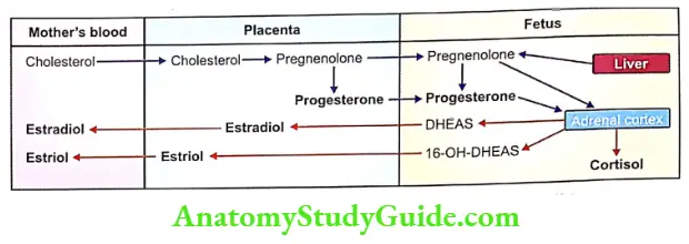
Placenta and fetus interact with each other in the synthesis of steroid hormones in the following manner:
- Cholesterol, which is the precursor for steroid hormones, is obtained by the placenta from the mother’s blood
- Placenta synthesizes pregnenolone from cholesterol
- From pregnenolone, progesterone is formed
- Some amount of the pregnenolone from the placenta enters the fetus. The fetal liver also produces a small quantity of pregnenolone
- The pregnenolone from the placenta and fetal liver forms the substrate for the formation of two substances in the adrenal gland of the fetus
- Dehydroepiandrosterone sulfate (DHEAS)
- (-I6-OH-DHEAS). Some of the DHEAS is also hydroxylated into 16- OH-DHEAS in the fetal liver
- DHEAS and 16-OH-DHEAS are transported back into the placenta to form estrogen
- Estradiol is synthesized from DHEAS and estriol from 16-OH-DHEAS. These two forms of estrogen enter the mother’s blood
- Some amount of the progesterone enters the fetus from the placenta,
- From this progesterone, cortisol, and corticosterone are synthesized in fetal adrenal glands.
Leave a Reply