Image Receptors X-Ray Film Intensifying Screens And Grids And Image Characteristics Short Notes
Question 1. Describe the composition of dental X-ray film.
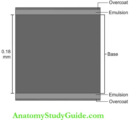
Answer: X-ray film is an image receptor and has four basic components:
Table of Contents
- Base
- Adhesive layer
- Emulsion
- Protective layer
Read And Learn More: Oral Medicine and Radiology Question And Answers
1. Base: The film base is 0.2 mm thick and is made up of flexible plastic (polyethylene terephthalate). It is transparent and exhibits a slight bluish tint to emphasize contrast and increase image quality. The base supports the emulsion and increases its strength. It also serves to tolerate heat, moisture, and chemical changes during processing.

2. Adhesive layer: A thin, transparent adhesive is added on the film base before the application of emulsion and functions to attach the emulsion on either side of the base.
3. Film emulsion: The emulsion is a coating attached by an adhesive layer on either side of the film. It is a homogenous blend of gelatine and silver halide crystals.
- Gelatine: It is used to suspend and disperse silver halide crystals over the film base. During film processing, gelatine absorbs the processing solutions and allows the chemicals to react with silver halide crystals.
- Silver halide crystals: Silver halide crystals are sensitive to X-rays and visible light. They are composed primarily of silver bromide and a lesser amount of silver iodide. The size of iodide crystals are larger than bromide hence their addition disturbs the regularity of bromide crystal structure and increases its sensitivity to X-ray radiation.
- The photosensitivity of silver halide crystals is further increased by the addition of small amounts of trace materials like sulfur or gold.
- The silver halide grains are flat, and tabular with a mean diameter of 1.6 pm in E speed film and are globular with 1 pm size in D speed film.
4. Protective layer: It is a thin, transparent coating placed over the emulsion. It protects the surface from manipulation, mechanical and processing damage.
Question 2. List and explain different types of intra-oral film.
(or)
List and explain different types of nonscreen films.
Answer: Intraoral film otherwise called the nonscreen film is used to capture the image of teeth and supporting structures. Three types of film are available which are as follows:
1. Periapical film: Peri = “around” apex = “terminal end of a tooth root” This film image is used to see the entire tooth and the supporting structures.
Radiology Film
Three sizes are available:
- Size 0: Smallest available size and is used for children. (22 x 35 mm)
- Size 1: To examine the anterior teeth in adults. (24 x 40 mm)
- Size 2: “Standard size adult film”—to examine anterior and posterior teeth in adults. (31 x 41 mm)
2. Bitewing film: It is used to examine the mandibular and maxillary posterior teeth crowns in one film. It has a “wing” or “tab” The patients bite on the wing to stabilize the film.
The indication for bitewing film is: To examine
- Interproximal surfaces (proximal caries)
- Crestal bone pattern and height.
Three sizes are available:
- Size 0: For posterior teeth in small children
- Size 2: For posterior teeth in adults and is a commonly used type
- Size 3: Used only for bitewing images. This film shows all the posterior teeth on one side of the arch in one film.
3. Occlusal film: The film is so named because the patient occludes or bites on the entire film during exposure. It is used to examine the larger areas of the maxilla or mandible. The film size is 57 x 76 mm.
Question 3. Intraoral film packet.
Answer:
The intraoral radiographic films are placed in a packet with ancillary materials to protect it from light and moisture. The film with its surrounding packaging is referred to as a “film packet” and is made up of four separate items:
- X-ray film: The dental radiographic film is a double emulsion film. A small, raised dot known as an identification dot is located on one corner of the film which is used to determine film orientation (right and left sides of the patient) after processing.
- Paper film wrapper: It is a protective sheet that covers the film and shields the film from light.
- Lead foil sheet: A single piece of lead foil is located behind the film in the packet. This sheet shields the film from backscattered radiation and prevents film fog.
- Outer wrapping: Soft vinyl or paper wrapper seals the film, protective paper, and lead foil sheet. This wrapper protects the film from exposure to light and saliva.
The outer wrapper of the film packet has two sides:
- Tube side: The tube side is white with a raised dot on one corner that corresponds to the identification dot on the film.
- Label side: Label side is color coded and contains a clinch. The packet should be unsealed only in the darkroom.
Radiology Film
Question 4. Describe film sensitivity.
(or)
Briefly explain film speed.
Answer:
- Film sensitivity or film speed determines the amount of radiation and exposure time to produce an image on the film with standard density. The silver halide crystals’ size, emulsion thickness, and radiosensitive dyes decide the film speed.
- Film speed is expressed as a reciprocal of the exposure needed to produce an optical density of 1. A fast film requires less exposure to produce a density of 1.
- Fast film: More sensitive films require fewer mAs and have greater film speed hence called fast films. The larger the silver halide crystals, the faster will be the film. Currently, group E film (Ektaspeed) and group F film are in use.
- Slow film: A film that requires more mAs are less sensitive to radiation and are called as slow film. Slow films are not in manufacture today. Film speed is slightly increased by increasing the temperature of the processing solution, but this may cause fog and graininess on film. A depleted processing solution slows down the effective speed.
Question 5. Write briefly about the extraoral film.
(or)
Write in brief about screen film.
Answer:
The majority of extraoral films are screen films. A screen film is one that uses indirect exposure. The energy of the X-ray beam is converted into light by intensifying screens. Intensifying screens reduce the X-ray dose to the patient.
- The film is sandwiched between the two intensifying screens in a cassette.
- When the cassette is exposed to X-rays, the screens convert the X-ray energy to light, which in turn exposes the film. The film is sensitive to fluorescent light rather than radiation.
- Silver halide crystals are sensitive to ultraviolet and blue light hence they are sensitive to screens that emit UV light, green, and blue light (300-500 nm). The appropriate screen film combination is mandatory.
- High contrast medium speed films are suitable for skull view, and less contrast wider latitude films are used for cephalo-metric radiography.
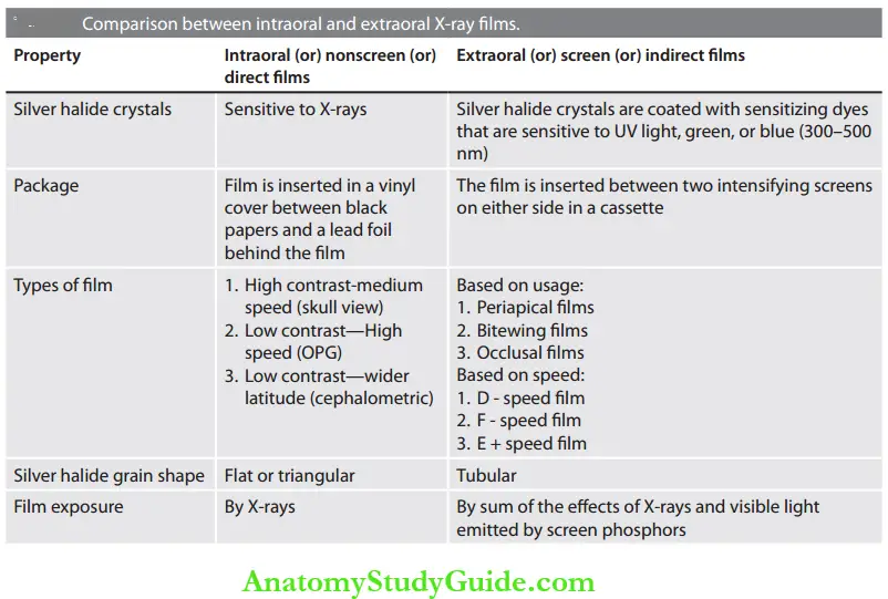
Question 6. Discuss the differences between screen and nonscreen films.
(or)
Discuss the differences between intraoral and extra-oral films.
Answer: The properties of intraoral and extraoral X-ray films differ in terms of sensitivity and shape of the silver halide crystals, type of package, film speed, and exposure.

Question 7. Describe the composition and uses of intensifying screens.
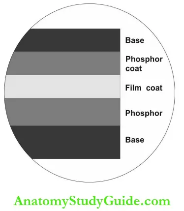
Answer: Intensifying screen is a device to convert X-ray energy to light energy.
- Intensifying screens Principle: X-rays cause certain materials like phosphors to fluoresce (emit UV or visible light radiation). This ability of phosphors to fluoresce when excited by X-rays makes the intensifying screens possible.
- Intensifying screens Composition: Base, phosphor layer, and protective plastic coat.

Intensifying screens Base:
- About 0.25 mm thick polyester plastic
- Provides mechanical support for the film
- It may also serve as a reflecting layer— reflects light emitted by the screen back to the film.
Intensifying screens Phosphor Layer:
- Composed of radiation-sensitive phosphorescent crystals supported in plastic material.
- Phosphor crystals are made up of rare earth elements like lanthanum and gadolinium with the addition of trace amounts of nio¬bium, thulium or terbium to increase the fluorescence (rare earth screens).
- Most dental extraoral radiography uses a screen-film combination having a speed of 250 or faster.
Radiology Film
Intensifying screens Coat: Up to 8 pm thick plastic is coated over phosphor to protect the phosphor layer and provide a surface that can be cleaned.
Intensifying screens Function:
- The presence of intensifying screens makes films 10-60 times more sensitive to X-rays than when they are used alone.
- Significant reduction in radiation dose to the patient.
Question 8. Describe the composition and functions of the grid.
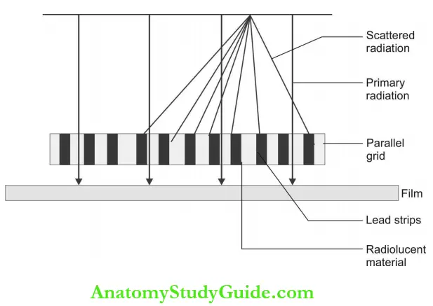
Answer: A grid is a material that reduces the scatter radiation from reaching radiographic film during exposure. It is employed in extraoral radiographic procedures to minimize the film fog caused by scatter rays and to increase the image contrast.
Grid Composition: A grid is composed of alternating strips of radiopaque (lead) and radiolucent (plastic) material.

Types of Grid:
- Focused grid
- Moving or Bucky grid
- Criss-cross grid
- Parallel grid
The grid is placed between the patient and the film. During exposure, the grid permits the passage of X-rays only between two lead stripes. Secondary photons generated in the subject are scattered toward the film, and are absorbed by the radiopaque material of the grid.
- Focused grid: In the focused grid, the lead strips are inclined gradually as they move from the center. Lines through each of these strips converge to a point and are hence known as a focused grid.
- Moving grid: Moves perpendicular to the direction of the grid lines during exposure.
- Criss-cross grid: Criss-cross grid is one in which two grids are sandwiched together so that the strips are both parallel and right angle to the long axis of the grid. Crossed grid removes more scattered radiation.
- Parallel grid: In parallel (or) linear grid all strips are vertical and arranged parallel to each other and also to the X-ray tube. The use of a parallel grid is recommended when the tilted tube technique is used.
- Grid lines: Grids are manufactured with varying numbers of line pairs of lead and plastic materials per inch. Grids with 80 or more line pairs are preferred because they do not show the grid lines on the image.
- Grid ratio: It is the ratio of the heights of the lead strips to the width of the radiolucent spacer. The higher the grid ratio more effective will be the removal of secondary photons. The various grid ratios available are 5:1, 6:1, 8:1, 10:1, and 12:1. Grid ratio of 8 or 10 is preferred.
Radiology Film
Question 9. Describe the Bucky grid.
(or)
Potter-Bucky grid.
Answer:
- The grid is an apparatus used in extraoral radiographic procedures to minimize the scatter rays and thereby decrease the film fog and enhance the image contrast. A grid is made by placing alternating strips of radiopaque (lead) and radiolucent (plastic) material.
- It is placed between the patient and the film. During exposure, the grid permits the passage of X-rays only through the radiolucent stripes. When secondary photons generated in the subject are scattered toward the film, they are absorbed by the radiopaque material of the grid.
- But the image of the radiolucent grid lines may be visible on the processed radiograph and interfere with diagnostic quality.
- To prevent the formation of a radiolucent grid line image, a special type of moving grid is used. The grid moves perpendicular to the direction of the grid lines during exposure and this movement has the blurring-out effect on radiolucent lines and does not interfere with absorption of scattered photons.
- The apparatus used for moving the grid is called as bucky and hence the name Bucky grid.
Question 10. Describe the label side of the film packet.
Answer:
The label of the packet contains a color-coded clinch that should be unsealed in the dark room. When the film is placed in the mouth, the color-coded side of the packet must face the tongue or palate.
The information carried by the label side includes:
- A circle that corresponds to the raised identification dot on the film
- The film speed
- The number of the films is enclosed.
Question 11. Describe the film duplication procedure.
Answer:
Armamentarium:
- Film duplicator—commercially available light source which provides diffuse light that in turn evenly exposes the duplicating film
- Duplicating film
- Films to be duplicated
- Darkroom.
Film duplication Procedure:
- Dental radiographs to be duplicated (original image) is placed on the light screen of the film duplicator by using film organizers.
- On the top of these radiographs, dupli¬cating films should be arranged with their emulsion side facing the image. To avoid blurring, good contact should be established between the original and dupli¬cating film.
- The film duplicator lid should be closed properly without disturbing the film’s position.
- After selecting the exposure time, the light source is activated to expose the duplicating film.
- The exposed film is then processed either by manual or automatic processing technique.
- The processed duplicate radiograph is then labeled with the patient’s name, right or left side, and date.
Question 12. What is the characteristic cure?
(or)
Explain H and D curves.
Answer:
- Hunter and Driffield introduced characteristic curves. They are constructed by taking a series of exposures of a certain film type, developing the film, and reading the density by use of a densitometer and plotting the density reading against a known exposure.
- In a typical H and D curve, there will be great variations in exposure at high and low levels which result in small changes in density.
- The characteristic curve will give useful information about the speed, contrast, and latitude of the film.
Question 13. Describe how intensifying screens reduce radiation dose.
Answer:
- A pair of intensifying screens absorb 60% of X-ray photons that reach the cassette after exposing the patient.
- The phosphor in the screen is efficient in converting X-ray photons to visible light.
- Each absorbed X-ray photon is converted into 4,000 low-energy, visible light (blue or green) photons which then expose the film. 1 X-ray photon = 4,000 visible light photons.
Question 14. What are intraoral measurement grids?
Answer:
- Intraoral measurement grids are used to quantify and evaluate bone levels. They are commercially available that superimpose thin radiopaque or radiolucent lines in the vertical or horizontal planes in 1-mm gradations.
- The marking grid is incorporated into the film packet or it is placed in front of the film packet during exposure. No increase in exposure is necessary, and the films are processed without changing the parameters.
- The measurement or marking grid should not be confused with grids used in extraoral radiography to absorb object scatter.
Question 15. What is radiographic noise?
Answer:
Radiographic noise is the appearance of the uneven density of the uniformly exposed film. It is seen in small areas of the film as localized variation in density. The primary causes of noise are radiographic mottle and radiographic artifacts.
- Radiographic mottle: Uneven densities resulting from the physical structure of the film and intensifying screens.
- Radiographic artifacts: Defects caused by errors in film handling, for example, fingerprints, bends on film, marks, or scratches.
In intraoral films, mottle may be seen as film graininess that is caused by the visibility of silver grains on the image when a high-temperature processing solution is used.
Image Receptors X-Ray Film Intensifying Screens And Grids And Image Characteristics Viva Voce
Question 1. Define radiograph.
Answer:
- A radiograph is a two-dimensional represen¬tation of a three-dimensional object. In conventional mode, a radiograph is an image produced by exposing the radiographic film to X-rays and then processing it to get a visible image.
- Radiopaque and radiolucent are the terms used to describe the appearance of all structures seen on a radiograph.
Question 2. What is a duplicate radiograph?
Answer: A duplicate radiograph is one that is identical to the original X-ray film and is mainly used for patient referrals to specialists, insurance claims, and for teaching-learning purposes.
Question 3. What is the duplicating film?
Answer: It is a type of photographic film used to make an identical copy of a radiograph (both intraoral and extraoral).
- The key differences between X-ray film and duplicating film:
- The duplicating film should not be exposed to X-rays and have to be used in a darkroom setting.
- The duplicating film should contain emulsion on one side only.
- When exposed to a longer period of light, the duplicating film appears lighter in contrast to X-ray film which appears darker on prolonged exposure to radiation.
- The emulsion-coated side seems dull and the other side appears shiny.
Duplicating films are available in three sizes:
- 5 x 12 inch,
- 6 x 12 inches, and
- 8 x 10 inches.
Question 4. What is two film packet in dental radio-logy?
Answer:
- An intraoral X-ray film packet may contain two films and produces two indistinguishable radiographs when exposed using the same exposure parameters required to produce a single radiograph.
- The two-film pocket is used when a duplicate record of a radiographic image is required (patient referral, insurance claims, etc.).
Question 5. What is film latitude?
Answer:
- Film latitude is the ability to distinguish different densities on a film. It refers to the range of exposures (mAs) to which an X-ray film will respond with a diagnostically acceptable range of densities (range from 0.5 to 2.5).
- Wide latitude film record a wide range of subject contrast but have lower contrast (long grayscale). High kVp produces an image with wide latitude and low contrast.
Question 6. What is radiographic noise?
Answer: Radiographic noise is the appearance of uneven densities on a uniformly exposed film.
Question 7. What is the purpose of using lead foil in intraoral films?
Answer:
- The lead foil placed in the back of the film prevents the radiation backscattered by the tissues from reaching the film and reduces film fog.
- It absorbs some of the radiation after they pass they penetrate the tissues and expose the film hence reducing secondary radiation to an extent.
- Lead foil contributes to the rigidity of the packet.
Question 8. Why intensifying screens are used in intraoral films?
Answer: The use of intensifying screens reduces the “resolution” of the image. So, we cannot get fine details.
Question 9. Why grids are not used in intraoral and panoramic radiography?
Answer:
- The function of the grid is to reduce the amount of scattered radiation originating in the object and thereby improve the contrast.
- In intraoral radiography, secondary radia¬tion does not lower the image quality due to small field size, hence do not require grids.
- In panoramic radiography, the usage of a small exposure field size does not require a grip
Question 10. What is radiographic blurring?
Answer: It is image unsharpness caused by:
- Image receptor blurring: Slow-speed films have fine silver grains hence fine sharpness. Faster films have larger grains and less sharp images.
- Motion blurring: Image unsharpness is due to the movement of the film, subject or X-ray source during exposure.
- Geometric blurring: Loss of image sharp¬ness occur due to larger focal spot size, reduced distance between the focal spot and an object, and increased distance between the film and object.
Question 11. What is parallax?
Answer:
- The image on each side of the double emul¬sion film will cause some loss of image unsharpness. Dental films are double-side emulsion coated, and the beam is divergent hence some unsharpness occurs when the image is recorded.
- Parallax is the apparent change in position or size of the subject when it is viewed from different sides of the film. In intraoral images, the effect of parallax is more apparent when wet films are viewed.
Image Receptors X-Ray Film Intensifying Screens And Grids And Image Characteristics Highlights
- Image receptors are materials used to capture and store images in radiography. In dentistry, film, film screen combinations are widely used. However, electronic sensors are used in the digital imaging system.
- Radiographic films are comparable to photographic films that are adapted for dental use. Dental radiology uses non screen films for intraoral purposes and screened films for extraoral radiography.
Leave a Reply