Describe the Rectus Sheath under the following headings:
- Rectus Sheath Definition
- Rectus Sheath Formation
- Rectus Sheath Contents and
- Rectus Sheath Applied anatomy
Answer:
1. Rectus sheath Definition:
It is an aponeurotic sheath enclosing the rectus abdominis muscle (and pyramidal if present). It has two walls: anterior and posterior.
Read And Learn More: Anatomy Question And Answers
2. Rectus sheath Formation:
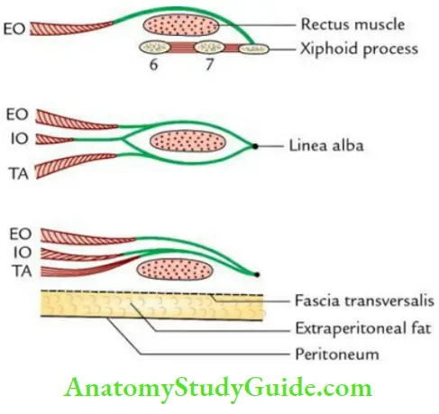
- It is formed by the aponeurotic layers of the external oblique, internal oblique, and transversus abdominis muscles.
- The formation of rectus sheath differs in its upper, middle, and lower one thirds

In the upper 1 /3rd (i.e. above the costal margin):
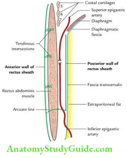
Anterior wall is formed by the external oblique (EO) aponeurosis alone. The posterior wall is absent. (Rectus muscle lies directly on the 5th, 6th, and 7th costal cartilages.)
In the middle 1 /3rd (i.e. between the costal margin and arcuate line):
- Anterior wall is formed by the aponeurosis of external oblique and the anterior lamina of the aponeurosis of the internal oblique.
- The posterior wall is formed by the aponeuroses of the transversus abdominis and the posterior lamina of the internal oblique.
In the lower 1 /3rd (i.e. below the arcuate line):
- Anterior wall is formed of the aponeuroses of the external oblique, internal oblique, and the transversus abdominis muscles.
- The posterior wall is absent. (Rectus muscle lies directly on the fascia transversalis).
Note: Arcuate line: The posterior wall of rectus sheath ends midway between umbilicus and pubic symphysis forming an arcuate line/linea semilunaris/fold of Douglas. This line is concave downward.
3. Rectus sheath Contents:
- 2 muscles: Rectus abdominis and pyramidalis (if present).
- 2 arteries: Superior and inferior epigastric arteries.
- 2 veins: Superior and inferior epigastric veins.
- 6 nerves: Terminal parts of lower 5 intercostal nerves and a subcostal nerve.

Note: The vessels and nerves lie posterior to the rectus abdominis muscle.
4. Rectus sheath Functions:
- It helps in maintaining the strength of the anterior abdominal wall.
- It prevents the recti abdominis from bowstringing during contraction of the anterior abdominal wall.
5. Rectus sheath Applied anatomy:
- The aponeurotic sheaths of the right and left recti fuse in the midline to form linea alba. In multiparous women, the upper part of linea alba gets stretched and gap leading to the divarication of recti abdominis.
- Knowledge of rectus sheath and its contents help the surgeon to do laparotomy by giving a paramedian incision, without cutting the rectus abdominis muscle.
Leave a Reply