Red Blood Cells Introduction
- Red blood cells (RBCs) are the non-nucleated formed elements in the blood. Red blood cells are also known as erythrocytes (erythros = red).
- The red color of the red blood cell (RBC) is due to the presence of the coloring pigment called hemoglobin. RBCs play a vital role in the transport of respiratory gases.
- RBCs are larger in number compared to the other two blood cells namely white blood cells and platelets.
Normal Value
The RBC count ranges between 4 and 5.5 million per cu mm of blood. In adult males, it is 5 million/cu mm and in adult females, it is 4.5 million/cu mm.
Table of Contents
Read And Learn More: Medical Physiology Notes
Morphology Of Red Blood Cells
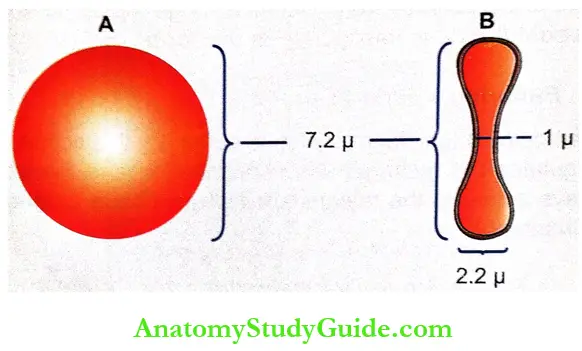
1. Normal Shape: Normally, the RBCs are disk-shaped and biconcave (dumbbell-shaped). The central portion is thinner and the periphery is thicker. The biconcave contour of RBCs has some mechanical and functional advantages.
Advantages of Biconcave Shape of RBCs
- It helps in equal and rapid diffusion of oxygen and other substances into the interior of the cell
- Large surface area is provided for the absorption or removal of different substances
- Minimal tension is offered on the membrane when the volume of cell alters
- Because of the biconcave shape, while passing through minute capillaries, RBCs squeeze through the capillaries very easily without getting damaged.
2. Normal Size
Diameter: 7.2 μ (6.9-7.4 μ).
Thickness: At the periphery, it is thicker with 2.2 μ and at the center it is thinner with 1 μ. The difference in thickness is because of the biconcave shape.
Surface area: 120 sq μ.
Volume: 85-90 cup.
3. Normal Structure
- RBCs are non-nucleated formed elements in the blood. The only mammal which has nucleated RBC is a camel.
- Because of the absence of a nucleus in human RBC, DNA is also absent.
- Other organelles such as mitochondria and Golgi apparatus also are absent in RBC. Because of the absence of mitochondria, the energy is produced from the glycolytic process.
- Red cell does not have insulin receptor and so the glucose uptake by this cell is not controlled by insulin.
- RBC has a special type of cytoskeleton which is made up of actin and spectrin.
- Both proteins are anchored to transmembrane proteins by means of another protein called ankyrin. The absence of spectrin results in hereditary spherocytosis.
- In this condition the cell is deformed, losses its biconcave shape and becomes globular (spherocytic). The spherocyte is very fragile and easily ruptured (hemolyzed) in hypotonic solutions.
Properties Of Red Blood Cells
1. Rouleaux Formation: When blood is taken out of the blood vessel, the RBCs pile up one above another like the pile of coins. This property of the RBCs is called rouleaux (pleural = rouleau) formation. It is accelerated by plasma proteins globulin and fibrinogen.
2. Specific Gravity: The specific gravity of RBC is 1.092 to 1.101.
3. Packed Cell Volume: Packed cell volume (PCV) is the proportion of blood occupied by RBCs expressed in percentage. It is also called the hematocrit value. It is 45% of the blood and the plasma volume is 55%.

4. Suspension Stability: During circulation, the RBCs remain suspended uniformly in the blood. This property of the RBCs is called suspension stability.
Lifespan Of Red Blood Cells
The average lifespan of RBC is about 120 days. After the lifetime the senile (old) RBCs are destroyed in reticuloendothelial system.
Determination of Lifespan of Red Blood Cells: The lifespan of the RBC is determined by the radioisotope method. The RBCs are tagged with radioactive substances like radioactive iron or radioactive chromium. The life of RBC is determined by studying the rate of loss of radioactive cells from circulation.
Fate Of Red Blood Cells
- When the cells become older (120 days), the cell membrane becomes more fragile. The diameter of the capillaries is less or equal to that of RBC. The younger RBCs can pass through the capillaries easily.
- However, because of its fragile nature, the older cells are destroyed while trying to squeeze through the capillaries.
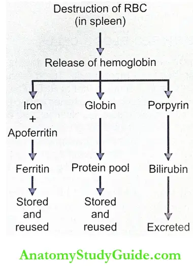
- The destruction occurs mainly in the capillaries of red pulp of spleen because the diameter of the splenic capillaries is very small. So, the spleen is called graveyard of RBCs.
- The destroyed RBCs are fragmented and hemoglobin is released from the fragmented parts. Hemoglobin is immediately phagocytized by macrophages of the body particularly the macrophages present in liver (Kupffer cells), spleen, and bone marrow.
- Hemoglobin is degraded into iron, globin, and porphyrin. Iron combines with the protein called apoferritin to form ferritin, which is stored in the body and reused later.
- Globin enters the protein depot for later use. The porphyrin is degraded into bilirubin which is excreted by the liver through bile.
- Daily 10% RBCs, which are senile, are destroyed in normal young healthy adults.
- It causes release of about 0.6 g/dl. of hemoglobin into the plasma. From this 0.9 to 1.5 mg/dL bilirubin is formed.
Functions Of Red Blood Cells
The major function of RBCs is the transport of respiratory gases. The functions of RBCs are:
1. Transport of Oxygen from the Lungs to the Tissues: Hemoglobin in RBC combines with oxygen to form oxyhemoglobin. About 97% of oxygen is transported in blood in the form of oxyhemoglobin.
2. Transport Carbon Dioxide from the Tissues to the Lungs:
- Hemoglobin combines with carbon dioxide and form carbhemoglobin. About 30% of carbon dioxide is transported in this form.
- RBCS contains a large amount of carbonic anhydrase. This enzyme is necessary for the formation of bicarbonate from water and carbon dioxide.
- Thus, it helps to transport carbon dioxide in the form of bicarbonate from tissues to lungs. About 63% of carbon dioxide is transported in this form.
3. Buffering Action in Blood: Hemoglobin functions as a good buffer. By this action, regulates the hydrogen ion concentration and thereby plays a role in the maintenance of acid-base balance.
4. In Blood Group Determination: RBCS carry the blood group antigens like A antigen. B antigen and Rh factor. This helps in determination of blood group and enables to prevention of reactions due to incompatible blood transfusion.
Variations In the Number Of Red Blood Cells
Physiological Variations
1. Increase in RBC Count: Increase in the RBC count is known as polycythemia. It occurs in both physiological and pathological conditions. When it occurs in physiological conditions it is called physiological polycythemia. The increase in number during this condition is marginal and temporary. It occurs in the following conditions:
- Age: At birth, the RBC count is 8-10 millions/cu mm of blood. The count decreases within 10 days after birth due to the destruction of RBCs causing physiological jaundice in some newborn babies. However, in infants and growing children, the cell count is more than the value in adults.
- Sex: Before puberty and after menopause in females the RBC I count is similar to that in males. During the reproductive period of females, the count is less than of males (4.5 millions/cu mm).
- High Altitude: Inhabitants of mountains (above 10,000 feet from mean sea level) have an increased RBC count of more than 7 millions/cu mm. It is due to hypoxia (decreased oxygen supply to tissues) in high altitudes. Hypoxia stimulates kidney to secrete a hormone called erythropoietin. The erythropoietin in turn stimulates the bone marrow to produce more RBCs.
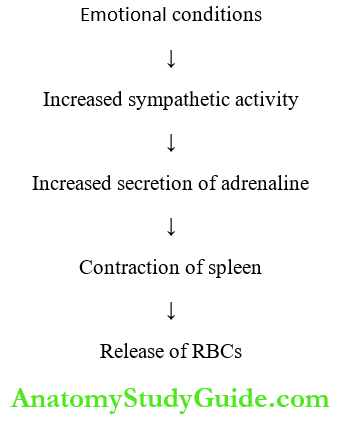
- Muscular Exercise: There is a temporary increase in RBC count after exercise. It is because of mild hypoxia and contraction of spleen. Spleen stores RBCs. Hypoxia increases sympathetic activity resulting in the secretion of adrenaline from the adrenal medulla. Adrenaline contracts spleen and RBCs are released into blood.
- Emotional Conditions: The RBC count increases during the emotional conditions such as anxiety. It is because of increase in sympathetic activity as in the case of muscular exercise.
- Increased Environmental Temperature: The increase in the atmospheric temperature increases RBC count. Generally increased temperature increases all the activities in the body including production of RBCs.
- After Meals: There is a slight increase in the RBC count after taking meals. It is because of the need for more oxygen for metabolic activities.

2. Decrease in RBC Count: Decrease in RBC count occurs in the following physio-logical conditions:
- High Barometric Pressures: At high barometric pressures as in deep sea, when the oxygen tension of blood is higher, the RBC count decreases.
- During Sleep: The RBC count decreases slightly during sleep and immediately after getting up from sleep. Generally, all the activities of the body are decreased during sleep including the production of RBCs.
- Pregnancy: In pregnancy, the RBC count decreases. It is because of increase in ECF volume. An increase in ECF volume, increases the plasma volume also resulting in hemodilu- tion. So, there is a relative reduction in the RBC count.
Pathological Variations – Pathological Polycythemia: Pathological polycythemia is the abnormal increase in the RBC count. The red cell count increases above 7 million/cu mm of the blood. Polycythemia is of two types, primary polycythemia, and secondary polycythemia.
- Primary Polycythemia – Polycythemia Vera
- Primary polycythemia is otherwise known as polycythemia vera. It is a disease characterized by persistent increase in RBC count above 14 millions/cu mm of blood.
- This is always associated with increased white blood cell count above 24,000/cu mm of blood. Polycythemia vera occurs in myeloproliferative disorders like malignancy of red bone marrow.
- Secondary Polycythemia: This is secondary to some of the pathological conditions Anisocytes occur in pernicious anemia. (diseases) such as:
- Respiratory disorders like emphysema
- Congenital heart disease
- Ayerza’s disease condition associated with hypertrophy of the right ventricle and obstruction of blood flow to lungs
- Chronic carbon monoxide poisoning
- Poisoning by chemicals like phosphorus and arsenic 6. Repeated mild hemorrhages.
All these conditions lead to hypoxia which stimulates the release of erythropoietin. Erythropoietin stimulates the bone marrow resulting in increased RBC count.
Anemia: The abnormal decrease in RBC count is called anemia.
Variations In Size Of Red Blood Cells
Under physiological conditions, the size of RBCs in venous blood is slightly larger than those in arterial blood. In pathological conditions, the variations in size of RBCs are:
- Microcytes-decrease in size
- Macrocytes-increase in size
- Anisocytosis cells without uniform size.
1. Microcytes: Microcytes are present in
- Iron deficiency anemia
- Prolonged forced breathing
- Increased osmotic pressure in blood.
2. Macrocytes: Macrocytes are present in
- Megaloblastic anemia
- Muscular exercise
- Decreased osmotic pressure in blood.
3. Anisocytes: Anisocytes occurs in pernicious anemia.
Variations In Shape Of Red Blood Cells
The shape of RBCs is altered in many conditions including different types of anemia.
- Crenation: Shrinkage as in hypertonic conditions
- Spherocytosis: Globular form as in hypotonic conditions
- Elliptocytosis: Elliptical shape as in certain types of anemia
- Sickle cell: Crescentic shape as in sickle cell anemia
- Poikilocytosis: Unusual shapes due to deformed cell membrane. The shape will be of flask, hammer, or any other unusual shape.
Variations In Structure Of Red Blood Cells
- Punctate Basophilism; The striated appearance of RBCS by the presence of dots of basophilic materials (porphyrin) is called punctate basophilism. It occurs in conditions like lead poisoning.
- Ring in RBC: Ring or twisted strands of basophilic material appear in the periphery of the RBCs. This is also called the Goblet ring. This appears in the RBCs in certain types of anemia.
- Howell-Jolly Bodies: In certain types of anemia, some nuclear fragments are present in the ectoplasm of the RBCs. These nuclear fragments are called Howell-Jolly bodies.
Leave a Reply