Definition And Significance Of Reflexes
- Reflex activity is the response to a peripheral nervous stimulation that occurs without our consciousness. It is a type of protective mechanism and it protects the body from irreparable damages.
- For example, when the hand is placed on a hot object, it is withdrawn immediately. When a very bright light is thrown into the eyes, eyelids are closed and pupil is constricted to prevent the damage of retina by the entrance of excessive light into the eyes.
Read And Learn More: Medical Physiology Notes
Table of Contents
Reflex Arc
The reflex arc is the anatomical nervous pathway for a reflex action. A simple reflex arc includes five components.
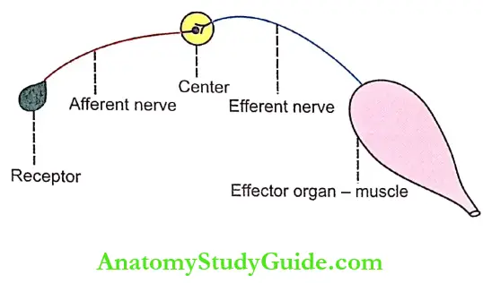
- Receptor: It is the end organ, which receives the stimulus. When the receptor is stimulated, impulses are generated in afferent nerve.
- Afferent Nerve: Afferent or sensory nerve transmits sensory impulses from the receptor to the center.
- Center: The center receives the sensory impulses via afferent nerve fibers and in turn, it generates appropriate motor impulses. The center is located in the brain or spinal cord.
- Efferent Nerve: Efferent or motor nerve transmits motor impulses from the center to the effector organ.
- Effector Organ: The effector organ is the structure such as the muscle or gland where the activity occurs in response to the stimulus.
- Afferent and efferent nerve fibers may be connected directly to the center. In some places, one or more neurons are interposed between these nerve fibers and the center. Such neurons are called connector neurons or internuncial neurons or interneurons.
Classification Of Reflexes
Reflexes are classified by five different methods depending upon various factor as given below:
- Whether inborn or acquired
- Situation of the center
- Purpose – functional significance
- Number of synapse
- Clinical basis.
Depending Upon Whether Inborn Or Acquired
- Unconditioned Reflexes or Inborn Reflexes:
- Unconditioned reflexes are the natural reflexes that are present since the time of birth hence the name inborn reflexes. Such reflexes do not require previous learning, training, or conditioning.
- The best example is the secretion of saliva when a drop of honey is kept in the mouth of a newborn baby for the first time. The baby does not know the taste of the honey but still saliva is secreted.
- Conditioned Reflexes or Acquired Reflexes: Conditioned or acquired reflexes are the reflexes that are developed after conditioning or training. These reflexes are not inborn but acquired after birth. Such reflexes need previous learning, training, or conditioning. An example is the secretion of saliva by the sight, smell, thought, or hearing of a known edible substance.
Depending upon the situation of the center
- Cerebellar Reflexes: Cerebellar reflexes are the reflexes that have the center in the cerebellum.
- Cortical Reflexes: Cortical reflexes are the reflexes that have the center in cerebral cortex.
- Midbrain Reflexes: Midbrain reflexes are the reflexes that have the center in midbrain.
- Bulbar or Medullary Reflexes: Bulbar or medullary reflexes are the reflexes which have the center in medulla oblongata.
- Spinal Reflexes: Reflexes having their center in the spinal cord are called spinal reflexes. Depending upon the segments involved, the spinal reflexes are divided into three groups:
- Segmental spinal reflexes
- Intrasegmental spinal reflexes
- Suprasegmental spinal reflexes.
Depending Upon The Purpose – Functional Significance
- Protective Reflexes or Flexor Reflexes: The protective reflexes are the reflexes that protect the body from nociceptive (harmful) stimuli. These reflexes are also called withdrawal reflexes or flexor reflexes. Protective reflexes involve flexion at different joints hence the name flexor reflexes.
- Antigravity Reflexes or Extensor Reflexes: Antigravity reflexes are the reflexes that protect the body against gravitational force. These reflexes are also called the extensor reflexes because the extensor muscles contract during these reflexes resulting in extension at joints
Depending Upon The Number Of Synapses
- Monosynaptic Reflexes: Reflexes having only one synapse in the reflex arc are called monosynaptic reflexes. The stretch reflex is the best example for a monosynaptic reflex and it is elicited due to the stimulation of muscle spindle.
- Polysynaptic Reflexes: Reflexes having more than one synapse in the reflex arc are called polysynaptic reflexes. Flexor reflexes (withdrawal reflexes) are the polysynaptic reflexes.
Depending Upon Clinical Basis: In clinical practice, the reflexes are classified into four types.
- Superficial reflexes
- Deep reflexes
- Visceral reflexes
- Pathological reflexes
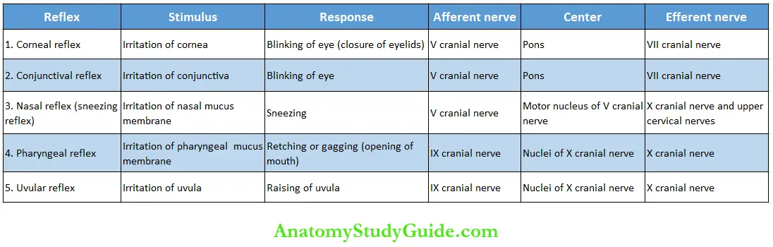
- Superficial Reflexes: Superficial reflexes are the reflexes, which are elicited from the surface of the body. The superficial reflexes are of two types, mucous membrane reflexes, and skin reflexes.
- Mucous Membrane Reflexes: The mucous membrane reflexes arise from the mucus membrane. Details of the mucus membrane reflexes are listed in Table.
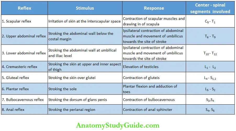
- Cutaneous Reflexes Or Skin Reflexes: Cutaneous reflexes are elicited from the skin by the stimulation of cutaneous receptors. The details of these reflexes are given in Table.
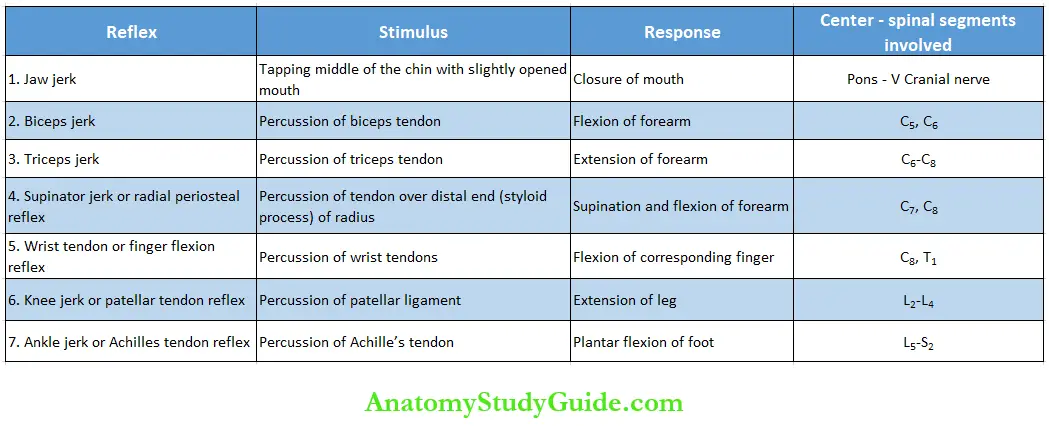
- Deep Reflexes: The deep reflexes are elicited from the deeper structures beneath the skin like tendon. These reflexes are otherwise known as tendon reflexes. The details of these are given in Table.
- Visceral Reflexes: Visceral reflexes are the reflexes arising from the pupil and the visceral organs.
- Visceral reflexes are:
- Pupillary reflexes
- Oculocardiac reflex
- Carotid sinus reflex.
- Pupillary Reflexes: Pupillary reflexes are the reflexes in which, the size of pupil is altered. Pupillary reflexes are
- Light reflex
- Accommodation reflex
- Ciliospinal reflex.
- Light Reflex: When the retina of the eye is stimulated by a sudden flash of light, constriction of pupil occurs. It is called light reflex. It is of two types:
- Direct light reflex: Stimulation of retina in one eye by a flash of light causes constriction of pupil in the same eye
- Indirect or consensual light reflex: Stimulation of the retina in one eye by the flash of light causes simultaneous constriction of pupil in the other eye also.
- Accommodation Reflex
While the eyes are fixed on a distant object, and d another object is brought in front of the eye (near the eye) the vision shifts from far object to near object. During that time some changes occur in the eyes. The changes during accommodation reflex are:- Constriction of pupil,
- Convergence of eyeball
- Increase in anterior curvature of lens.
- Ciliospinal Reflex: Ciliospinal reflex is the dilatation of pupil due to stimulation of skin over the neck.
- Light Reflex: When the retina of the eye is stimulated by a sudden flash of light, constriction of pupil occurs. It is called light reflex. It is of two types:
- Oculocardiac Reflex: It is the reflex in which heart rate decreases due to the pressure applied over eyeball.
- Carotid Sinus Reflex: Carotid sinus reflex is the decrease in heart rate and blood pressure caused by pressure over the carotid sinus in neck due to a tight collar.
- Pupillary Reflexes: Pupillary reflexes are the reflexes in which, the size of pupil is altered. Pupillary reflexes are
- Visceral reflexes are:
- Pathological Reflexes: Pathological reflexes are the reflexes that are elicited only in pathological conditions. Three pathological reflexes are well-known:
- Babinski’s sign
- Clonus
- Pendular movements.
- Babinskfs Sign
- The abnormal plantar reflex is called Babinski’s sign. It is also called Babinski’s reflex or phenomenon. It is named after the discoverer Joseph Babinski.
In the normal plantar reflex, a gentle scratch over the outer edge of the sole of the foot causes plantar flexion and adduction of all toes. But in Babinski’s sign, there is dorsiflexion of great toe and fanning of other toes. - When Babinski’s reflex is present, the condition is commonly called Babinski positive sign and when it is negative, the condition is called Babinski’s negative sign.
- Babinski’s sign is present in the upper motor neuron lesion. Physiological conditions when Babinski’s sign is present are infancy and deep sleep. It is present in infants because of nonmyelination of pyramidal tracts.
- The abnormal plantar reflex is called Babinski’s sign. It is also called Babinski’s reflex or phenomenon. It is named after the discoverer Joseph Babinski.
- Clonus
- Clonus is a series of rapid and repeated involuntary jerky which occur while eliciting a deep reflex. When a deep reflex is elicited in a normal person, the contractions of a muscle or group of muscles are smooth and continuous.
- But clonus occurs when the deep reflexes are exaggerated due to hypertonicity of muscles in pyramidal tract lesion. Clonus is well seen in calf muscles producing ankle clonus and quadriceps producing patella clonus.
- Ankle Clonus:
- Ankle clonus is the rhythmical contractions of calf muscles caused by sudden dorsiflexion of the foot.
- The repeated rhythmical contractions of calf muscles lead to a series of rhythmic plantar flexion at ankle joint. The sudden dorsiflexion of foot is done by supporting the knee of the patient in a slightly flexed position.
- Patellar Clonus: Patellar clonus is the rhythmic jerky movements of patella produced by grasping it between thumb and index finger of the examiner and pushing it down forcibly towards the foot. It is caused by clonic contractions of quadriceps muscle.
- Ankle Clonus:
- Pendular Movements:
- Pendular movements are the slow oscillatory movements (instead of brisk movements) that are developed while eliciting a tendon jerk. Unlike clonus, the pendular movements occur because of the hypotonicity of muscles.
- The pendular movements are very common while eliciting the knee jerk or patellar tendon reflex in patients affected by cerebellar lesions.
- A tap on the patellar tendon, when the leg is hanging freely, causes a brisk extension of the leg due to the contraction of the quadriceps muscle (knee jerk).
- In normal conditions, after the extension, the leg returns back to the resting position immediately. In cerebellar lesions, the leg swings forwards and backwards several times before coming to rest. Such movements are similar to movements of the pendulum of a clock hence the name pendular movements.
- Babinskfs Sign
Properties Of Reflexes
- One-Way Conduction (Bell- Magendie Law): During any reflex activity, the impulses are transmitted in only one direction through the reflex arc as per Bell- Magendie law. The impulses pass from receptors to the center and then from center to effector organ.
- Reaction Time: Reaction time is the time interval between application of stimulus and the onset of reflex. It depends upon the length of afferent and efferent nerve fibers, velocity o- impulse through these fibers and central delay. Central delay is the delay at the synapse. It is also called synaptic delay.
- Summation: The summation in reflex action is of two types.
- Spatial Summation: When two afferent nerve fibers supplying a muscle are stimulated separately with subliminal stimulus, there is no response. But the muscle contracts when both the nerve fibers are stimulated together with the same strength of stimulus. It is called spatial summation.
- Temporal Summation: When one nerve fiber is stimulated repeatedly with subliminal stimuli, these stimuli are summed up to give response in the muscle. It is called temporal summation.
- Thus, both spatial summation and temporal summation play an important role in the facilitation of responses during the reflex activity.
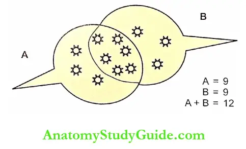
- Occlusion
- It is demonstrated in a flexor reflex involving a muscle, which is innervated by two motor nerves. The nerves can be called as A and B. When both the nerves, A and B nerves are stimulated simultaneously, the tension developed by the muscle is less than the sum of the tension developed when each nerve is stimulated separately.
- For example, if nerve A is stimulated alone, the arbitrary unit of tension developed is 9. If nerve B is stimulated the 9 units of tension developed. So, the sum of tension developed when the nerves A and B are separately stimulated = 9 + 9 = 18 units.
- But, when both A and B are stimulated together, the tension produced is (A+B) = 12 units. Thus, the tension here is less than sum of tension produced when A and B were stimulated separately. This phenomenon is called occlusion. The occlusion is due to the overlapping of the nerve fibers during the distribution.
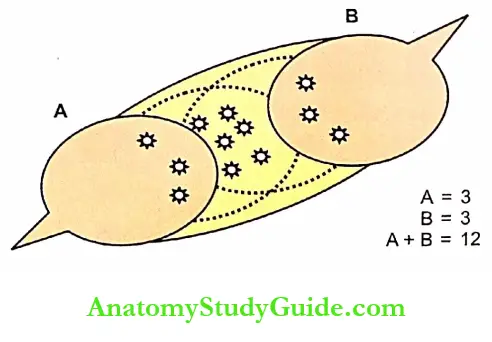
- Subliminal Fringe
- In some reflexes involving the muscle with two nerve fibers, the tension developed by simultaneous stimulation of two nerves is greater than the sum of tension produced by the stimulation of these nerves separately.
- For example, if nerve A is stimulated alone, the arbitrary unit of tension developed by muscle = 3 units. If nerve B is stimulated alone, the tension produced = 3 units. So, the sum of tension developed, I nerves A and B are stimulated separately = 3+3 = 6 units. When both nerves A and B were stimulated together, the tension developed is (A + B) = 12 units.
- Thus, the tension here is greater than the sum of tension produced if A and B are separately stimulated. This phenomenon is called subliminal fringe. It is due to the effect of spatial summation.
- Recruitment
- Recruitment is defined as the successive activation of additional motor units with progressive increase in force of muscular contraction.
- When an excitatory nerve is stimulated for a long time, there is a gradual increase in the response of reflex activities. It is due to the activation of more and more motor neurons. Recruitment is similar to the effect of temporal summation.
The indefinite increase in response does not produce unlimited recruitment. - A plateau is reached. Thus, there is a limit to the number of motor neurons, which are recruited. So, beyond certain limit, the prolongation of stimulation does not increase the response.
- After Discharge
- After discharge is the persistence or continuation of response for some time even after cessation of stimulus. When a reflex action is elicited continuously for some time, and then the stimulation is stopped, the reflex activity (contraction) will be continued for some time even after the stoppage of the stimulus.
- It is because of the discharge of impulses from the center even after the stoppage of stimulus. The internuncial neurons which continue to transmit afferent impulses even after stoppage of stimulus are responsible for after discharge.
- Rebound Phenomenon: The reflex activities can be forceful for some time. But, when the inhibition is suddenly removed, the reflex activity becomes more forceful than before inhibition. It is called the rebound phenomenon. The reason for this state of over-excitation is not known.
- Fatigue: When a reflex activity is continuously elicited for a long time, the response is reduced slowly and at one stage, the response does not occur. This type of failure to give response to the stimulus is called fatigue. The center or the synapse of the reflex arc is the first seat of fatigue.
Reciprocal Inhibition And Reciprocal Innervation
Reciprocal Inhibition
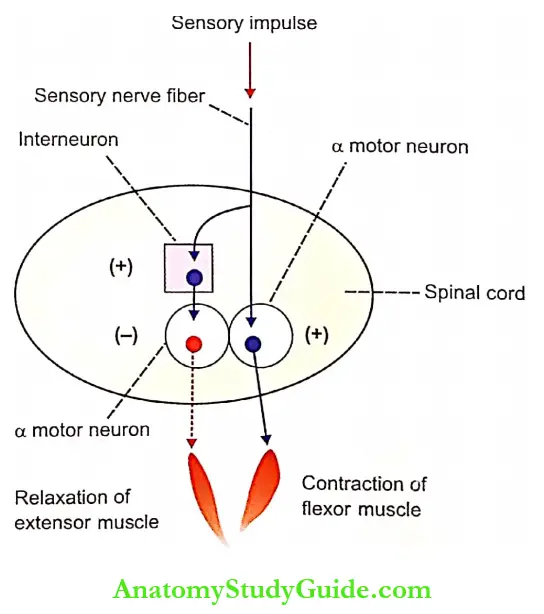
- Reciprocal innervation is one of the important features of both flexor and extensor reflexes. Usually, the excitation of one group of muscles is associated with inhibition of another, i.e. antagonistic group of muscles on the same side.
- For example, when a flexor reflex is elicited, the flexor muscles are excited (contracted) and the extensor muscles are inhibited (relaxed) on that side. This phenomenon is called reciprocal inhibition. Reciprocal inhibition occurs because of reciprocal innervation.
Reciprocal Innervation
- The neural mechanism involved in reciprocal inhibition was postulated by Sherrington. Hence, it is called Sherrington’s law of reciprocal innervation.
- According to this law, reciprocal inhibition is due to the segmental arrangement of afferent and efferent connections in the spinal cord.
- The afferent nerve fibers, which evoke flexor reflex in a limb have connections with motor neurons supplying flexors and the motor neurons supplying the extensors of the same side. The afferent nerve fibers excite the motor neurons supplying flexors.
- Simultaneously, it also inhibits the motor neurons supplying extensors through an interneuron. Accordingly, the flexor muscles contract and extensor muscles relax resulting in flexion of the limb.
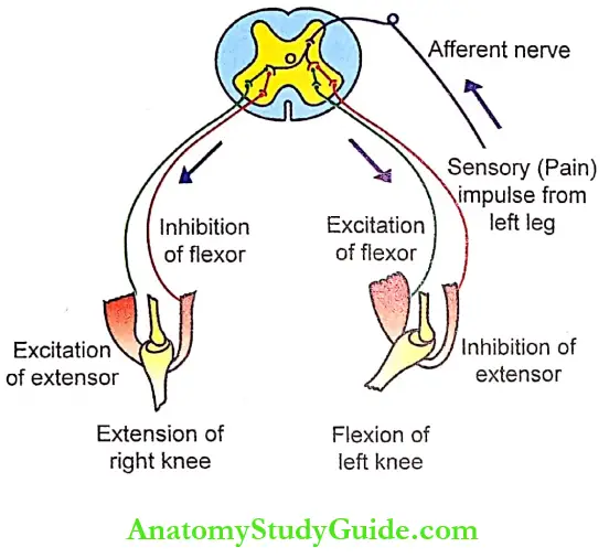
Crossed Extensor Reflex
- Crossed extensor reflex is the withdrawal reflex in which the flexors of the withdrawing limb are excited (contracting) and extensors are inhibited (relaxed) white the opposite occurs in the other limb.
- For example, while eliciting a flexor reflex activity in a limb, that limb is flexed. Simultaneously the opposite limb is extended. The flexors are excited and extensors are inhibited in this limb but in the opposite limb, the flexors are inhibited and extensors are excited.
- This type of crossed extensor reflex is because of reciprocal inhibition. It occurs in upper motor neuron lesion.
- Crossed extensor reflex is demonstrated in a spinal animal. When one limb of this animal is pinched, it is withdrawn, i.e. flexed. But the opposite limb is extended.
Significance Of Reciprocal Inhibition: Reciprocal inhibition and reciprocal innervation are very important in spinal reflexes, which are involved in locomotion. It helps in the forward movement of one limb while causing the backward movement of the opposite limb.
Reflexes In Motor Neuron Lesion
Upper Motor Neuron Lesion: Dicing upper motor neuron lesion, all the superficial reflexes are lost. The deep reflexes are exaggerated and ine Babinski’s sign is positive.
Lower Motor Neuron Lesion: During lower motor lesions, all the superficial and deep reflexes are lost.
Leave a Reply