Renal Circulation Introduction
Blood vessels of kidneys are highly specialized to facilitate the functions of the nephrons in the formation of urine. In the adults, during resting conditions both the kidneys receive 1,300 mL of blood per minute or about 26% of the cardiac output. Kidneys are the second organs to receive maximum blood flow, the first organ being the liver which receives 1,500 mL per minute. The maximum blood supply to kidneys has got functional significance.
Table of Contents
Renal Blood Vessels
- Blood is supplied to the kidney by the renal artery, which arises directly from the abdominal aorta and enters the kidney through the hilus.
- While passing through the renal sinus, the renal artery divides into many segmental arteries, which subdivide into interlobar arteries
Read And Learn More: Medical Physiology Notes

- Each interlobar artery passes in between the medullary pyramids. At the base of the pyramid, it turns and runs parallel to the base of the pyramid forming an arcuate artery.
- Each arcuate artery gives rise to interlobular arteries. The interlobular arteries run through the renal cortex perpendicular to an arcuate artery.
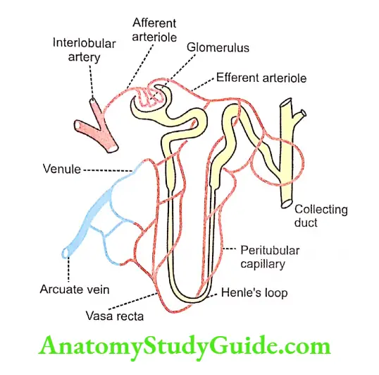
- From each interlobular artery, numerous afferent arterioles arise.
- The afferent arteriole enters the Bowman’s capsule and forms a glomerular capillary tuft.
- The afferent arteriole divides into 4 or 5 large capillaries. Each large capillary divides into small capillaries, which form the loops.
- And, the capillary loops unite to form the efferent arteriole. which leaves the Bowman’s capsule.
- The efferent arterioles form a second capillary network surrounding the tubular portions of the nephrons.
- This second set of capillaries is called peritubular capillaries.
- Thus, the renal circulation forms a portal system through the presence of two sets of capillaries – glomerular capillaries and peritubular capillaries.
- The network of peritubular capillaries supplies the tubular portion of cortical nephrons only.
- The tubular portion of juxtamedullary nephrons are supplied by some specialized capillaries called vasa recta.
- Vasa recta arise directly from the efferent arteriole of the juxtamedullary nephrons and run parallel to the renal tubule into the medulla and ascend up towards the cortex.

- The peritubular capillaries and vasa recta drain into the venous system.
- The venous system starts with peritubular venules and the other vessels are interlobular veins, arcuate veins, interlobar veins, segmental veins, and finally the renal vein.
- The renal vein leaves the kidney through the hilus and joins the inferior vena cava.
Measurement Of Renal Blood Flow
The blood flow to the kidneys is measured by using plasma clearance of para-aminohippuric acid
Regulation Of Renal Blood Flow
- The regulation of blood flow to the kidneys is independent of the nerves innervating renal blood vessels.
- The renal blood flow is regulated mostly by means of autoregulation.
Autoregulation
- The intrinsic ability of an organ to regulate its own blood flow is called autoregulation.
- Autoregulation is present in some vital organs in the body such as the brain, heart, and kidneys.
- It is highly significant and more efficient in kidneys.
Renal Autoregulation
- Renal autoregulation is important to maintain the glomerular filtration rate (GFR).
- Blood flow to kidneys remains normal even when the mean arterial blood pressure varies widely thus maintaining the normal GFR.
- Renal blood flow is constant till the mean arterial pressure falls as low as 60 mm Hg or increases as high as 180 mm Hg.

Two mechanisms are involved in renal autoregulation:
- Myogenic response
- Tubuloglomerular feedback.
1. Myogenic Response
- Whenever the blood flow to the kidneys increases, it stretches the elastic wall of the afferent arteriole.
- The stretch of the vessel wall increases the flow of calcium ions from the extracellular fluid into the cells.
- The influx of calcium ions leads to the contraction of smooth muscles in the afferent arteriole which causes constriction and an increase in the resistance in the afferent arteriole. So, the blood flow is controlled.
2. Tubuloglomerular Feedback
Macula densa plays an important role in tubuloglomerular feedback which controls the renal blood flow and GFR.
Special Features Of Renal Circulation
The renal circulation has some special features to cope with the functions of the kidneys.
Such special features are:
- The renal arteries arise directly from the aorta. The pressure in the aorta is very high and it facilitates a high blood flow to the renal parenchyma.
- Both kidneys receive about 1,300 mL of blood per minute, i.e. about 26% of cardiac output. Kidneys are the second organ to receive maximum blood flow, the first organ being the liver which receives 1,500 mL per minute, i.e. about 30% of cardiac output.
- The whole amount of blood that flows to the kidney has to pass through the glomerular capillaries before entering the venous system. Because of this, the blood is completely filtered at the renal glomeruli.
- Renal circulation has a portal system, i.e. a double network of capillaries. The afferent arteriole enters the Bowman’s capsule forming a glomerular capillary tuft.
- And, the glomerular capillaries unite to form the efferent arteriole, which leaves the Bowman’s capsule.
- The efferent arteriole gives rise to the renal portal system, i.e. it forms a second capillary network called peritubular capillaries, which surrounds the tubular portions of the nephrons. Both capillary systems are functionally important.
- Renal glomerular capillaries form a high-pressure bed with a pressure of 60-70 mm Hg. It is much greater than the capillary pressure elsewhere in the body, which is only about 25-30 mm Hg.
- High pressure maintains the glomerular capillaries because the Geter of afferent arteriole is more than that of ellorum arteriole. The high capillary pressure augments glomerular filtration.
- The peritubular capillaries form a low-pressure bed with a pressure of 8-10 mm Hg. This low pressure helps tubular reabsorption.
- The autoregulation of renal blood flow is well established. It maintains the renal blood flow when the mean arterial blood pressure varies within the range of 60 mm Hg and 180 mm Hg. The autoregulation is mainly due to the activity of macula densa.
Leave a Reply