Restoration Of Endodontically Treated Tooth
Rationale For Use Of Post
If the coronal tooth structure is lost due to fracture or severe carious lesion and the remaining tooth structure is not sufficient to provide enough support to the prosthesis then the post is used to achieve additional support from the root.
Table of Contents
The post is inserted into the root canal to get support from the root and restorative material is supported on the coronal part of the post which is known as the “core”. The main function of the post is to support the core material. If more than 25% of anterior tooth structure and 50% of posterior tooth structure is lost then post and core with crown prosthesis is recommended treatment.
Classification Of Post
Various types of posts are available to match different treatment needs. Broadly they can be classified as under.
Procedure For Post And Core Treatment
Evaluation of Endodontically Treated Tooth:
Once the root canal treatment is completed, the operator should observe the endodontically treated tooth for some time. The tooth should be asymptomatic after root canal treatment. There should be no pain or sensitivity during chewing or any other function.
Read and Learn More: Preclinical Prosthodontics Notes
Selection of Post and Core System:
If the tooth root diameter and canal diameter are small and the use of prefabricated posts needs more widening of canals then it results in additional loss of tooth structure. In such cases, custom-made post and core are used. Where wider canals are available prefabricated posts are used.
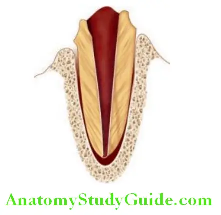
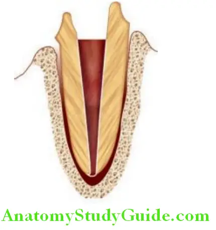
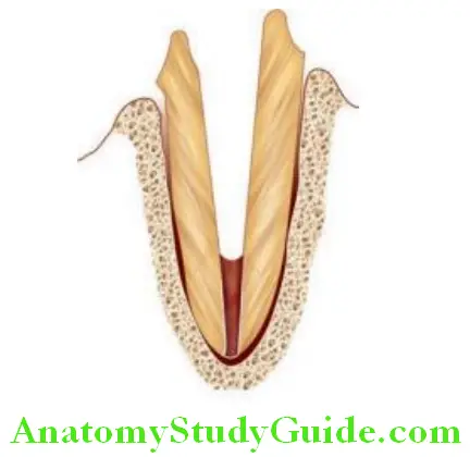
Preparation of Post Space:
After endodontic treatment, the root canals are filled with restorative materials, most commonly the gutta-percha. For post-space preparation, the gutta-percha is removed from the filled canals using Peeso Reamers or Gates Glidden drills. The apical seal of filled root canals should be kept intact. A minimum of 5 mm of gutta-percha is kept in the root canals to maintain an apical seal. Post space is modified after the removal of gutta-percha from the canals.
- Use of prefabricated post: Prefabricated post is used when the root canal diameter is wider and needs less canal preparation to accommodate prefabricated post.
- Use of custom post: When the canal diameter is smaller and needs more canal preparation for receiving prefabricated post, in such cases custom post is fabricated after making an impression of narrow post space (canal space). In such cases usually post and core are single units.
Ferrule:
When part of the coronal tooth structure is lost, the remaining tooth structure is encircled with part of the crown prosthesis. This remaining natural crown structure encircled by crown prosthesis is known as “ferrule”. A minimum of 2 mm of the ferrule is indicated for the longevity of the prosthesis.
Procedure And Pictorial Representation Of Post And Core Treatment
A fractured tooth or grossly carious tooth with more than 25% tooth structure loss is recommended for post and core treatment. Endodontic treatment is recommended in such cases where the pulp is exposed due to a fracture of tooth structure or carious lesion. After removing the pulp tissue, the root canals are widened and shaped to receive the inert restorative material in place of the pulpal tissue.
The coronal gutta-percha is removed keeping the apical seal intact to prevent infection. Apical gutta-percha of 3–5 mm is kept in the root canal to preserve the apical seal. Appropriate inlay wax or pattern resin is inserted into the root canal and a wax pattern/resin pattern is made of the canal. The same material is extended on the crown portion to

shape it like a prepared tooth. The extended material forming a crown-like portion is called as the core. The post and core wax pattern is cast and converted to custom post and core
and cemented into post space. The impression is made after cementation and crown prosthesis are prepared. The prepared crown prosthesis is cemented over the core to restore the fractured tooth.
Leave a Reply