Scalp Temple And Face Important Questions With Answers
Question 1: Why do infections of the superficial fascia of the scalp cause more pain?
Answer: Superficial fascia has abundant fibrous tissue and a rich nerve supply.
Hence, infection of the superficial fascia irritates the nerve and one gets severe pain.
Question 2:Why are sebaceous cysts and seborrhoea more frequently associated with the scalp?
Answer: The scalp has plenty of hair and sebaceous glands.
The ducts of sebaceous glands are prone to infection and get damaged by combing.
For this reason, the scalp is a common site for sebaceous cysts. Hence, sebaceous cysts and seborrhoea are more common.
Read and Learn More Head Anatomy
Question 3: What is the “dangerous area of the scalp” and why is it called so?
Answer: The third layer of the scalp is loose areolar tissue. It is a dangerous area of the scalp.
An accumulation of blood in this area will not spread in the following directions.
1. It cannot descend in the neck because of the firm attachment of epicranial aponeurosis to the superior nuchal line.
2. It cannot descend laterally because of its attachment to the superior temporal line.
The only way it can spread is anteriorly. Hence, it accumulates deep in the eyelid and results in a black eye.
This results in damage to the eye hence it is called a dangerous area.
Question 4: What is “safety valve hematoma”? How the haemorrhage from the blood vessels of the scalp is arrested?
Answer: 1. Safety valve hematoma occurs due to the following reasons:
1. Fracture of the bone of the scalp, and
2. Intracranial bleeding (usually due to birth trauma in the newborn).
The symptoms of intracranial bleeding are delayed due to leakage of blood.
It spreads to the sub-aponeurotic layer from the fracture site.
It expands to accommodate large quantities of blood.
Since this hematoma delays the onset of symptoms of a serious condition, it is called safety valve hematoma.
2. By compressing at the site of injury, bleeding is arrested since underneath the scalp is the cranium which is a hard structure.
Question 5: Why do the wounds on the face bleed profusely?
Answer: The scalp has a profuse blood supply. It is supplied by 5 paired arteries.
These arteries pass through the dense connective tissue.
Ruptured arteries of any part of the body are constricted by the contraction of smooth muscles present in walls of the vessel.
The vessels in the scalp pass through the dense connective tissue.
The arteries of the scalp cannot overcome the resistance of the tough dens deep fascia.
Hence, they are kept open and they bleed profusely.
Question 6: What are the modifications of palpebral fascia?
Answer: The palpebral fascia of the two eyelids forms the orbital septum.
1. In upper eyelid becomes thick and forms tarsal plates or tarsi.
Tarsi are thin plates of condensed fibrous tissue located near the lid margins.
They give stiffness to the lids.
The upper tarsus receives two tendinous slips from the levator palpebrae superioris. I
- One from the voluntary part, and
- Another from involuntary part.
2. At the angles, it forms palpebral ligaments.
Question 7: What is stye (hordeolum)?
Answer: 1. Definition: It is a suppurative inflammation of one of the glands of Zeis.
It is a large sebaceous gland.
2. Clinical features
- The gland is swollen, hard, and painful.
- The lid is oedematous.
- Pus points near the base of one of the roots (follicle) of an eyelash.
Question 8: What is chalazion?
Answer: Definition: It is inflammation of a tarsal gland, causing a localized swelling pointing inward.
Question 9:Why do the wrinkles of face tend to gap?
Answer: As the person ages, the skin loses its elasticity (resilience) which results into wrinkles , on the skin.
If the skin incision is not parallel to these cleavage or wrinkle lines (Langer lines), it has tendency to gap.
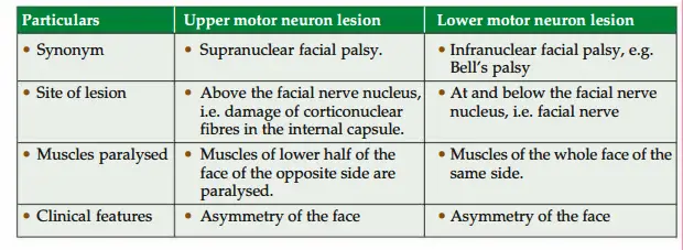
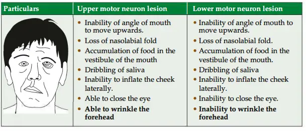
supranuclear lesion of facial nerve, only the lower part of the face is – paralysed.
Question 10:Why the upper part of face is spared?
Answer: The supranuclear lesions are also called upper motor neuron lesions.
upper part of face affects muscles of the lower½ of the face only.
The muscles of the upper½ of the face are spared because muscles of the upper½ of the face (muscles of forehead and eyebrows) are supplied by both cerebral hemispheres.
This is called bilateral cortical innervation.
Muscles of Face
1. Muscles of Face Action,
2. Muscles of Face Nerve supply, and
3. Muscles of Face Applied anatomy
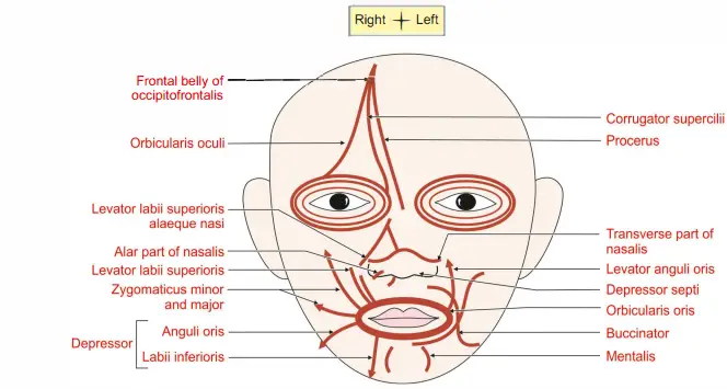
1. Muscles of Face Action: The muscles of facial expression can be grouped as (mimetic muscles)
Muscles acting on the orifice of the orbit: These are subgrouped as constrictor (sphincters) and dilators.
Frontal belly of occipitofrontalis is responsible for elevation of eyebrows as in an expression of surprise and it also contracts in looking upwards.
- The action is antagonistic to the orbital part of orbicularis oculi.
- Corrugator supercilii drags the eyebrow medially and downward and protects the eye from bright sunlight.
- It produces vertical wrinkles of the forehead.
- Orbicularis oculi has three parts.
- Palpebral part closes the eye gently as in sleep and blinking.
- Orbital part closes the eye firmly as in dust storm.
- Lacrimal part dilates the lacrimal sac.
- Muscles acting on the orifice of the nose
- Procerus (extended, tall). It is extended part of frontalis. It produces transverse wrinkles across bridge of nose as in frowning.
- Nasalis has two parts.
- Transverse part called compressor naris. It compresses the nasal aperture.
- Alar part called dilator naris. It dilates the anterior nasal aperture in deep inspiration.
- Depressor septi dilates anterior nasal aperture in anger.
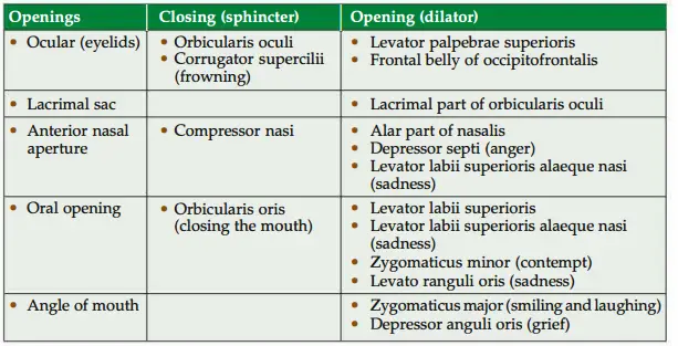
- Muscles acting on the orifice of the mouth
- Closure: Orbicularis oris closes the mouth.
- Dilators
- Subcutaneous layer: Risorius (risus-to laugh).
- Superficial layer:
- Zygomaticus major draws the angle of mouth upward and laterally as in laughing.
- Zygomaticus minor elevates and everts upper lip.
- Levator labii superioris alaeque nasi elevates and everts the upper lip and dilates the nostril.
- Middle layer
- Depressor anguli oris draws the angle of mouth downward.
- Levator anguli oris
- Depressor labii inferioris draws angle of mouth downward and somewhat laterally as in expression of irony.
- Levator labii superioris elevates the lip.
- Deeper layer
- Mentalis protrudes the lower lip.
- Buccinator flattens the cheek and forcibly expels the air between the lips (whistling).
Muscles of Face Nerve supply!: The muscles of the face are developed from 2nd pharyngeal arch and the nerve of the 2nd pharyngeal arch is facial nerve.
Hence, all the muscles are supplied by facial nerve.
Muscles of Face Applied anatomy
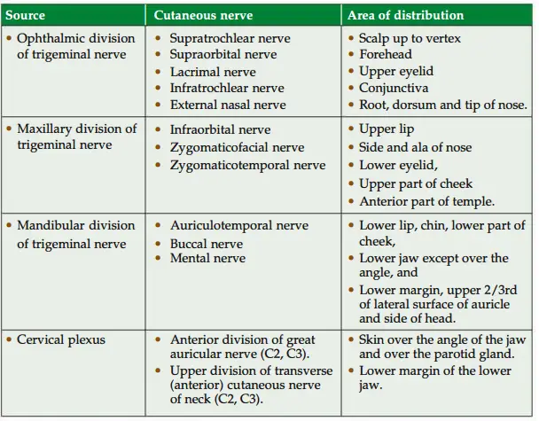
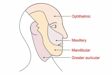
Describe Facial Vein under the following headings:
1. Formation; 2. Relations; 3. Tributaries; 4. Termination; 5. Applied anatomy
1. Formation: The angular vein receives superior labial vein and continues as facial vein.
2. Facial Vein Relations
- It lies deep to
- Zygomaticus major,
- Riorius, and
- Platysma.
- From above downwards, it lies superficial to
- Buccinator,
- Body of mandible,
- Masseter,
- Posterior belly of digastric, and
- Stylohyoid muscle.
- At termination, it crosses: a. Internal carotid artery, b. External carotid artery,
- Hypoglossal nerve, and d. Loop of lingual artery.
3. Facial Vein Tributaries
- Superior ophthalmic vein,
- Vein from alar nasi,
- Superior labial vein,
- Buccal vein,
- Deep facial vein from pterygoid plexus,
- Inferior labial vein,
- Masseteric vein,
- Tonsillar vein,
- Submental vein, and
- Submandibular vein
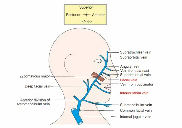
4. Facial Vein Termination: Common facial vein terminates into internal jugular vein.
5. Facial Vein Applied anatomy
Infection of the face can spread to intracranial venous sinus. Hence, the veins draining Upper lip Septum of nose, and Adjoining nose lying between angular and deep facial veins forms the dangerous area of the face.
I. ..A is the dangerous area of face.
Following are the routes for the spread of infection.
Angular vein + superior ophthalmic vein + cavernous sinus.
Deep facial vein + pterygoid venous plexus + emissary vein + cavernous sinus.
The spread of septic emboli from the infected area to cavernous sinus can cause serious complications because of following reasons.
Veins of the face do not have valves.
- Veins of the face lie on facial muscles.
- There is no deep fascia on the face.
- The movements of the facial muscles may facilitate the spread of septic emboli to cavernous sinus.
Question 11:Why the facial muscles are called “muscles of facial expression”?
Answer: The muscles of face are not inserted on bone but in the skin.
Since there is no deep fascia in the face, contraction of these muscles causes contraction of some part of skin (on the face. It acts as a medium to express the emotional feelings.
Hence, facial muscles are called muscles of facial expression.
Question 12: What is the nerve supply of facial muscles?
Answer: 1. All the muscles on the face are supplied by facial nerve except levator palpebrae superioris which is supplied by oculomotor nerve.
2. Majority of the muscles on the face are muscles of facial expression.
However, there are exceptions.
These are buccinator and platysma.
They are supplied by facial nerve.
3. The muscles present on the face but included as muscles of mastication are temporalis, masseter, medial andlateral pterygoid.
These aresupplied by mandibular nerve, branch of trigeminal nerve (Vth cranial nerve).
Question 13:What are the constituents of lacrimal apparatus?
Answer: It consists of
1. Lacrimal gland
- Orbital part, and
- Palpebral part
2. Conjunctiva! sac
3. Punctum
4. Canaliculus
- Superior, and
- Inferior canaliculus
5. Lacrimal sac
Nasolacrimal duct
Question 14: What are the structural diffrences between lacrimal gland and serous salivary gland?
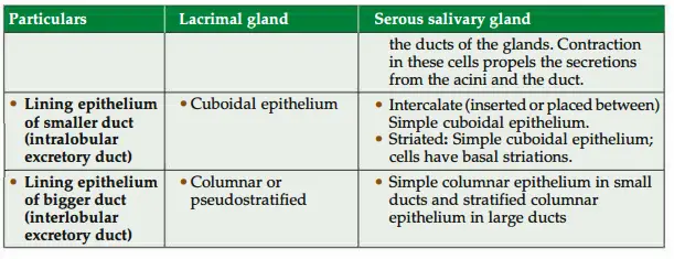
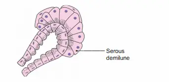
Question 15: microscopic structure of serous demilune
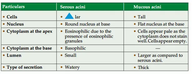
Question 16: Enumerate the diffrence between serous and mucus acini.
Question 17: What are serous demilunes?
Answer:
- Some of the mucous acini are capped with serous cells.
- They are arranged as a half-moon.
- Hence, they are called serous demilunes.
Where do we get myoepithelial cells in the body? How will you identify – them?

- Site
- Ducts: Between the epithelium and the basement membrane of the ducts.
- Sweat gland: There are cuboidal secretory cells of the sweat gland.
- At the base of the secretory cells, there are numerous myoepithelial cells.
- Identifying features :
- The cells are thin and spindle-shaped.
- They are located at the base of the secretory cells
Question 18: What are the functions of saliva?
Answer: 1. The saliva lubricates the luminal surface of th upper digestive and respiratory tracts.
2. Saliva moistens the food to help in deglutination.
3. Saliva initiates the digestion of carbohydrates by the enzyme salivary amylase.
4. Saliva contains lysozyme and irnmglobulin. They are bactericidal in nature.
Question 19: What is dacryocystitis?
Answer: Inflammation of the lacrimal sac is called dacrocystitis.
Question 20: What is the nature of the lacrimal gland?
Answer: It is a serous gland.
Question 21:What are the parts of the lacrimal gland?
1. Orbital part
2. Palpebral part
Leave a Reply