Somatosensory System
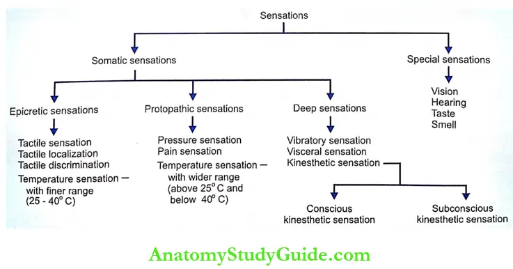
Definition And Types Of Sensations: Somatosensory system is defined as sensory system associated with different parts of the body. It is also defined as the faculty of bodily perception of various sensations. Sensations are of two types
Table of Contents
- Somatic Sensations: Somatic sensations are the sensations arising from skin, muscles, tendons, and joints. These sensations have specific receptors, which respond to a particular type of stimulus.
- Special Sensations: Special sensations are complex sensations for which the body has some specialized sense organs. The special sensations are usually called special senses. Sensations of vision, hearing, taste, and smell are special sensations.
Read And Learn More: Medical Physiology Notes
Types Of Somatic Sensations: Generally, somatic sensations are classified into three types
- Epicretic Sensations: Epicretic sensations are mild or light sensations. Such sensations are perceived more accurately. Epicritic sensations are
- Fine touch or tactile sensation
- Tactile localization
- Tactile discrimination
- Temperature sensation with finer range between 25 and 40°C.
- Protopathic Sensations: Protopathic sensations are crude sensations. These sensations are primitive type of sensations. Protopathic sensations are
- Pressure sensation
- Pain sensation
- Temperature sensation with a wider range, i.e. above 40°C and below 25°C.
- Deep Sensations; Deep sensations are the sensations arising from the deeper structures beneath the skin and the visceral organs. The deep sensations are classified into three types
- The sensation of vibration or pallesthesia
- Kinesthetic sensation or kinesthesia: Sensation of the position and movements of different parts of the body. This sensation arises from the proprioceptors present in muscles, tendons, joints, and ligaments. Proprioceptors are the receptors, which give response during various movements of a joint. The kinesthetic sensation is of two types.
- Conscious kinesthetic sensation
- Subconscious kinesthetic sensation. The impulses of this sensation are called non-sensory impulses.
- Visceral sensations arising from viscera
Sensory Pathways: The nervous pathways of the sensations are called the sensory pathways. These pathways carry the impulses from the receptors in different parts of the body to the centers in brain. The sensory pathways are of two types:
- Pathways of the somatosensory system
- Pathways of viscerosensory system.
- The pathways of the somatosensory system convey information from the sensory receptors in the skin, skeletal muscles, and joints. The pathways of this system are constituted by somatic nerve fibers called somatic afferent nerve fibers.
- The pathways of the viscerosensory system convey the information from the receptors of the viscera. The pathways of this system are constituted by visceral or autonomic fibers.
- Somatosensory Pathways: Each sensory pathway is constituted by two or three groups erf neurons
- First order neurons
- Second order neurons
- Third-order neurons.
- The pathways of some of the sensations like kinesthetic sensation have only first and second-order neurons.
- The details of the pathways are given in Table. The diagrams of the pathways are given in the previous chapter along with ascending tracts of the spinal cord.
Sensory Fibers Of Trigeminal Nerve; Trigeminal nerve carries somatosensory information from the face, teeth, periodontal tissues (tissues around teeth), oral cavity, nasal cavity, cranial dura mater, and major part of scalp to sensory cortex. It also conveys proprioceptive impulses from the extrinsic muscles of the eyeball.
- Sensory Fibers Of Trigeminal Nerve Origin
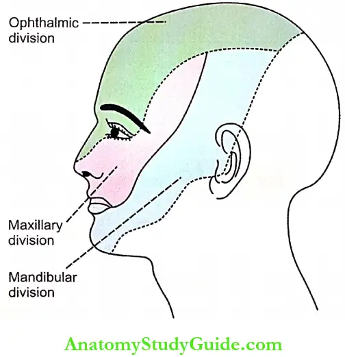
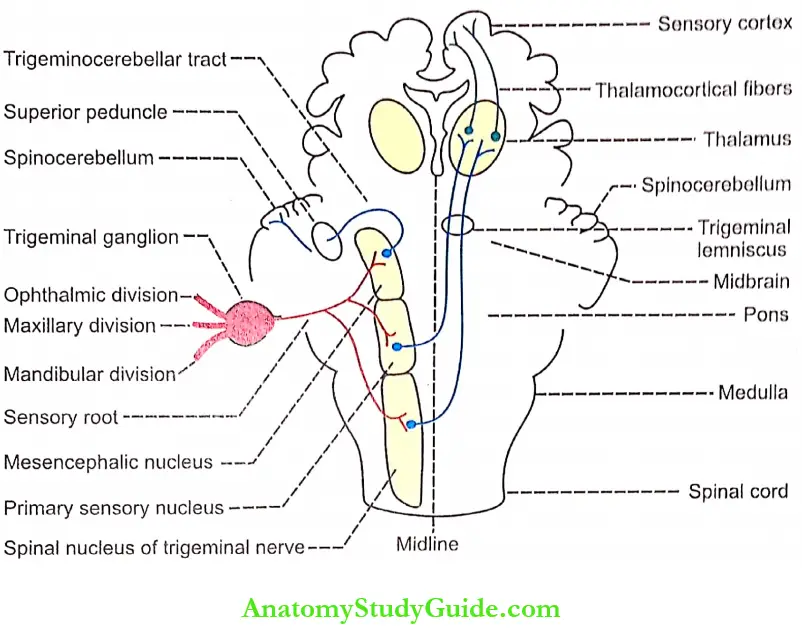
- The sensory fibers of trigeminal nerve arise from the trigeminal ganglion situated near the temporal bone. The peripheral processes of neurons in this ganglion form three divisions of trigeminal nerve namely, ophthalmic, mandibular, and maxillary divisions.
- The cutaneous distribution of the three divisions of trigeminal nerve is shown. The central processes from the neurons of the trigeminal ganglion enter the pons in the form of sensory root.
- Sensory Fibers Of Trigeminal Nerve Termination
- After reaching the pons, the fibers of sensory root divide into two groups namely, descending fibers and ascending fibers. The descending fibers terminate on primary sensory nucleus and spinal nucleus of trigeminal nerve. The primary sensory nucleus is situated in pons.
- The spinal nucleus of the trigeminal nerve is situated below the primary sensory nucleus and extends up to the upper segments of spinal cord.
- The ascending fibers of the sensory root terminate in the mesencephalic nucleus of trigeminal nerve situated in the brainstem above the level of primary sensory nucleus.
- Sensory Fibers Of Trigeminal Nerve Central Connections
- The majority of fibers from the primary sensory nucleus and spinal nucleus of the trigeminal nerve ascend in the form of the trigeminal lemniscus and terminate in the ventral posteromedial nucleus of thalamus in the opposite side.
- The remaining fibers from these two nuclei terminate on the thalamic nucleus of the same side. From thalamus, the fibers pass via superior thalamic radiation and reach the somatosensory areas of the cerebral cortex.
- The primary sensory nucleus and the spinal nucleus of the trigeminal nerve relay the sensations of touch, pressure, pain, and temperature from the regions mentioned above.
- The fibers from the mesencephalic nucleus form the trigerninocerebellar tract that enters the spinocerebellum via the superior cerebellar peduncle of the same side. This nucleus -.cnveys the proprioceptive impulses from the facial, muscles of mastication and ocular muscle.

Lemnisci: Prominent bundle of sensory nerves in the brain is called the lemniscus or fillet. The brain has four types of lemniscus.
- Spinal lemniscus: It is formed by spinothalamic tracts in the medulla oblongata.
- Lateral lemniscus: It is formed by the fibers carrying the sensation of hearing from cochlear nuclei to the inferior colliculus and medial geniculate body.
- Medial lemniscus: The medial lemniscus is formed by fibers arising from nucleus cuneatus and nucleus gracilis.
- Trigeminal lemniscus: It is formed by fibers from sensory nuclei of trigeminal nerve. This lemniscus carries general senses from the head, neck, face, mouth, eyeballs, and ears.
Applied Physiology: Lesions or other nervous disorders in sensory pathways affect the sensory functions of the body
- Anesthesia: Loss of all sensations
- Hyperesthesia: Increased sensitivity to sensory stimuli
- Hypoesthesia: Reduction in the sensitivity to sensory stimuli
- Hemiesthesia: Loss of all sensations in one side of the body
- Paresthesia: Abnormal sensations such as tingling, burning, prickling and numbness
- Hemiparesthesia: Abnormal sensations in one side of the body
- Dissociated anesthesia: Loss of some sensations while other sensations are intact
- General anesthesia: Loss of all sensations with loss of consciousness produced by anesthetic agents
- Local anesthesia: Loss of sensations in a restricted area of the body
- Spinal anesthesia: Loss of sensations, due to lesion in spinal cord or induced by anesthetic agents injected beneath the coverings of spinal cord
- Tactile anesthesia: Loss of tactile sensations
- Tactile hyperesthesia: Increased sensitivity to tactile stimuli
- Analgesia: Loss of pain sensation
- Hyperalgesia: Increased sensitivity to pain stimulus
- Paralgesia: Abnormal pain sensation
- Thermoanesthesia or thermanesthesia or therm-analgesia: Loss of thermal sensation
- Pallanesthesia: Loss of sensation of vibration
- Astereognosis: Loss of ability to recognize any object with closed eyes due to loss of cutaneous sensations
- Illusion: Mental depression due to misinterpretation of a sensory stimulus
- Hallucination: Feeling of a sensation without any stimulus.
Somatomotor System
Motor Activities Of The Body: The motor activities of the body depend upon different groups of tissues of the body. Motor activities are generally divided into two types
- The activities of skeletal muscles which are involved in the posture and movement
- The activities of smooth muscles, cardiac muscles, and other tissues, which are involved in the functions of various visceral organs.
- The activities of the skeletal muscles (voluntary functions) are controlled by the somatomotor system, which is constituted by the somatic motor nerve fibers.
- The activities of tissues or the visceral organs (involuntary functions) are controlled by the visceral or autonomic nervous system, which is constituted by the sympathetic and parasympathetic systems.
Somatomotor System: The movements of the body depend upon the different groups of skeletal muscles. Various types of movements or motor activities brought about by these muscles are
- Execution of smooth, precise, and accurate voluntary movements
- Coordination of movements responsible for skilled activities
- Coordination of movements responsible for maintenance of posture and equilibrium.
- The voluntary actions and the postural movements are carried out by not only the simple contraction and relaxation of skeletal muscles but also the adjustments of tone in these muscles.
- The execution, planning, coordination, and adjustments of the movements of the body are under the influence of different parts of the nervous system, which are together called the motor system. The sensory system of the body also plays a vital role in the control of the movements.
- Spinal reflexes are responsible for most of the movements concerned with voluntary actions and posture Stimulation of the receptor activates the motor neuron in spinal cord leading to the contraction of the muscle innervated by the spinal motor neuron.
- Apart from these reflexes, the signals for voluntary motor activities are also sent from different areas of the brain particularly, the cerebral cortex to spinal motor neurons.
The coordination and control of movements initiated by the cerebral cortex depend upon two factors:
- Feedback signals from the proprioceptors in muscle and other sensory receptors
- Interaction of other parts of the brain such as the brainstem, cerebellum, and basal ganglia.
- Thus, the motor system includes spinal cord and its nerves, cranial nerves, brainstem, cerebral cortex, cerebellum, and basal ganglia. The neuronal circuits between these parts of the nervous system which are responsible for the motor activities are called the motor pathways.
- The classification of motor pathways is given at the later part of this chapter because the knowledge of role of different parts of the nervous system is essential to understand the classification of the motor pathways.
Spinal Cord And Cranial Nerve Nuclei
- Motor Neurons
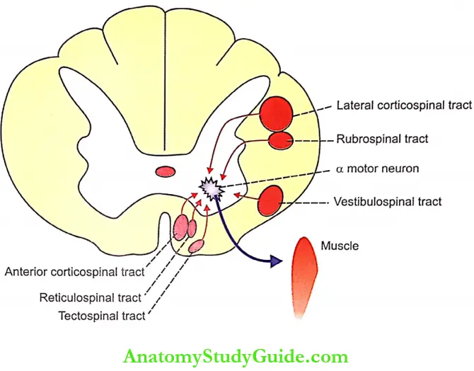
- The activities of skeletal muscles are executed by the impulses discharged from the alpha motor neurons situated in ventral (anterior) gray horn of spinal cord and the nuclei of many of the cranial nerves present in brainstem.
- The alpha motor neurons in the spinal cord, which innervate the intrafusal fibers of the skeletal muscles are responsible for the contraction of muscles in the upper limbs, trunk, and lower part of the body.
- The gamma motor neurons, which innervate the intrafusal fibers of the muscle, are responsible for the maintenance of muscle tone.
- The motor neurons of the cranial nerve nuclei situated in the brainstem send their signals to the muscles of neck and upper part of trunk via cranial nerves.
- Final Common Pathway
- The activities of a particular skeletal muscle depend upon the excitation of alpha motor neuron (also known as lower motor neuron) in the spinal cord or cranial nerve nuclei.
- This is the only pathway through which the signals from other parts of the nervous system reach the muscles. Hence, the alpha motor neurons are called the “Final common pathway” of the motor system.
- Functions of Motor Neurons
- The motor neurons responsible for the contraction of skeletal muscles are arranged topographically in the ventral gray horn of the spinal cord. The neurons situated in the medial part of the ventral gray horn innervate the muscles near the midline of the body called the axial muscles and the muscles in the proximal portions of limbs called primal muscles.
- These two types of muscles are involved in the adjustment of posture and gross movement. The motor neurons in the lateral part of ventral gray horn innervate the muscles in distal portions of the limbs called the distal muscles. The distal muscles are involved in well-coordinated skilled voluntary movements.
- The motor neurons in the cranial nerve nuclei of the brainstem innervate the extrinsic muscles of the eyeball and the muscles of the face, tongue, neck, and upper part of the trunk.
- These muscles are concerned with ocular movements and the movements of facial expressions, chewing, swallowing, and the movements of head and shoulder. The motor neurons are situated in the nuclei of cranial nerves 3, 4, 5, 6, 7, 9, 10, 11, and 12.
Cerebral Cortex
- The cortical areas concerned with origin of motor signals are the primary motor area, premotor area, supplementary motor area in frontal lobe, and sensory area in the parietal lobe.
- The cortical areas send their output signals to the spinal cord via corticospinal tracts and to brainstem via corticobulbar tracts.
- About 30% of the fibers forming corticospinal and corticobulbar tracts take their origin from primary and supplementary motor cortex, 30% from the premotor area, and remaining 40% from parietal lobe particularly from the somatosensory area.
Cerebellum
- Cerebellum plays an important role in planning, programming, and integrating the skilled voluntary movements. It is also concerned with the maintenance of muscle tone, posture, and equilibrium.
- Cerebellum receives impulses from proprioceptors of muscle, vestibular apparatus, cerebral cortex, brainstem, and basal ganglia. It interprets these impulses and sends signals to the motor cortex, reticular formation, and nuclei of the brainstem.
Basal Ganglia: Basal ganglia play an important role in the coordination of skilled movements, regulation of automatic associated movements, and control of muscle tone by sending output signals to motor cortex, reticular formation, and spinal cord.
Classification Of Motor Pathways: There are two methods to classify the motor pathways. In the first method of classification, the motor pathways are divided into pyramidal and extrapyramidal tracts. By the second method, the motor pathways are classified into Bvieral and medial systems.
- Pyramidal and Extrapyramidal Pathways: The motor pathways are classified into pyramidal and extrapyramidal tracts depending upon situation of their fibers in medulla oblongata.
- Pyramidal Tracts: The pyramidal tracts are those fibers, which form the pyramids in the upper part of medulla. Pyramidal tracts are the anterior and lateral corticospinal tracts. These tracts control the voluntary movements of the body.
- Extrapyramidal Tracts: Motor pathways other than pyramidal tracts are known as extrapyramidal tracts.
- Extrapyramidal tracts:
- Medial longitudinal fasciculus
- Anterior and lateral vestibulospinal tracts
- Reticulospinal tract
- Tectospinal tract
- Reticulospinal tract
- Rubrospinal tract
- Olivospinal tract.
- The extrapyramidal tracts are concerned with the regulation of tone, posture, and equilibrium.
- Depending upon the location or termination, the motor pathways are divided into two categories namely, the lateral system or pathway and the medial system or pathway. The lateral motor system is phylogenetically new and medial motor system is old.
- Lateral Motor System: The fibers of this system terminate in the motor neurons situated in the lateral part of ventral gray horn in the spinal cord (directly or via interneurons) and on the equivalent motor neurons of cranial nerve nuclei in the brainstem.
- Components of the lateral system
- Lateral corticospinal tract: The lateral corticospinal tract arises from different areas of the cerebral cortex and terminates in the alpha motor neurons situated in lateral part of ventral gray horn of spinal cord.
- Rubrospinal tract: This tract arises from the red nucleus in the midbrain.
- Corticobulbar tract: The fibers of this track take origin from different areas of the frontal and parietal lobes of the cerebral cortex along with corticospinal tracts. The part of the corticobulbar tract belonging to lateral motor system terminates on the nucleus of the hypoglossal nerve and the motor nucleus of facial nerve. The fibers from the hypoglossal nerve innervate the muscles of the tongue. The fibers from motor nucleus of facial nerve innervate the muscles of lower part of the face.
- Functions of the lateral motor system
- The lateral corticospinal tract activates the muscles of distal portions of limbs and regulates skilled voluntary movements.
- The rubrospinal tract facilitates the tone in the muscles, particularly the flexor muscles.
- The corticobulbar fibers of the lateral system are concerned with movements of expression in lower part of the face and movements of tongue.
- Medial Motor System: The fibers of the medial motor system terminate on the motor neurons situated in the medial part of ventral gray horn of the spinal cord (via interneurons) and on the equivalent motor neurons of cranial nerve nuclei situated in brainstem.
- Functions of the lateral motor system
- Components of medial motor system
- Anterior corticospinal tract: It arises from different areas of the cerebral cortex and terminates in the alpha motor neurons situated in the medial part of the ventral gray horn of spinal cord.
- Corticobulbar tract: This tract also takes origin from different areas of the frontal and parietal lobes of cerebral cortex along with corticospinal tracts. The fibers of the corticobulbar tract belonging to medial motor system innervate the muscles of trunk and limbs, muscles of the jaw, and muscles of the upper part of the face.
- Lateral and medial vestibulospinal tracts: These tracts arise from the lateral vestibular nucleus and medial vestibular nucleus respectively.
- Reticulospinal tract: It arises from reticular formation in brainstem.
- Tectospinal tract: The fibers of this tract take origin from superior colliculus of midbrain.
- Functions of the medial motor system
- The fibers of anterior corticospinal tract are responsible for the maintenance of posture and equilibrium
- The fibers of the corticobulbar tract belonging to medial motor system innervate the axial muscles of upper part of trunk and are involved in the maintenance of posture and equilibrium. In addition, these fibers are also involved in the movements of chewing and movements of eyebrow
- The vestibulospinal tract is concerned with the adjustment of the position of head and body during angular and linear acceleration
- The pontine fibers of the reticulospinal tract facilitate the tone of extensor muscles and regulate the postural reflexes. However, the medullary fibers of this tract inhibit the tone of the muscles involved in postural movements
- The tectospinal tract is responsible for the movement of head in response to visual and auditory stimuli.
- Functions of the medial motor system
- Components of the lateral system
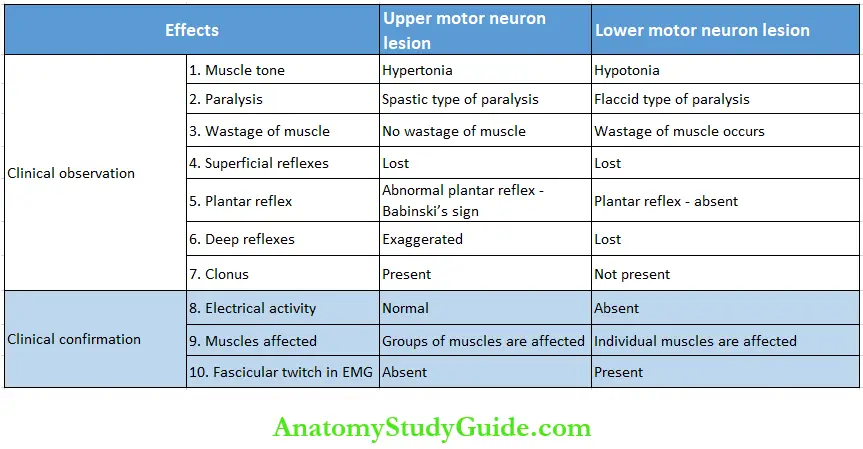
Upper Motor Neuron And Lower Motor Neuron; The neurons of the motor system are divided into upper motor neurons and lower motor neurons depending upon their location and termination.
- Upper Motor Neuron: Upper motor neurons are the neurons in the higher centers of the brain, which control the lower motor neurons. There are three types of upper motor neurons:
- Motor neurons in the cerebral cortex. The fibers of these neurons form corticospinal (pyramidal) and corticobulbar tracts
- Neurons in the basal ganglia and brainstem nuclei
- Neurons in the cerebellum.
- The motor neurons in the cerebral cortex, which give origin to pyramidal tracts belong to the pyramidal system and the remaining motor neurons belong to the extrapyramidal system.
- Some controversy exists in including the neurons of the extrapyramidal system under the category of upper motor neurons. However, considering in terms of the definition, the neurons other than the lower motor neurons are to be named as upper motor neurons.
- Lower Motor Neuron
- Lower motor neurons are the anterior gray horn cells in the spinal cord and the motor neurons of the cranial nerve nuclei situated in the brainstem, which innervate the muscles directly.
- Thus, the lower motor neurons constitute the “Final common pathway” of the motor system. The lower motor neurons are under the influence of the upper motor.
Applied Physiology
Effects of Lesion of Motor Neurons; The effects of lesions of upper motor neurons and lower motor neurons are given in Table. The effects of lower motor neuron lesion are the loss of muscle tone and flaccid paralysis. The effects of upper motor neuron lesions depend upon the type of neuron involved.

- Effects of upper motor neuron lesion: The lesion in the pyramidal system causes hypertonia and spastic paralysis. Spastic paralysis involves only one group of muscles, particularly the extensor muscles
- Lesion in the basal ganglia produces hypertonia and rigidity involving both flexor and extensor muscles
- The lesion in the cerebellum causes hypotonia, muscular weakness, and incoordination of movements.
- Paralysis: Paralysis is defined as the complete loss of strength and functions of a muscle group or a limb.
- Causes for paralysis: Common causes for paralysis are trauma, tumor, stroke, cerebral palsy (a condition caused by brain injury immediately after birth) multiple sclerosis, and neurodegenerative diseases.
- Types of paralysis: The- paralysis of the muscles in the body depends upon lie type and location of motor neurons affected by the lesion. Different types of paralysis are given in Table.
Leave a Reply