Spinal Cord Anatomy Questions And Answers
Give the extent of the spinal cord.
Table of Contents
- Extent
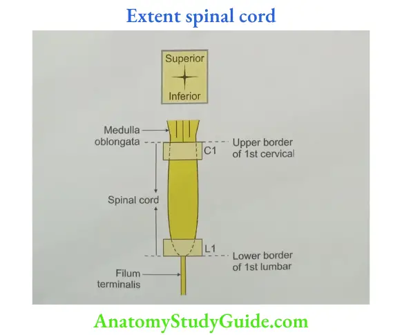
- Adult: It extends from the upper border of the atlas vertebra to the lower border of 1st lumbar vertebra.
- In children, it extends up to the 3rd lumbar vertebra.
- Continuation
- Superiorly, it is continuous with the medulla oblongata.
- Inferiorly, it terminates as conus medullaris.
- Vertebral column: It occupies the upper two-thirds of the vertebral canal and is enclosed in 3 meninges.
Read And Learn More: Anatomy Important Question And Answers
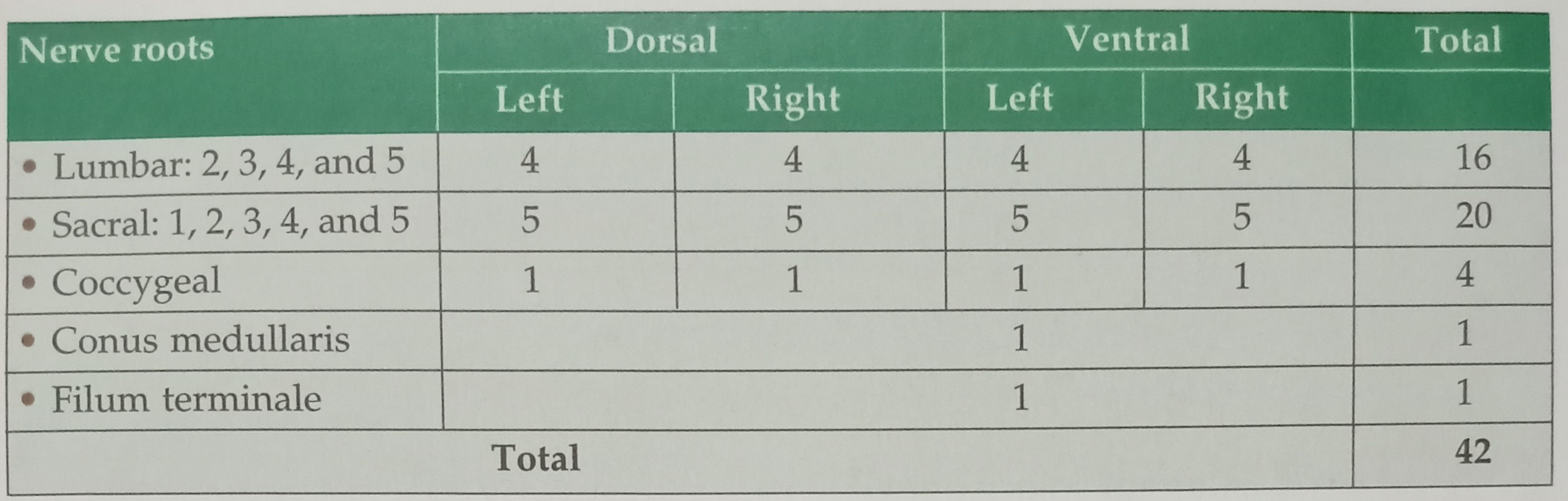
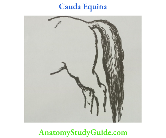
What is cauda equina?
- The word “cauda” means tail and “equina” means horse
- It constitutes
What is a tract?
The smallest bundle of fibers connecting any two masses of grey matter is called tract. It has same origin, course and destination.
What is a lemniscus?
Lemniscus (pl. lemnisci): The larger bundle of fibres connecting various masses of grey matter is called lemniscus. Many tracts combine together to form lemniscus

What is a peduncle?
Peduncle: The biggest bundle of fibers connecting various grey matters is called a peduncle. Many lemnisci combine together to form a peduncle.

What is the ascending tract? Name the ascending tracts.
- Ascending tracts: Fibres arising from the nuclei of the spinal cord and reaching the nuclei of the brainstem, cerebellum, and/or nuclei of the cerebrum, are called ascending tracts.
- Ascending tracts are
- Spinothalamic,
- Anterior spinothalamic
- Lateral spinothalamic
- Spinocerebellar
- Anterior spinocerebellar
- Posterior spinocerebellar
- Spino-olivary.

What is the descending tract? Name the descending tracts.
- Descending tracts: Fibres arising from the grey matter of the cerebrum, nuclei of cerebrum, cerebellum, and brainstem, and reaching nuclei of the spinal cord are called descending tracts.
- Examples of descending tracts
- Corticospinal,
- Rubrospinal,
- Vestibulospinal,
- Olivospinal, and
- Reticulospinal.

What is the basis of the nomenclature of tracts?
The nomenclature of the tracts depends upon
- Origin and destiny of the tract: The 1st word of the tract indicates the origin and the last word indicates the destiny of the tract: They are divided into ascending and descending tracts.
- Examples of descending tracts
- Corticospinal: It arises from the grey matter of the cerebral cortex and ends in the nuclei of the spinal cord.
- Rubrospinal: It arises from a red nucleus of the midbrain and ends in nuclei of the spinal cord.
- Vestibulospinal: It arises from the vestibular nuclei of the medulla oblongata and ends in the nuclei of the spinal cord.
- Olivospinal: It arises from inferior olivary nuclei of the brainstem and ends in nuclei of the spinal cord
- Reticulospinal: It arises from the reticular nuclei of the brainstem and ends in nuclei of the spinal cord.’
- Examples of ascending tracts
- Spinothalamic tract: It arises from the nuclei of the spinal cord and ends in the nuclei of the thalamus.
- Spinocerebellar tract: It arises from nuclei of spinal cord and ends in nuclei of cerebellum.
- Examples of descending tracts
- Course of the tract: The first word of the tracts signifies the course of the tract.
For example,
Lateral spinothalamic tract courses through the lateral column of the spinal cord.
Anterior spinothalamic tract courses through the anterior column of the spinal cord. - Relation with other tracts of same destiny:
The words prefixing the tract having the same destiny indicate the relation of the tract with each other. For example,- Anterior spinocerebellar tract: It is anterior to posterior spinocerebellar tract.
- Posterior spinocerebellar tract: It is posterior to anterior spinocerebellar tract.
- Depending upon the level of consciousness
- The fibres reaching the grey matter of spinal cord and action taking place at the level of spinal cord is called local reflex action.
- The fibres reaching cerebellum and response given without knowledge of brain is called unconscious proprioceptive tract.
- The fibres reaching cerebrum and the response given by analysing the situation with previous experience is called conscious proprioceptive tract.
- Shape of target nucleus.
- The word ‘gracilis’ means long and slender. The fibres of fasciculus gracilis end in gracilis tubercle that is long and slender. Hence, it is called fasciculus gracilis.
- The fibres of fasciculus cuneatus end into nucleus cuneatus. The word ‘cuneatus’ means wedge shaped. The fibres of fasciculus cuneatus end in cuneate tubercle that is wedge shaped. Hence, it is called fasciculus cuneatus.
- Scientists who have worked on it: The posterior column tract is named Goll and Burdach because it is studied by Goll and Burdach.
How the ascending tracts are grouped?
Grouping of tracts
- Ascending tracts can be grouped as tracts reaching up to
- Thalamus, and
- Cerebellum.
- All tracts reaching the cerebrum must first reach thalamus except olfactory tract. The tracts reach in VPL nucleus of thalamus. VPL is Ventro-Postero-Lateral VPL begins with the name of the nucleus of thalamus. The 1st letter of tract ending into VPL nucleus of thalamus.
Ventral spinothalamic tract
Posterior column, and
Lateral spinothalamic tract. - Tracts reaching up to cerebellum are
- Anterior spinocerebellar tract, and
- Posterior spinocerebellar tract.
How the descending tracts are grouped?
Descending tracts are divided into
- Pyramidal, and
- Extrapyramidal.
- Pyramidal tracts begin from pyramidal cells of cerebral cortex and form decussation which resembles a pyramid. They include
- Corticospinal, and
- Corticonuclear tracts
- Extrapyramidal tracts arise other than pyramidal cells of cerebral cortex. They arise from basal nuclei or nuclei of brainstem and reach spinal cord.
Please note,
- The fibres of extrapyramidal tract do not pass through the pyramid of medulla. They are not direct fibres from cerebral cortex to anterior horn cells of spinal cord. Hence, they are called indirect corticospinal tract.
- Most important extrapyramidal tracts are
- Reticulospinal, and
- Vestibulospinal tracts.
- The other extrapyramidal tracts are
- Rubrospinal,
- Reticulospinal, C. Tectospinal,
- Vestibulospinal, and
- Olivospinal.
Honorable Padmashree, Late Dr Mahdi Hasan, used the following easy method to remember the names of extrapyramidal tract:
He used to say, “The letter “P” indicates pyramidal.
- After “P,” drop the next alphabet “Q”.
- The letter “Q” is followed by the letter “R”
- Pick up the alternate alphabet after “R”.
- Each alternate letter after the letter “P” gives the name of the tract.
- They are “T” and “V”.
- The letters “R”, “T” .and “V” indicate
- “R”-Rubrospinal, Reticulospinal
- “T”-Tectospinal
- “V”-Vestibulospinal
- “P” is preceded by “O” which indicates Olivospinal
Draw and label a transverse section of the spinal cord showing external features.

Draw and label a transverse section of spinal cord showing grey matter.

Draw and label a transverse section of the spinal cord showing the main ascending and descending tracts.


Lateral lemniscus
- Lateral lemniscus Lateral lemniscus Definition: It is bigger bundle of white matter forming auditory pathway.
- Features
- It is 3rd order neuron of auditory pathway.
- It starts from superior olivary nucleus.
- The fibres decussate and go to opposite side of dorsal part of pons.
- It forms trapezium shaped structure called trapezoid body.
- It terminates at inferior colliculus of midbrain.
- It reaches medial geniculate body through inferior brachium.
- The nucleus of the lateral lemniscus consists of small groups of neurons (18,000 to 24,000).
- Lateral lemniscus Relations: It is located very close to the inferior colliculus.
Medial lemniscus
- Medial lemniscus Introduction: It is a bigger bundle of the axons, which arises from gracile and cuneate nuclei. It consists of 2nd order sensory neurons.
Course: Their axons cross the midline. They run ventrally and medially. They are called internal arcuate fibres. The crossing fibres of the two sides constitute the sensory decussation. - Medial lemniscus Formation: Medial lemniscus formed by
The fibres of the 2nd order neurons of posterior column, and
The fibres of the anterior spinothalamic tract. - Location: It is formed in the middle of the medulla oblongata.
- Representation
- Somatotopic representation

- Modality representation: The fibres are arranged in the posterior column. They are arranged from dorsal to ventral as follows
- Pressure,
Vibration,
Movement,
Position, and
Touch.The touch fibres include- Tactile localization,
- Tactile discrimination, and
- Stereognosis.

- Somatotopic representation
- Position of medial lemniscus at different levels

- Applied anatomy
- Lesion before formation of medial lemniscus: Loss of conscious proprioceptive sensations on the same side.
- Lesion after formation of medial lemniscus: Loss of conscious proprioceptive sensations of the opposite side.
LAQ-1 Describe extrapyramidal tracts under following heads Tracts, Functions, and Features
Extrapyramidal Definition: Fibres passing from the cerebral cortex to spinal cord via other parts of the cerebrum, and nuclei of brainstem are called extrapyramidal tracts.
- Through extrapyramidal tracts, the cerebral cortex has indirect effects on the motor cells of the brainstem, and spinal cord.
- These pathways form part of an extensive system, known as the extrapyramidal system.
- Since these pathways do not pass through the pyramid in the medulla oblongata, they are called extrapyramidal tracts. They are also called indirect
- corticospinal tract. C. The most important extrapyramidal tracts are
- Rubrospinal
- Tectospinal
- Reticulospinal
- The lateral reticulospinal tract arises from the medullary part of the reticular formation. The fibres course through the lateral white column. They are largely intermingled with corticospinal fibres. Its influence on anterior horn cells is facilitatory.
- The medial reticulospinal tract comes from cells in the pontine reticular formation. It descends in the anterior white column, to have an inhibitory action on motor neuron.

- Vestibulospinal tracts: The lateral vestibulospinal tract arises from the lateral vestibular nucleus present in the medulla. It runs down the cord approximately in the region of anterior nerve root region.
The vestibulospinal tract primarily affects trunk and limb girdle musculature. It is of great importance for posture and balance. The reticuloand vestibulospinal fibres synapse with interneurons which in turn project to the motor neurons.
- Functions: They are mainly concerned to regulate the tone and posture. It involves complex movements of more automatic nature, such as theMaintenance of balance during movements, Right muscle tone is necessary for normal movements, etc.
- Features
- Extrapyramidal fibres end in relation to gamma neurons. They are present in anterior horn cells of spinal cord.
- The control of extrapyramidal system is in premotor area no. 66. It is situated in posterior parts of superior, middle and inferior frontal gyri.
The corticospinal and rubrospinal tracts are described as being facilitatory to flexors and inhibitory to extensors, while the vestibulospinal tract is said to have the opposite effect. The medial reticulospinal tract is generally regarded as facilitatory and the lateral tract as inhibitory.
Note: We use the rubrospinal tract while sitting. We use the vestibulospinal tract while standing.
LAQ-2 Describe the corticospinal tract or pyramidal tract under the following heads Origin, Course, Termination, and
Applied anatomy
- Corticospinal Tract Origin
- Types of cells: The axons of the pyramidal tract arise from pyramidal cells of cerebral cortex.
- Area of origin of fibers
- One-third fibers arise from upper two-thirds of precentral gyrus (area no. 4).
- One-third fibres arise from premotor cortex (area no. 6).
- One-third fibres arise from post central gyrus (somatosensory cortex area 3, 1, 2).
- Remaining fibres arise from adjacent parietal cortex (area no. 5).
- Types of fibres: Myelinated and relatively low conducting small fibres.
- Distribution of fibres:
- 55% of the corticospinal fibres are concerned with the muscles of the upper limb.
- 20% fibres are concerned with the muscles of the trunk.
- 25% fibres are concerned with the muscles of the lower limb.
- Corticospinal Tract Course: After arising from different areas of cerebral cortex, they pass through
- Corona radiata of cerebral cortex.
- Anterior two-thirds of posterior limb of internal capsule.
- Middle two-thirds of crus cerebri of cerebral peduncle of midbrain.
- Basilar part of pons through pontine nuclei.
- Anterior part of medulla oblongata.
- The fibres start decussating in the upper part of medulla oblongata and decussation is completed at lower of medulla oblongata.
- The crossing of fibres forms a bulging which resembles a pyramid.
- Each pyramid contains about a million axons of varying diameter. The majority are myelinated. The diameter varies.
- Most have a diameter of 1-4u.
- 10% have diameters of 5-10μ.
- Very few have 11-22μ. The largest diameter axons arise from giant pyramidal neurons.
- Ninety percent or fibres decussate and travel through lateral column of spinal cord and hence called lateral corticospinal tract.
- Almost 98% fibres end by synapsing with interneurons, which in turn project to alpha and gamma motor neurons of the anterior horn.
- The 2% fibres that synapse directly with motor neurons are those which originate from the giant Betz cells.
- Corticospinal Tract Termination
- Details of termination
- Most of the fibres terminate contralaterally on interneurons in the
- Lateral parts of lamina of IV-VI of spinal grey matter.
- Both medial and lateral parts of laminae VII.
- These fibres are connected to the alpha and gamma motor neurons of the lamina IX.

Fibres from the frontal cortex, terminate mostly on interneurons in laminae V-VIII.
-
- Somatotopic representation: It is represented as follows
- In various parts of cerebral cortex
- On paracentral lobule (present on medial surface of cerebral cortex)-leg.
- Superomedial border-knee joint
- Precentral gyrus-upper limb.
Representation depends upon the skilled activities of the muscles, not in proportion to the bulk of the muscle.
- In internal capsule, it occupies anterior two-thirds of posterior limb.
- Upper limb: Near genu,
- Lower limb: Posteriorly.
- In midbrain, it occupies middle two-thirds of crus cerebri
- Upper limb: Medially,
- Lower limb: Laterally.
- In pons, through the pontine nuclei of basilar part.
- The decussation of the fibres starts at the upper part and completes at lower part of medulla oblongata.
- 85 to 90% fibres cross and occupy lateral column-as lateral corticospinal tract.
- 10 to 15% of fibres do not cross and occupy anterior column and are called as anterior corticospinal tract.
- In spinal cord, the arrangement in lateral corticospinal tract is as follows:
- Leg: Laterally L for L
- Upper limb: Medially.
- In various parts of cerebral cortex
- Somatotopic representation: It is represented as follows
- Applied anatomy: Effects of lesion of corticospinal tract are
- The lesion anywhere above the synapse with the motor nuclei of lamina IX is called upper motor neuron lesion and the lesion anywhere after the synapse is called lower motor neuron lesion.
- The most common site for the upper motor neuron is lesion in the internal capsule. The most common cause for the lower motor neuron is poliomyelitis.
- The lesion above the level of decussation results into paralysis of the muscles of the upper and lower limbs on the opposite side.
- The lesions below the decussation results in the paralysis of the muscles of the limbs on the same side of the body.
- Characteristic features of upper motor neuron lesions are
- Muscle weakness: A pattern of weakness in the extensors (upper limb) or flexors (lower limb). It is known as pyramidal weakness.
- Disease control of active movement-particularly slowness.
- Spasticity-velocity dependant change in muscle tone.
- Clasp-knife response-initial higher resistance to movements followed by lesser resistance.
- Babinskin sign-+ve.
- Increased deep reflex.
- Pronator drift.
- The muscles are not paralyzed but are weak and control of the muscles is lost. Since upper motor neurons do not innervate muscles directly, there is no loss of muscle tone and no wasting of the affected muscles.
- The lower motor neurons are released from cortical control. As a result, they become hyperactive leading to:
- Flaccid paralysis,
- Fibrillation, fasciculations caused by increased receptor concentration on muscle to compensate for lack of innervation.
- Hyporeflexia.
LAQ-3 Describe corticospinal tract under following heads
Somatofapic representation,
Demerton of fibres,
Position of fibres after decussation,
Somatotopic representation after decussation,
At thoracic region, and
At lumbar region.
- Somatotopic representation: It is representation of the various body parts in the primary motor cortex. The word somatotopy is the point-for-point correspondence of an area, of the body, to a specific point, on the central nervous system. Such an arrangement is called a motor homunculus. It is a Latin word which means “a tiny person”.
- The leg area is located close to the midline.
- The body is represented on the lateral, convex surface of the primary motor cortex. The representation is from top to bottom. They are the
- Buttock,
- Trunk,
- Shoulder,
- Elbow,
- Wrist,
- Fingers, and
- Thumb.
- The area for arm and hand is the largest and occupies the part of precentral gyrus between the leg and face areas.
- These areas are not proportionate to their size in the body.
- The fibres arising from these areas converge towards the internal capsule.
- The converging fibres are called corona radiata fibres.
- They descend through the anterior two-thirds of posterior limb of internal capsule.
- The fibres of the lower limb are placed away and that of the upper limb are near the genu of the internal capsule.
- They continue in the lateral part of the middle two-thirds of crus cerebri of the midbrain.
- The fibres of the body rotate and arrange themselves.
- After rotation, the fibres of lower limb are placed laterally and the fibres of the upper limb medially. Remember “L” for “L”. Leg, for Lateral.
- They traverse through the basilar part of pons.
- The somatotopic representation of the body in midbrain, pons and medulla remains same, i.e. the fibres of the upper limb are medially and that of lower limb laterally.
- Seventy-five to 90% fibres of corticospinal tract decussate in the medulla oblongata, i.e. the fibres cross to the opposite side.
- Decussation of fibres
- The fibres decussate in the most caudal part of anterior median fissure of the medulla oblongata.
- This cross-over point is called the pyramidal decussation.
- Position of fibres after decussation
- The decussated fibres occupy the lateral column as the lateral corticospinal tract.
- The remaining 10 to 25% do not decussate and continue in the anterior column of spinal cord as uncrossed anterior corticospinal tract.
- Somatotopic representation after decussation
- The fibres of the upper limb are placed medially and that of the lower limb are placed laterally.
- At the cervical part of spinal cord, the fibres of the crossed, lateral corticospinal tract enter the intermediate zone of grey matter of spinal cord.
- They synapse with another neuron in the ventral horn of lamina IV, V, VI and
- The ventral horn neuron is considered as a second-order neuron or lower motor neuron.
- They are not part of the corticospinal tract. They terminate in motor endings of the muscles of forearm and hand.
- At thoracic region, the fibres terminate in motor endings of intercostal and segmental back muscles.
- At lumbar region, the fibres terminate in motor endings of muscles of lower limb.
LAQ-4 Describe corticonuclear tract under following heads
Origin of corticonuclear tract,
Organisation of fibres in internal capsule,
Functions,
Modes of innervation of cranial nerves,
Course, and
Somatotopic representation of corticonuclear tract.
- Origin of corticonuclear tract: The fibres of corticonuclear tract arise from the giant pyramidal cells in layer 5 of the cerebral cortex. They arise from
- Area no. 6, 8. They are present on the caudal portion of frontal eye field. They occupy middle frontal gyrus.
- Pinpoint distribution of Brodmann’s area.
- Area no. 4 is situated on precentral gyrus.
- Area no. 3, situated on anterior wall of post-central gyrus.
- Area no. 1, situated on post-central gyrus, and
- Area no. 2, situated on posterior wall of post-central gyrus.

- Organisation of fibres in internal capsule: The fibres of corticospinal tract arising from various areas converge to form the corona radiata. Those arising from
- Area no. 4, occupy the genu of the internal capsule,
- Area no. 8 and 6 of frontal eye field occupy caudal portion of the anterior limb,
- Fibres arising from areas numbered 3, 1, and 2 may occupy the most rostral portions of the posterior limb of internal capsule.
- Functions: They control the muscles of the head, face and neck. Motor nuclei of cranial nerves present in brainstem, control the movements of
- Head, neck, face,
- Jaw,
- Eyeball,
- Tongue,
- Pharynx, and
- Larynx.
- Modes of innervation of cranial nerves: The corticobulbar tract innervates the motor nuclei of cranial nerves in three ways.
- 1st by direct innervation,
- 2nd through the nucleus ambiguus, and
- 3rd through nuclei of medial longitudinal fasciculus or bundle.
- The cranial nerves which are directly innervated are
- Facial,
- Accessory, and
- Hypoglossal nerves
- The cranial nerves which are innervated through the nucleus ambiguus are
- Glossopharyngeal, and
- Vagus nerves
- The cranial nerves innervated through nuclei of medial longitudinal fasciculus or bundle are
- Oculomotor,
- Trochlear, and
- Abducent nerves
- All the motor cranial nerves are innervated bilaterally except the lower part of facial nerve, and hypoglossal nerve which are innervated unilaterally.
- Depending upon the innervation, the synapse may be unilateral or bilateral.
- Bilaterally innervated nuclei have a slightly stronger connections contralaterally than ipsilaterally.
- Course
- Superior colliculus of midbrain
- The fibres that arise in areas 8 and 6 terminate in the rostral interstitial nuclei of the medial longitudinal bundle, and in the nuclei of reticular formation present in the paramedian part of pons.
- The post-synaptic fibres of these nuclei synapse with oculomotor nuclear complex.
- The fibres arising from the oculomotor nuclear complex constitute the oculomotor nerve.
It courses through the cerebral peduncle of midbrain, and passes through the tegmentum, penetrates the red nucleus and substantia nigra.
It lies in the oculomotor sulcus of crus cerebri and emerges through interpeduncular fossa of the midbrain.
- Inferior colliculus of midbrain
- The fibres of the corticonuclear tract descend in the lower part of midbrain.
- They synapse with nuclei of medial longitudinal fasciculus, and nuclei present in the paramedian pontine reticular formation.
- The post-synaptic fibres form trochlear nerve.
- It decussates with the nerve of the opposite side and runs posteriorly and courses on dorsal surface of midbrain.
- It winds on external surface of midbrain and emerges through interpeduncular fossa.
- The trochlear nerve is the only cranial nerve emerging from dorsal aspect of brainstem.
- Pons
- The fibres descend from the upper part of pons.
- They synapse with mesencephalic nucleus of the trigeminal nerve in midbrain, motor and sensory nuclei of the trigeminal nerve present in pons, and spinal nucleus of trigeminal nerve in medulla oblongata.
- The postsynaptic fibres form trigeminal nerve.
- It emerges from ventral aspect of brainstem at the junction of pons and middle cerebellar peduncle.
- Pontomedullary junction
- Some of the fibres of corticonuclear tract descend in the lower part of pons, and synapse with the nucleus of abducent nerve.
- The post-synaptic fibres form abducent nerve.
- It emerges at the junction of pons and pyramid of medulla oblongata. d. Some of the corticonuclear fibres also synapse with nucleus of facial nerve in pons.
- The axons of the facial nerve wind the abducent nucleus and form a bulging in the floor of IVth ventricle called facial colliculus.
- Facial nerve emerges at the junction of pons and olive as motor part of facial nerve.
- The remaining fibres descend from the upper part of medulla oblongata, and synapse with nucleus ambiguus.
- The axons form the vagus nerve which emerges from post-olive sulcus.
- The part of the fibres of the corticonuclear tract synapses with motor nucleus of hypoglossal nerve.
- The post-synaptic fibres form hypoglossal nerve which emerges from preolive sulcus of medulla oblongata.
- Medulla oblongata
- A few fibres of the corticonuclear tract descend in the lower part of medulla oblongata.
- The fibres after synapse form the cranial part of accessory nerve which emerges at post-olive sulcus of medulla oblongata.
- Superior colliculus of midbrain
- Somatotopic representation of corticonuclear tract
- Lower part of the precentral gyrus is occupied by the fibres of corticonuclear tract.
- The fibres are arranged from top downward as the fibres of the head, larynx and pharynx.
OLA-8 Enumerate the sensations carried by posterior column.
- From skin of the body below neck
- Tactile localizations, B. Tactile discrimination, C. Stereognosis, and
- Vibration sensation.
- From joints of the body below neck
- Joint sensation, and
- Position of joint.
- From muscles of the body below neck
- Internal organs
- Bladder distension
- Bowel distension
Nomenclature of posterior column
Synonymous of posterior column
- It occupies the dorsal column of spinal cord. Hence, it is called dorsal or posterior column.Association memory
The muscle gracilis is present in medial compartment of thigh and nucleus gracilis is present on medial side of medulla oblongata. Please get used to the word ‘gracilis.’ The gracilis muscle is present in lower limb and the fasciculus gracilis carry the sensations from lower limb. - The dorsal column tract carries fine sensations. They carry to the sensory cortex of brain for interpretation. Hence, it is called conscious proprioceptive tract. The word ‘conscious’ refers to an awareness of one’s environment and one’s own existence, sensations and thoughts. The word ‘proprio’ means “oneself” and ‘ceptive’ means “sensations”.
- As it is named by Goll and Burdach, it is also called tract of Goll and Burdach.
Pathway of posterior column
- The peripheral processes of the fasciculus gracilis and cuneatus start from appropriate receptors. The cell bodies are present in the dorsal root ganglion of the respective spinal nerves. The central processes occupy the posterior column.
- All the tracts are 2nd order neurons except the central processes of posterior column which are 1st order neurons. Here, the fasciculus gracilis representing lower limb is placed medially and fasciculus cuneatus representing upper limb is placed laterally.
- The fasciculus gracilis synapses in nucleus gracilis which is present in posteromedial part of medulla oblongata. The fasciculus cuneatus synapses in nucleus cuneatus that is present in posterolateral part of medulla oblongata. The fibres of the leg are placed medially and that of the upper limb are placed laterally.
- Postsynaptic fibres form the 2nd order neurons, cross the midline and go to the opposite side. This decussating or crossing takes place in the substance of medulla oblongata. And it is in the form of an arc. Hence, it is called internal arcuate fibre.
- After crossing, the fibres of the lower limbs are placed anteriorly and that of the upper limb are placed posteriorly. The fibres of upper and lower limbs are joined to form a big bundle called medial lemniscus.
SAQ-3 Medial lemniscus
- Formation: Medial lemniscus is formed by
- The 2nd order neurons of posterior column of upper limb and lower limb, and
- The fibres of anterior spinothalamic tract.

- The keyword to remember the fibres of medial lemniscus is “PAST” Wherein P stands for fibres of Posterior column and AST for fibres of Anterior Spino-Thalamic tract.
- Site of formation: Medial lemniscus is formed in medulla oblongata at the level of sensory decussation. It is situated posterior to the pyramid.
- Course: It ascends upwards through tegmental part of pons and midbrain. It reaches the thalamus. The fibres of the medial lemniscus synapse in the Ventro-Postero-Lateral (VPL) nucleus of thalamus.
Please remember it is VPI nucleus, not BPL.
The word VPL acts as mneumonic.
V stands for ventral spinothalamic tract,
P represents posterior column, and
L signifies lateral spinothalamic tract
Medial lemniscus ends in VPL nucleus of thalamus.
Somatotopic arrangement of fibres of posterior column in thalamus and brain
Somatotopic arrangement
- In thalamus: The body is represented in ventro-posterolateral nucleus of thalamus (Ref Fig. 3.25B).
- In the brain: It is represented in the sensory area of the brain in the form of a little person. Hence, it is also called a sensory homunculus.
-
- The leg area is located close to the midline.
- The lateral, convex surface of the primary motor cortex is arranged from top to bottom in the areas that correspond to the buttock, trunk, shoulder, elbow, wrist, fingers and thumb.
- The area for the arm and hand is the largest and occupies the part of precentral gyrus between the leg and face area.
Modality arrangement of fibres of posterior column in the spinal cord.
Modality arrangement: In the spinal cord, the fibres of different modalities are arranged in posterior column as pressure, vibration, movement position and touch.
You can remember it by the keyword “not prompt but by pre vimpt”.
Pressure ViMP is Touch
The p represents pressure,
Vi represents vibration,
M signifies movement,
P denotes position, and T represents touch.
The arrangement is from posterior to anterior. It is equally true for both the limbs.
Anterior spinothalamic tract
- Functions: It carries crude touch and pressure from limbs of opposite half of the body.


- Course
- The peripheral processes start from these receptors, the cell body of which is situated in dorsal root ganglion.
- They are thinly myelinated or unmyelinated.
- The central process enters on the dorsal horn of the spinal cord.
- They have one or more synapses with neurons in the dorsal horn or nucleus proprius or lamina III to IV.
- The axons of the projecting neurons then cross in the ventral white commissure of the spinal cord and ascends as anterior spinothalamic tract.
- In medulla oblongata, it joins with the fibres of posterior column and forms medial lemniscus.
- Somatotopic representation: The fibres of the sacral segment are placed superficially and that of cervical segment are placed deep.
- Modality representation: The sensation for pressure is placed medially and touch is placed laterally.
Lateral spinothalamic tract
- Functions: It carries pain and temperature from upper and lower limbs of opposite half of the body. The key word is “PaTeL”:
- The 1st two letters “Pa”represent pain
- The letters “Te”represent temperature, and
- The letter “L”indicates lateral spinothalamic tract.
- Receptors responsible are
- Free nerve endings for pain
- Krasue receptor and Ruffini corpuscles for temperature (r for red)
- Modality representation
- In spinal cord, the fibres of the temperature are placed medially and that of the pain are placed laterally.
- In thalamus, the pain fibres are placed in the most posterior cells of ventroposterior lateral nucleus of thalamus.
LAQ-5 Describe lateral spinothalamic tract/describe the pathway of pain and temperature under following heads
Receptors,
Origin,
Course,
Somatotopic representation, and
Applied anatomy
Introduction: It is an ascending tract, carries sensations of pain and temperature from upper limb, lower limb and trunk (Fig. 3.17).
- Receptors
- Pain: The receptors for pain are free nerve endings.
- Temperature:
- The receptors for cold are end bulbs of Krause (c for Krause for cold).
- The receptors for hot are organs of Ruffini (r for red)

- Origin: The fibres arise from the respective receptors.
- Course
- 1st order neuron: The peripheral processes start from the respective receptors. The cell body is situated in the dorsal root ganglion. The central process of the neurons passes through the dorsal nerve roots and enters the spinal cord and synapse with the 2nd order neuron.
- 2nd order neuron: It is located in the laminae I to VI of the grey matter of the spinal cord. The axons cross and go to the opposite side. It forms the lateral spinothalamic tract. It ascends through the lateral white column of the spinal cord and enters the brainstem. In the medulla oblongata, it forms a bigger bundle and is called spinal lemniscus. The tract ends in the ventro-posterolateral nucleus of thalamus.
- 3rd order neuron: It lies in the ventro-posterolateral nucleus of thalamus. The fibres arising from the nucleus pass through the posterior one-third of posterior limb of internal capsule. The fibres ascend through corona radiata and reach post-central gyrus of the cerebral cortex (area 3).
- Somatotopic representation
- Thalamus: In thalamus, the fibres of the upper limb and lower limb are arranged
Cerebral cortex: The fibres end in post-central gyrus on superolateral surface of cerebral cortex. They are arranged in upside down. The fibres are arranged in a manner in the following sequence on.

- Lower limb: Paracentral lobule.
Trunk: Superomedial border.
Upper limb: On upper part of post-central gyrus.
The area of representation is according to density of receptors. - Applied anatomy
- Syringomyelia: This condition is characterized by the presence of elongated cavities involving the central canal of the spinal cord resulting in its dilatation. As the central canal dilates, it destroys the decussating spinothalamic fibres in the anterior white commissure.
- Although other sensations, viz. touch and proprioceptive, etc. are preserved, pain and temperature sensations are bilaterally affected, resulting in what is called ‘dissociated sensory loss’.
- Syringomyelia: This condition is characterized by the presence of elongated cavities involving the central canal of the spinal cord resulting in its dilatation. As the central canal dilates, it destroys the decussating spinothalamic fibres in the anterior white commissure.
This lesion commonly involves the lower cervical and upper thoracic spinal segments. Therefore, analgesia and thermo-anaesthesia is usually seen in both upper limbs.

Spinocerebellar tract
- Definition: Spinocerebellar tracts connect spinal cord to the cerebellum. They are
- Anterior spinocerebellar,
- Posterior spinocerebellar,
- Rostral spinocerebellar, and
- Cuneocerebellar tract.
- Functions
- All these tracts carry unconscious proprioceptive sensations. They are also helpful in co-ordination of muscles controlling the posture of the body.
- These pathways also carry extreoceptive impulses.
- Anterior and posterior spinocerebellar tracts contain large diameter myelinated fibres. They are more in the posterior spinocerebellar tract.
- Anterior spinocerebellar tract is associated with finer caliber fibres. It terminates predominantly in the paleocerebellum.
- Axons of all the spinocerebellar tracts and the cuneocerebellar tract form part of the ‘mossy-fibre system’.
- They end in the cerebellar cortex, in a highly organized, somatotopical and functional pattern.
- Anterior spinocerebellar tract conveys information from the entire lower limb and lower part of body.
- Posterior spinocerebellar tract conveys the information from the individual muscles of lower limb.
- However, the cuneocerebellar tract carries proprioceptive sensations from upper limb.
Please note, the homologous tract of anterior spinocerebellar in upper limb is rostral spinocerebellar or superior spinocerebellar tract.

Course of anterior spinocerebellar tract.
- It starts from Golgi tendon organ, the cell body of which is situated in dorsal root ganglion. The central process goes through the dorsal root of spinal nerve and synapses to the cells of the lamina Vth and VIth in the lumbar and sacral segments.
- The 2nd order neuron fibres cross to opposite side. These ascend in the peripheral part of lateral white column of spinal cord. They lie anterior to the fibres of posterior spinocerebellar tract. They pass through the medulla oblongata.
- In the lower part of the pons, they lie ventromedial to the inferior cerebellar peduncle.
- In the upper part of the pons, they are seen within the superior cerebellar peduncle. These fibres finally curve along lateral aspect of superior cerebellar peduncle, and recross with peduncle to regain their original site of origin.
- They take an unexpectedly long route to the cerebellum.

Course of posterior spinocerebellar tract.
Course: It begins at the level of 3rd lumbar segment of spinal cord.
- The 1st order neuron fibres are the central processes of dorsal root ganglia.
- These relay in the dorsal nucleus.
- It is also called thoracic or Clarke’s column.
- It lies on the medial side of the base of posterior grey column.
- The relay gives rise to 2nd order neuron which form dorsal spinocerebellar tract.
- The uncrossed tract ascends in the lateral column of white matter of spinal cord.
- Here it is situated as a flattened band at the posterior region of lateral column. Medially, it is in contact with lateral corticospinal tract.
- It lies in lateral margin of the lower part of medulla oblongata.
- It passes through inferior cerebellar peduncle and reach the cerebellum.
- The axons of the posterior spinocerebellar tract are the largest in the central nervous system measuring 20μm in diameter.

Course of Cuneocerebellar Tract
- It is also called posterior external arcuate fibres. It is uncrossed tract. It is functionally similar to the posterior spinocerebellar tract.
- From cervical nerves, impulses destined for the cerebellum do not travel by the anterior and posterior spinocerebellar tracts. Because the thoracic nucleus does not extend above T1 level.
- It reaches the accessory cuneate nucleus by the cuneate tract.
- The tract enters the ipsilateral cerebellar hemisphere through the inferior cerebellar peduncle.
Fig. 3.23: Cuneocerebellar tract - The anterior and posterior spinocerebellar tracts give collateral to the interposite nucleus of cerebellum.
- They terminate ipsilaterally in the vermis, and dining region of the anterior lobe of cerebellum.

LAQ-6 Describe posterior column/tract of Goll and Burdach/fasciculus gracilis and cuneatus/tract of conscious proprioceptive impulses under follo owing heads
Functions,
Origin,
Somatotopic representation,
Modality representation, and
Applied anatomy
Introduction: It is an ascending tract present in the posterior column of spinal cord. It carries sensations from upper limb, lower limb and trunk.
- Functions: It carries following sensations.

- Origin: The fibres of the posterior column arise from respective receptors present in the skin.
- 1st order neuron
- The peripheral process (of posterior column) begins from respective receptors and goes to cell body situated in dorsal root ganglion.
- The central process enters the spinal cord and terminate in the nucleus gracilis and cuneatus located in medulla oblongata.
- 2nd order neuron
- 1st order neuron
It begins from nucleus gracilis and fibres decussate in the medulla oblongata and form medial lemniscus, this is called sensory decussation.

-
-
- Pons: The fibres of the medial lemniscus ascend through tegmental part of pons and lie medial to the trigeminal lemniscus and lateral to trapezoid body
- Midbrain
- At the level of inferior colliculus: The medial lemniscus occupies tegmental part of midbrain. It lies lateral to tegmental decussation and medial to trigeminal lemniscus.
- At the level of superior colliculus, the red nucleus appears. It pushes medial lemniscus laterally, and a band of lemniscus is placed anteroposteriorly.
- Thalamus: VPL The fibres reach to ventral-posterior-lateral nucleus of thalamus.
- 3rd order neuron
-
The fibres start from ventro-postero-lateral nucleus of thalamus and pass through posterior one-third of posterior limb of internal capsule and reach to post-central gyrus of cerebral cortex (area 3, 1 and 2)

- Somatotopic representation
- Spinal cord
- The fasciculus gracilis carries fibres from lower limb, perineum and lower trunk.
- The fasciculus cuneatus carries fibres from upper limb and upper part of trunk.
- The fasciculus gracilis lies medially and fasciculus cuneatus lies laterally.
- Medulla oblongata and pons: In medial lemniscus, fibres of lower limb are placed anteriorly and the fibres of upper limb are placed posteriorly.
- Spinal cord
Thalamus: In thalamus, the fibres of the upper limb and lower limb are arrangedVentroposteromedial and ventroposterolateral nucleus of thalamus
-
- Cerebral cortex: The fibres end in post-central gyrus. The fibres of the upper limb, lower limb, head, face and neck are arranged in upside down. The arrangement is in the following sequence.
- Lower limb: Paracentral lobule.
- Trunk: Superomedial border.
- Upper limb: On upper part of post-central gyrus.
- Hand, thumb, face, larynx, pharynx: In the lower part of post-central gyrus, the representation is according to density of receptors.
- Cerebral cortex: The fibres end in post-central gyrus. The fibres of the upper limb, lower limb, head, face and neck are arranged in upside down. The arrangement is in the following sequence.
- Modality representation: In spinal cord, the fibres of the different modalities are arranged from posterior to anterior as follows:Pressure,
Vibration,
Movement,
Position, andTouch- Tactile localization,
- Tactile discrimination, and
- Stereognosis.
- Applied anatomy
- Certain degenerative disorders selectively destroy the gracile and cuneate funiculi.
- These include
- Vitamin B deficiency (subacute combined degeneration), etc.
- These include
- Characteristic features
- There is impairment of proprioceptive sensibility. The patient loses the sense of tactile discrimination, vibration, passive movement and appreciation of posturae.
- There is also an inability to maintain balance when the eyes are closed (Romberg’s sign).
- Light and crude touch is not affected as this modality is also served by the spinothalamic tract.
- There will be difficulty with walking in the dark or keeping the balance when washing the face with the eyes shut.
- Tabes dorsalis: The syphilis a sexually transmitted disease. It affects the spinal cord. The condition is called tabes dorsalis. (Tabes-wasting, dorsalisdorsal column). The organisms responsible for syphilis cause selective destruction of dorsal nerve root fibres at the point of their entrance into the spinal cord, especially in the lower thoracic and lumbosacral regions.
- Impaired joint position sense. It results in
- Stumbling and difficulty in walking, especially in the dark, when the visual compensation is imperfect. Loss of appreciation of posture or passive movements of the limbs, especially the legs.
- Jerks: Loss of knee and ankle jerk.
- Muscles: Hypotonia and ataxia.
- Sensations:
- Paraesthesia with numbness in the lower limbs.
- Hypersensitivity of skin to touch, heat and cold.
- Loss of cutaneous sensation in parts of trunk and lower limbs.
- Stabbing pain in the lower limbs.
- Certain degenerative disorders selectively destroy the gracile and cuneate funiculi.
Bladder and bowel: Loss of awareness that the urinary bladder is full.

LAQ-7 Describe pathway of unconscious proprioceptive impulses under following heads
Dorsal spinocerebellar tract,
Ventral spinocerebellar tract, and
Cuneocerebellar tract (posterior external arcuate fibres)
The unconscious proprioceptive impulses are carried by dorsal and ventral spinocerebellar tracts and they are described as follows:
- Dorsal spinocerebellar tract
- Origin: This tract begins in the dorsal nucleus (Clarke’s column cells) in the spinal segments C8 to L3.
- Function: It is responsible for the unconscious proprioceptive impulses from the lower limb and caudal part of the body. The tract is described as
- The fibres arise from the respective receptors. The peripheral process starts from the receptors and goes to the cell bodies, which are situated in the dorsal root ganglion. The central process synapses in the cells present in the laminae to VI of grey matter of the spinal cord.
- Crossing of the fibres: It is an uncrossed tract and lies at the periphery of the posterior 1/2 of the lateral funiculus.
- Course
- It ascends through the lateral column of the spinal cord, dorsal to the ventral, spinocerebellar tract.
- The fibres ascend up to medulla oblongata and pass through the inferior cerebellar peduncle.
- Termination: The fibres terminate in the nucleus interpositus of the cerebellum.
- Ventral spinocerebellar tract: This tract is predominantly crossed and lies at the periphery of the anterior 1⁄2 of the lateral funiculus. The tract continues through the medulla and pons to the midbrain. It enters the cerebellum via the superior cerebellar peduncle. It terminates in the nucleus interpositus of the cerebellum.Both the dorsal and ventral spinocerebellar tracts carry proprioceptive impulses mainly from the lower limb; while the dorsal tract is concerned with movements of individual limb muscle and fine coordination of muscles controlling posture. The ventral tract is concerned with movement or posture of the whole limb.
- Cuneocerebellar tract (posterior external arcuate fibres): It arises in the medulla from the accessory cuneate nucleus and conveys proprioceptive impulses from the upper limb and neck to the cerebellum. It reaches the cerebellum as a component of inferior cerebellar peduncle.Since Clarke’s column cells are not present above C8 level, cervical spinal fibres carrying proprioceptive impulses for cerebellum from the level above C8 travel with the fasciculus cuneatus and relay in the accessory cuneate nucleus in the medulla.Cuneocerebellar tract is regarded as the tract of upper limb equivalent of the dorsal spinocerebellar tract of the lower limb. In the cat, a rostral spinocerebellar tract has been described as the forelimb equivalent of the ventral spinocerebellar tract
Development of spinal cord
- Source: The spinal cord is developed from the caudal cylindrical part of the neural tube.
- Neural tube: Initially, the cavity is in the form of a dorsoventral cleft.
- The lateral walls are thick, but the roof (dorsal) and the floor (ventral) are thin.
- The wall of the tube subdivided into the
- Matrix cell or ependymal layer, the
- Mantle layer, and the
- Marginal layer.
The mantle zone grows faster in the ventral part of the neural tube and becomes thicker, than in the dorsal part. As a result, the ventral part of the lumen of the neural tube becomes compressed.
- Sulcus limitans: The line separating the compressed ventral part, from the dorsal part, is called the sulcus limitans. With its formation, the lateral wall of the developing spinal cord can be divided into a
- Dorsal part, called the dorsal or alar lamina or plate. It develops into structures that are motor in function.
- Ventral part, called the ventral or basal lamina or plate. It develops into structures that are sensory in function.
- Posterior median septum: With continued growth in thickness of the mantle layer, the spinal cord gradually acquires its definitive form. With growth of the alar lamina, the dorsal part of the cavity within the cord becomes obliterated, the posterior median septum is formed in this situation.
- Anterior median fissure: The ventral part of the cavity remains as the central canal. Further enlargement of the basal lamina causes it to project forwards on either side of the midline. It leaves a furrow, called the anterior median fissure. It os present between the projecting basal laminae of the two sides.
- Neurons: The nerve cells that develop in the mantle zone of the basal lamina become the neurons of the anterior grey column. The axons of these cells grow out of the ventrolateral angle of the spinal cord to form the anterior nerve roots of the spinal nerves. The nerve cells that develop in the mantle layer of the alar lamina form the neurons of the posterior grey column. These are sensory neurons of the second order. Their axons travel predominantly upwards in the marginal layer to form the ascending tracts of the spinal cord. Many of these cells form interneurons.
- The dorsal nerve roots are formed by the axons of cells that develop from the neural crest. Groups of these cells collect on the dorsolateral aspect of the developing spinal cord to form the dorsal nerve root ganglia (or spinal ganglia). The axons of these cells divide into two processes. The central processes migrate towards the spinal cord and establish contact with the dorsolateral aspect of the latter, thus forming the dorsal nerve roots. These axons finally synapse with neurons of the posterior grey column developing in the alar lamina.
- Sensory nerves: The peripheral processes of the cells of the dorsal nerve root ganglia grow outwards to form the sensory components of the spinal nerves. The axons of neurons in the posterior grey column enter the marginal layer, to form the ascending tracts of the spinal cord. At the same time, axons of cells developing in various parts of the brain grow downwards to enter the marginal layer of the spinal cord and form its descending tracts. These ascending and descending tracts form the white matter of the spinal cord. As the mantle layer takes on the shape of the anterior and posterior grey columns, the white matter becomes subdivided into anterior, lateral and posterior white columns. The spinal cord at first extends throughout the length of the developing vertebral canal.
- End of spinal cord: Subsequently, however, the vertebral column becomes much longer than the spinal cord, with the result that at full term the lower end of the cord is at the level of the 3rd lumbar vertebra. This process of recession of the spinal cord continues after birth. This results into adult position of spinal cord that ends at the level of the lower border of the 1st lumbar vertebra.
- Effect of recession: Intervertebral foramina are no longer lie at the level at which the corresponding spinal nerves emerge from the spinal cord. The nerves have, therefore, to follow an oblique downward course to reach the foramina. This obliquity is least for the cervical nerves, and greatest for the sacral and coccygeal nerves.

Neural Crest
Neural Crest Introduction: The ectoderm overlying notochordal process thickens and is called neural plate.
- Neural crest: The specialized cells situated at the junction of surface ectoderm and neural plate are called cells of neural crest.
- Derivatives: It gives rise to following structures (Fig. 3.28):
- Skin
- Melanoblast
- Related to nervous system
- Cranial nerve ganglion
- Connective tissue-glial cells
- Nerve Schwann cells
- Arachnoid and pia mater (leptomeninges)
- Ganglion of
- Cranial nerve
- Dorsal root ganglion
- Sympathetic chain and preaortic ganglion
- Parasympathetic ganglion of GIT
- Head, face and neck
- Dermis in face and neck
- Connective tissue and bones of face and skull
- Cells of teeth: Odontoblast
- Thyroid gland: C cells
- Related to heart: Conotruncal septum of heart
- Related to abdomen: Adrenal medulla

- Skin
Neural tube
The central nervous system is developed from a hollow dorsally placed neural tube. It is derived from neuroectoderm.
The formation of neural tube is described under following heads.
- Chronological age: The closure of the neural tube starts on 21st day, in the middle part.
- Germ layer: Neuroectoderm.
- Site: Dorsal part of the notochord.
- Source: The formation of the neural tube passes through three stages, namely neural plate, neural groove and neural tube.
The part of the ectoderm dorsal to the notochord becomes thick and is called neural plate. It deepens along the midline and a neural groove is formed. The groove becomes further deep and the 2 edges of the neural plate come together and the neural groove is converted into neural tube. The closure of neural tube begins in the cervical region.
It proceeds cranially and caudally. The neural tube is opened cranially and caudally. The opening at cranial and caudal end is called cranial and caudal neuropore.
Normally cranial and caudal neuropore closes at 25th and 28th day, respectively.

- Anomalies: Most defects of spinal cord result from abnormal closure of neural folds in the 3rd and 4th weeks of development. The resulting abnormalities may involve the meninges, vertebrae, muscles and skin. Failure to close caudal neuropore results in spina bifida anomalies.
- Anencephaly: Failure to close the cranial neuropore. In anencephaly, baby is not able to swallow amniotic fluid. It is called decreased swallow reflex. It results in polyhydramnios. Alpha-foetoprotein levels will be high in anencephaly and spina bifida anomalies.
Spina bifida, a splitting of the vertebral arches.

Spina bifida occulta: It is a defect in the vertebral arches that is covered by skin with tuft of hair. It usually does not involve underlying neural tissue.
Spina bifida cystica: It is a severe neural tube defect in which neural tissue and/or meninges protrude through a defect in the vertebral arches and skin to form a cystic sac.
-
- C. Arnold-Chiari malformation: It is cerebellomedullary malformation. There is herniation of caudal part of vermis through foramen magnum. It results in obstructive hydrocephalus.
- Dandy-Walker syndrome: There is a cyst of the posterior cranial fossa. It results in
Atresia of foramen Magendie and Lushka.
Agenesis of vermis of cerebellum.
- Aqueductal stenosis: It is the most common cause of congenital hydrocephalus. NEET is transmitted by X-linked trait.
- Foetal alcohol syndrome: It includes
- Growth retardation
- Microcephaly
- Congenital heart anomalies.
Leave a Reply