The Arches Of The Foot
Applied anatomy of arches of foot
1. Absence of arches is called flat foot. It is also called pes planus.
2. The exaggeration of longitudinal arch of the foot is known as pes cavus.
3. The deformities of foot
- Clubfoot (a congenital deformity of the foot. which is twisted out of shapes or position)
- Talipes calcaneus: Person walks on heel (talipes—clubfoot).
- Talipes equinus: Person walks on toes.
- Talipes varus: The foot is bent inwards, persons walk on the outer border, and the foot is inverted and adducted.
- Talipes valgus: Bent outwards, person walks on inner border of foot, i.e. foot everted and abducted.
- Talipes equinovarus: Clubfoot, inverted, adducted and plantar flexed. It is associated with spina bifida.
Read And Learn More: Anatomy Notes And Important Question And Answers
Enumerate Functions of the Foot
1. Acts as a pliable platform to support the body weight during standing position. This is achieved by elastic arches. The arches are segmented to sustain the stress of weight. They thrust the weight at the optimum level.
2. It acts as a lever to propel the body forward during walking, running, and jumping.
Name the inverters of foot
1. Tibialis anterior,
2. Tibialis posterior,
3. Flexor digitorum longus, and
4. Flexor hallucis longus.
Talipes Equinovarus—Clubfoot
1. It is the most common type of foot deformity.
2. Foot is inverted, adducted and plantar flexed.
3. The condition may be associated with spina bifida.
4. Talipes (clubfoot) may be of two types
- Talipes calcaneovalgus: Foot is dorsiflexed at ankle joint, everted at midtarsal joints.
- Talipes equinovarus: Foot is plantar flexed at ankle joint and inverted at midtarsal joints.
5. High medial longitudinal arch.
Supports of Arches
1. The medial arch is supported by
- Spring ligament: It supports the head of the talus.
- Plantar aponeurosis: It acts as a tie beam.
- Abductor hallucis and flexor digitorum brevis: They act as spring ties.
- Tibialis anterior: It lifts the centre of the arch. It also forms a stirrup-like support with the help of peroneus longus muscle.
- Tibialis posterior: It adducts the midtarsal joint and supports the spring ligament.
- Flexor hallucis longus: It extends between the anterior and posterior ends of the arch and supports the head of talus.
2. Lateral longitudinal arch is supported by the structures forming the tie beam. They are
- Long plantar ligament,
- Short plantar ligament,
- Plantar aponeurosis,
- Flexor digitorum brevis,
- Flexor digiti minimi, and
- Abductor digiti minimi.
3. Peroneus longus, peroneus brevis and peroneus tertius support the arch.
Describe medial longitudinal arch under following heads
1. Functions,
2. Formations of the arch,
3. Factors maintaining arch, and
4. Applied anatomy.
1. Medial Longitudinal Arch Functions:
The main functions of the arch are,
1. Helps to
- Absorb shock,
- Propel the body in walking, running and jumping, and
- Walk on the uneven surfaces.
2. Increases pliability,
3. Brings resilience,
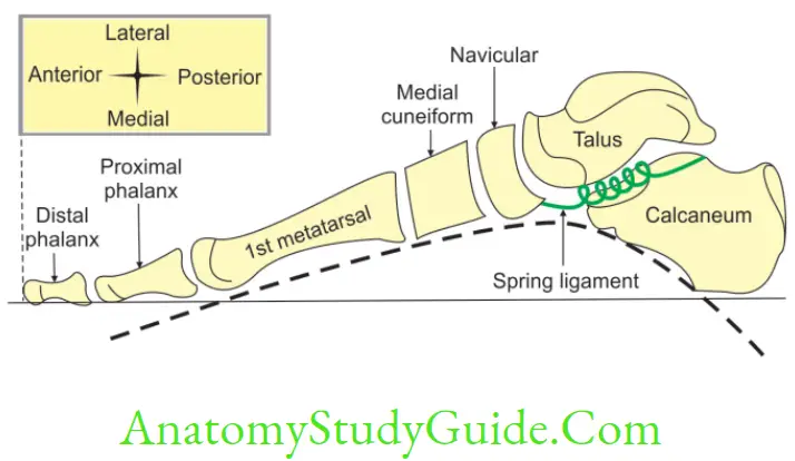
2. Medial Longitudinal Arch – Formation of the arch
1. Ends
- Anterior end is formed by heads of 1st, 2nd and 3rd metatarsals.
- Posterior end is formed by medial tubercle of calcaneum (it is short and strong).
2. Summit is formed by superior articular facet of body of talus.
3. Pillar
- Anterior pillar is formed by three cuneiform bones, navicular and talus.
- Posterior pillar is formed by medial ½ of the calcaneum.
4. Joint: Talocalcaneonavicular.
5. Classification of joints: Ball and socket type of synovial joint.
3. Medial Longitudinal Arch Factors Maintaining arch
1. Bones: They are not responsible for maintaining the stability of the arch. The bones are mostly wedge shaped with narrower edge facing plantar surface. The head of talus acts as keystone.
2. Tie beam (bow string)
- Plantar aponeurosis: It is a most important ligament stretching like a tie beam. By the contraction, it increases the height of the arch.
- Flexor hallucis longus: It also acts as tie beam. It extends from sustantaculum tali to great toe. It gives slip to its weakest sister flexor digitorum longus, which acts on the medial 3 digits. It is most efficient means of maintaining an arch as it ties the two pillars. This is the bulkiest of the calf muscles, which is multipennate.
3. Intersegmental ties (staples)
- Spring ligament: It extends from sustantaculum tali of calcaneus to tuberosity of navicular bone. It is next important ligament for maintenance of medial longitudinal arch.
- Flexor hallucis brevis also acts as staples.
4. Slings: They are
- Tendon of tibialis anterior, and
- Tendon of peroneus longus. They are inserted into the same two bones (medial cuneiform and 1st metatarsal bone). Tibialis anterior increases the height of medial longitudinal arch. Peroneus longus decreases height of the medial longitudinal arch.
5. Tibialis anterior and posterior have a significant influence on the medial longitudinal arch as they bring out inversion.
4. Medial Longitudinal Arch Applied anatomy
1. Absence of arches is called flat foot. It is also called pes planus.
2. The exaggeration of longitudinal arch of the foot is known as pes cavus.
3. The deformities of foot.
- Clubfoot (a congenital deformity of the foot, which is twisted out of shapes or position)
- Talipes calcaneus: Person walks on heel (talipes—clubfoot).
- Talipes equinus: Person walks on toes.
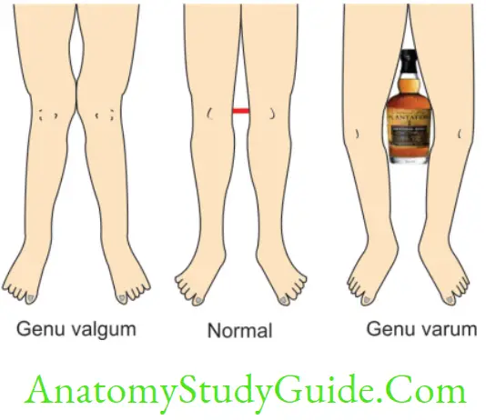
- Talipes varus: The foot is bent inwards, persons walk on the outer border, and the foot is inverted and adducted.
- Talipes valgus: Bent outwards, person walks on inner border of foot, i.e. foot everted and abducted.
- Talipes equinovarus: Clubfoot, inverted, adducted and plantar flexed. It is associated with spina bifida.
Describe lateral longitudinal arch under following heads
1. Functions,
2. Formations of the arch,
3. Factors maintaining arch, and
4. Applied anatomy.
1. Lateral Longitudinal Arch Functions:
The lateral longitudinal arch is formed by a few bones and joints, therefore, rigidity is more and helps in transmission of weight and thrust.
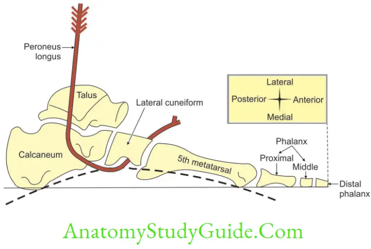
2. Lateral Longitudinal Arch Formations of Arch
1. Ends
- Anterior end is formed by cuboid and by the heads of 4th and 5th metatarsal bones.
- Posterior end is formed by lateral part of calcaneum, which is short and strong.
2. Summit is formed by articular facet of superior surface of calcaneum.
3. Pillar
- Anterior pillar is formed by 4th, 5th metatarsals and cuboid.
- Posterior pillar is formed by lateral 1/2 of the calcaneum.
4. Joint involved is calcaneocuboid.
5. Classification of joint: Saddle type of synovial joint.
3. Lateral Longitudinal Arch Factors Maintaining Arch
1. Bones: Bones do not play any important role in maintenance of the arch. A lar projection of cuboid, called calcanean angle, occupies the lower part of anterior surface of calcaneum. This bony projection maintains the upward tilt of the long axis of calcaneus.
2. Tie beam or bow string
1. Lateral part of plantar aponeurosis is a main structure acting as tie beam.
2. The other structures forming the tie beam are
- Tendons of flexor digitorum longus of the 4th and 5th toes,
- The lateral ½ of flexor digitorum brevis, and
- Abductor digiti minimi.
3. Intersegmental tie or staples
- Short plantar ligament is very thick. It fills the concavities of calcaneus and cuboid.
- Long plantar ligament is thin and extends from the heel to both ridges of cuboid. It helps to maintain the concavity of the arch.
4. Sling: Peroneus longus by contraction increases the height of lateral longitudinal arch. It is the most important single factor in maintaining its integrity.
4. Lateral Longitudinal Arch Applied Anatomy
1. Absence of arches is called flat foot. It is also called pes planus.
2. The exaggeration of longitudinal arch of the foot is known as height of pes cavus
3. The deformities of foot
- Clubfoot: A congenital deformity of the foot, which is twisted out of shapes or position.
- Talipes calcaneus: Person walks on heel (talipes—Clubfoot).
- Talipes equinus: Person walks on toes.
- Talipes varus: The foot is bent inwards, persons walk on the outer border, and the foot is inverted and adducted.
- Talipes valgus: Bent outwards, person walks on inner border of foot, i.e. foot everted and abducted.
- Talipes equinovarus: Clubfoot, inverted, adducted and plantar flexed. It is associated with spina bifida.
Leave a Reply