Axilla Enumerate The Muscles Acting In Raising The Arm Above The Head
- Serratus anterior, and
- Trapezius.
Axillary Anatomy
Table of Contents
Question-1: Describe Axilla Under The Following Heads
1. Axilla Boundaries,
2. Axilla Walls,
3. Axilla Contents, and
4. Axilla Applied anatomy.
Upper Part Of The Arm Introduction:
It is pyramid ![]() shaped area. It is present on the medial side of the upper part of the arm.
shaped area. It is present on the medial side of the upper part of the arm.
1. Axilla Boundaries
- .Apex is also called cervicoaxillary canal and is formed
- Medially: By the outer border of 1st rib.
- Laterally: By the superior border of the scapula.
- Anteriorly: By the posterior surface of the clavicle.Base is formed by
Read And Learn More: Anatomy Notes And Important Question And Answers
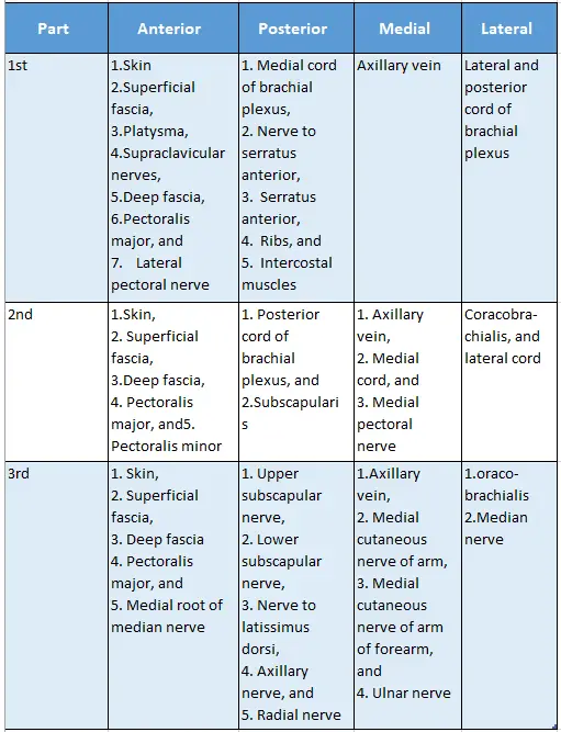
- Skin,
- Superficial fascia, and
- Deep fascia extending from anterior axillary fold to posterior axillary fold.
2. Axilla Walls
The anterior wall is formed by the following structures.
- The superficial layer is formed by the pectoralis major.
- The deep layer is formed by the subclavius, clavipectoral fascia, pectoralis minor
- And suspensory ligament of the axilla from above downwards.
- The anterior axillary fold is usually the level of the 5th rib.
Axillary Artery Branches
2. Posterior wall is formed by
- Subscapularis muscle,
- Latissimus dorsi muscle, and
- Tere major muscle.
- The posterior axillary fold is at the lower fold and corresponds to the inferior edge of
- serratus anterior.
3. Lateral wall is formed by
- Intertubercular sulcus of the shaft of the humerus which contains the long head of biceps brachii, and
- Coracobrachialis and short head of biceps.
4. Medial wall is broad and formed by
- Upper 4 to 5 ribs and their intercostal muscles,
- The upper part of the serratus anterior is covered by a strong fascia,
- Long thoracic nerve, and
- Intercostobrachial nerve (T2).
Axillary Anatomy
3. Contents of Axilla
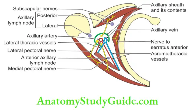
1. Axillary sheath
2. Axillary artery and its branches
- Superior thoracic artery,
- Acromiothoracic artery,
- Lateral thoracic artery,
- Subscapular artery,
- Posterior circumflex humeral artery, and
- Anterior circumflex humeral artery
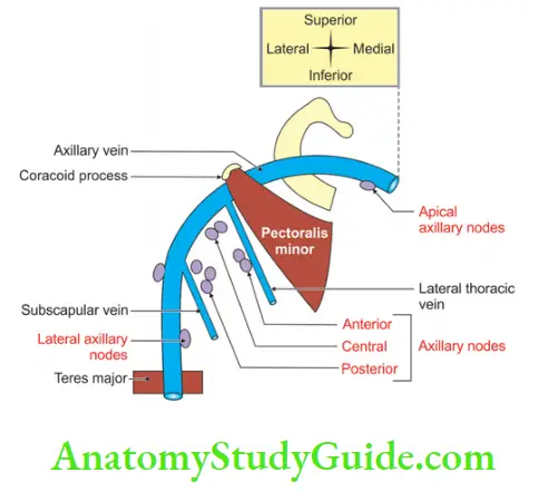
3.Axillary vein and its tributaries,
- Superior thoracic vein,
- Acromiothoracic vein,
- Lateral thoracic vein,
- Subscapular vein,
- Posterior circumflex humeral vein, and
- Anterior circumflex humeral vein
Contents Of Axilla
4. Cords Of Brachial Plexus And Their Branches
- Lateral cord and its branches
-
- Lateral pectoral nerve,
- The lateral root of the median nerve, and
- Musculocutaneous nerve.
2. Medial cord and its branches
-
- The medial root of the median nerve,
- Medial pectoral nerve,
- The medial cutaneous nerve of the arm,
- The medial cutaneous nerve of the forearm, and
- Ulnar nerve.
3. Posterior cord and its branches
-
- Upper subscapular,
- Lower subscapular,
- Nerve to latissimus dorsi,
- Axillary nerve, and
- Radial nerve
Note: The two subscapular nerves and the nerve to latissimus dorsi are present on the posterior wall.
4. Long thoracic, and
5.Intercostobrachial nerves.
Axillary lymph nodes
-
- Anterior,
- Posterior,
- Lateral,
- Central, and
- Apical.
6. Axillary fat
7. Axillary tail of breast
Axillary Artery Branches
8. Muscles
-
- The proximal part of the biceps brachii, and
- Coracobrachialis.
Contents Of Axilla
4. Axilla Applied Anatomy
1. Abscess in superficial or deep to clavipectoral fascia.
-
- Edge of anterior axillary fold, or
- Deltopectoral groove.
2. Between the pectoralis major and minor
Deep to pectoralis minor muscle. Here pus would surround vessels and nerves and ascend into the neck or may track along the vessels in the arm.
3. Axillary abscess is drained by putting the knife at the base of the axilla midway between the anterior and posterior axillary fold and moving towards on thoracic side.
4. One should be careful of the relations of vessels while removing lymph nodes.
Axillary Anatomy
Axilla Applied Anatomy Axillary Fascial ‘Tent’
The axillary fascial ‘tent’: The axillary lymph nodes are enclosed by layers of fascia that resemble a tent lying on its side. This ‘tent’ can be described as follows:
1. Axillary Fascial ‘Tent Boundaries
-
- Anterior wall: Clavipectoral fascia including the pectoralis minor muscle.
- Postero-inferior wall: Deep fascia covering the chest wall upper ribs and intercostal muscles.
- Antero-superior wall: Fascia covering the subclavian vessels.
- Apex: Points upwards and medially where these layers of fascia come into contact with each other.
- Base: Points downwards and laterally and is open.
2. Axillary Fascial ‘Tent Content
-
- The lymph nodes are enclosed in this fascial ́tent’.
3. Axillary Fascial ‘Tent Applied Anatomy
-
- Any operation designed for a block dissection of the axillary lymph nodes should excise the ‘tent’ intact and should not enter the ‘tent’ which may result in the liberation of malignant cells.
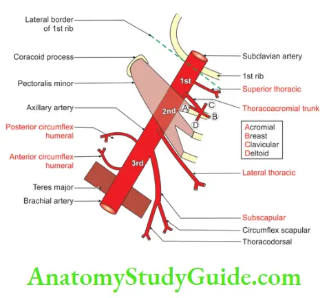
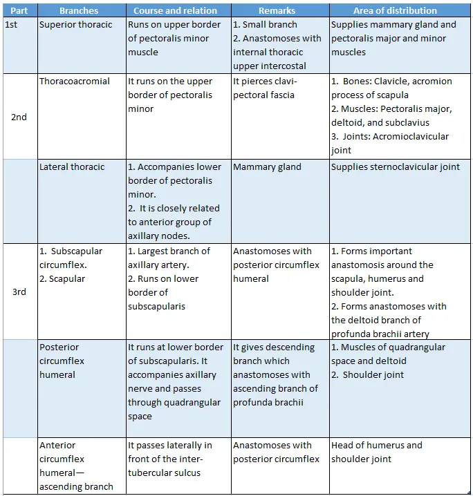
Name The Branches That Arise From Each Of The Three Parts Of The Axillary Artery
1. The axillary artery is divided into three parts by pectoralis minor.
The first part gives one branch.
The second part gives two branches.
The third part gives three branches. The branches from each part are
- 1st part (medial to pectoralis minor): Superior thoracic
- 2nd part (deep to pectoralis minor)
1. Acromiothoracic which gives branches as. (ABCD)
Acromial,
Breast,
Clavicular, and
Deltoid.
2. Lateral thoracic
3. 3rd part
- Subscapular,
- Anterior circumflex humeral artery, and
- Posterior circumflex humeral artery.
Contents Of Axilla
Axillary Anatomy
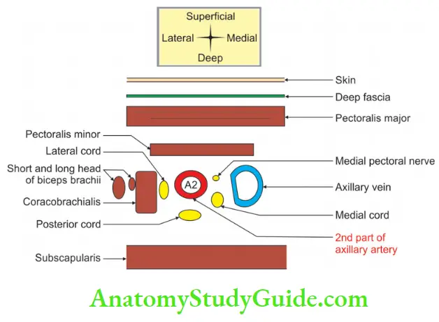
2nd Part Of Axillary Artery Introduction: It is deep to pectoralis minor.
1. Relations of 2nd part of the axillary artery.
1. 2nd Part Of Axillary Artery Anterior
- Skin,
- Superficial fascia,
- Deep fascia,
- Pectoralis major, and
- Pectoralis minor.
2. 2nd Part Of Axillary Artery Posterior
- A posterior cord of the brachial plexus, and
- Subscapularis.
3. 2nd Part Of Axillary Artery Lateral
- A lateral cord of the brachial plexus, and
- Coracobrachialis.
4. 2nd Part Of Axillary Artery Medial
- A medial cord of the brachial plexus,
- Medial pectoral nerve, and
- Axillary vein.
2. 2nd Part Of Axillary Artery Branches
1. Acromiothoracic artery which gives branches as
- Acromial for acromial part of clavicle,
- Breast to the upper part of the mammary gland,
- Clavicular supplies clavicle, and
- Deltoid to the deltoid muscle.
Contents Of Axilla
2. Lateral thoracic which is the main artery of the lateral half of the mammary gland.
Question-2: Describe the Axillary Artery Under the Following Heads
1. Axillary Artery Origin,
2. Axillary Artery Course and relations,
3. Axillary Artery Branches, and
4. Axillary Artery Applied anatomy.
Answer:
Axillary Artery Introduction: It is the artery of the axilla.
1. Origin
It is the continuation of 3rd part of the subclavian artery.
Extent: It extends from the outer border of 1st rib to the lower border of the teres major.
2. Axillary Artery Course and relations
The axillary artery is divided into three parts by the pectoralis minor muscle
- 1st part: Medial to pectoralis minor.
- 2nd part: Deep to pectoralis minor.
- 3rd part: Lateral to pectoralis minor.
Relations of the axillary artery
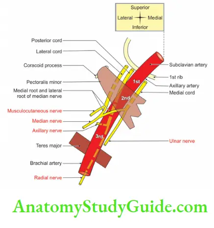
Clue: Let us understand relations from the third part of the axillary artery.
The branches of the respective cords of the brachial plexus are related to the 3rd part of the axillary artery.
The cords of the brachial plexus are related to the respective positions of 2nd part of the axillary artery.
relations of the axillary artery with the text below.
To get the relations of the cords of the brachial plexus to 1st part of the axillary artery, one needs to understand the following things.
1. Lateral cord lies on the lateral side of 1st part of the axillary artery.
2. To get the relations of the medial and posterior cords of the brachial plexus, one needs to
- Rotate the posterior cord and medial cord.
- The rotation should be 90° and in a clockwise direction.
- The rotation should be from relations of 2nd part of the axillary artery.
3. The final position will be: The medial cord is related on the posterior side and the posterior cord is related on the lateral side of 1st part of the axillary artery.
3. Axillary Artery Branches
Branches of the axillary artery and their distribution
4. Axillary Artery Applied anatomy
- The axillary artery can be effectively compressed against the humerus, along the inner edge of the coracobrachialis.
- The axillary artery is likely to rupture during the reduction of an old dislocated head of the humerus.
Axillary Lymph Nodes
- Anterior axillary lymph nodes,
- Lateral axillary lymph nodes,
- Posterior axillary lymph nodes,
- Central axillary lymph nodes, and
- Apical axillary lymph nodes
Axillary Lymph Nodes
The lymph nodes of the axilla are described in the following ways
Location of axillary lymph nodes
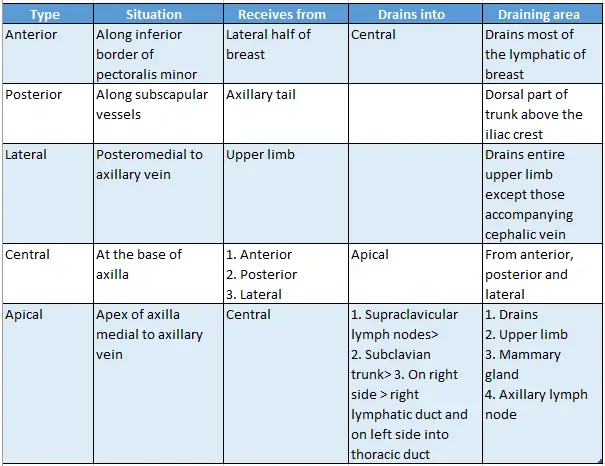
Applied Anatomy:
Anterior axillary lymph nodes are enlarged in malignancy of lateral
1/2 of the mammary gland.
Leave a Reply