The Permanent Mandibular Canine
There are two mandibular canines, one in each arch, and are located distal to the mandibular lateral incisor.
Table of Contents
- They resemble the maxillary canine with the following major differences:
- The crown is mesiodistally narrower; the length may be as long or longer by 0.5–1 mm.
- The root is shorter; the labiolingual dimension is lesser by a fraction of a mm.
- The cingulum is not well developed; marginal ridges are not as prominent and thus the lingual surface resembles the mandibular lateral incisor.
Read And Learn More: Oral Anatomy Notes
- The cusp is not as well developed; cusp ridges are thinner labiolingually.
- The cusp tip is in line with the centre or lingual to a line bisecting the root labiolingually.
- The morphology of this tooth will be described from the labial, lingual, mesial, distal and incisal aspects.
- The nomenclature, chronology and measurement of the mandibular canine are given in tables.
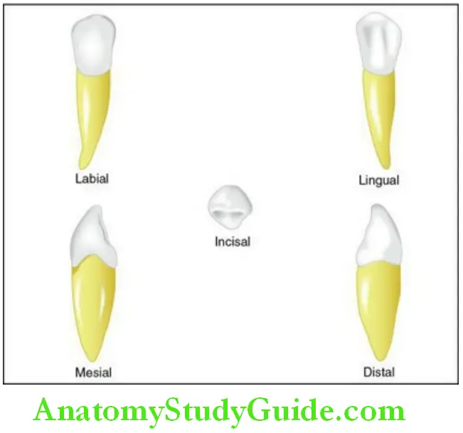
Nomenclature: Mandibular canine:
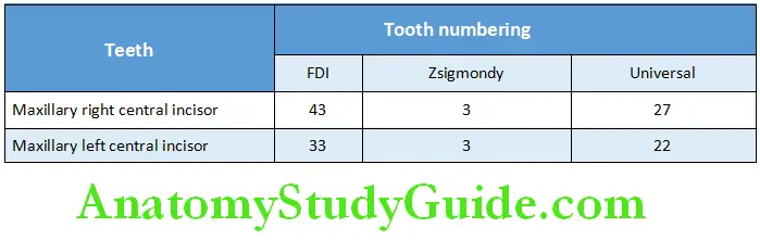
Mandibular canine: Chronology and measurements:
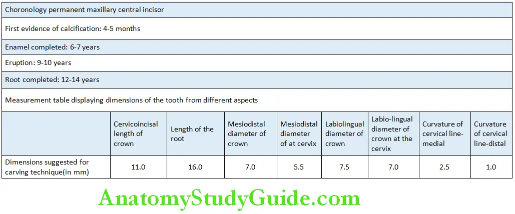
Labial aspect:
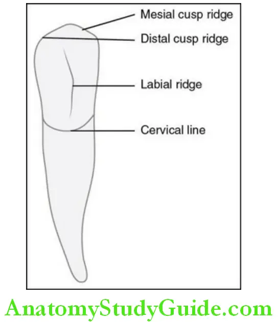
Features of the crown:
The mesiodistal diameter of the crown is less than the maxillary canine by about 1 mm, but the length of the crown might be longer.
The crown seems to be twisted distally on the root base but this feature of the crown is best appreciated from the incisal aspect.
Shape and surface:
- The shape is trapezoidal.
- The surface is convex; a labial ridge is present extending from the Cervical third to the cusp tip but is less prominent than that of the Maxillary canine.
Mesial outline:
- The mesial outline is in line with the mesial outline of the root and is Straight.
- The mesial contact area is located within the incisal third near the Mesioincisal line angle.
Distal outline:
- The distal outline is almost parallel but more rounded than the mesial Outline.
- The distal contact area is within the incisal third but cervical to the Mesial contact area.
Cusp tip:
- The cusp tip is formed by the union of the mesial and distal slopes Meeting at an obtuse angle.
- The distal slope is longer than the mesial slope.
Cervical line:
- The cervical line is semicircular and curved apically towards the root.
Features of the root:
- The root is shorter than the maxillary canine by 1–2 mm and the apex is more pointed.
- The mesial outline is straight to the middle third and then tapers to the apex.
- The distal outline is slightly convex till the middle third and then tapers.
- The curvature of the root if present is in a mesial direction.
Lingual aspect:
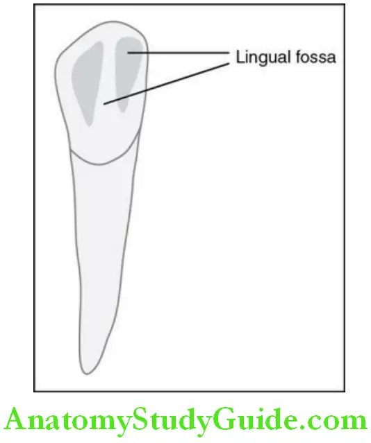
Features of the crown:
Shape and surface:
- The surface is flat, smooth and regular as in the mandibular incisor; the Shape is the same as the labial side.
Cingulum:
- The cingulum is smooth, poorly developed and less prominent.
- The cingulum is generally distally placed.
Lingual fossa and marginal ridges:
- The lingual fossa is divided by the lingual ridge into the mesial lingual Fossa and distal lingual fossa; the lingual ridge runs from the cingulum to The cusp tip.
- The lingual fossa appears flatter as the marginal ridges are less Prominent.
- The mesial marginal ridge is longer than the distal marginal ridge due To the distal placement of the cingulum.
Features of the root:
- The root is narrower than the maxillary canine due to increased lingual Convergence.
Mesial aspect:
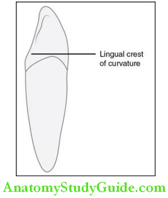
Features of the crown:
Shape and surface:
- The crown is wedge or triangular-shaped with the apex at the cusp tip.
- The surface is convex.
Labial and lingual outline:
- The labial outline is less curved from the cervical line to the crest of Curvature than the maxillary canine; from the crest curvature to the Cusp tip, it is straight but lingually inclined.
- The labial crest of curvature is located in the cervical third.
- The lingual outline is less convex than the maxillary canine in the Cervical third due to the less prominent cingulum; it is concave from The cervical third to the cusp tip.
- The lingual crest of curvature is located in the cervical third.
Cusp tip:
- The cusp tip is generally lingually placed or maybe in line with the
Centre of the root
Cervical line:
The cervical line is more curved in an incisal direction than in the
Maxillary canine.
Features of the root:
- The root is conical with a more pointed apex than in the maxillary canine.
- The developmental depressions are more prominent and deeper.
Distal aspect:
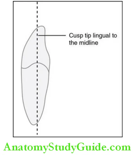
Features of the crown:
The distal aspect is similar to the mesial aspect except for the following differences:
- Distolingual twist of the crown
- Lesser curvature of the cervical line
Incisal aspect:
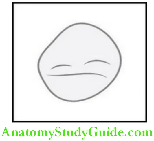
- The mesiodistal dimension is less than the labiolingual dimension.
- The cusp tip is lingual to the centre and mesially located.
- The mesial cusp ridge is lingually inclined.
- The distal half of the labial outline is more lingually inclined due to the distolingual twist.
The features that can help in the identification of the mandibular canine are given in the box.
Features to identify the mandibular canine:
- The distal cuspal slope is longer than the mesial.
- The incisal ridge is lingual to the long axis of the tooth from the proximal aspect.
- Distally placed less prominent cingulum.
- The Crown is tilted distally on the root base.
Differentiation Between The Right And Left Mandibular Canine
- Distolingual twist
- Distally placed cingulum
- The distal cuspal slope is longer than the mesial.
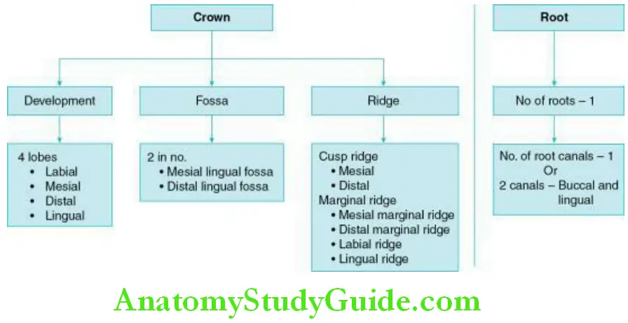
Development And Landmarks: Crown And Root Of The Mandibular Canine
Variation And Developmental Anomalies
- The canine may exhibit a long or a short root.
- Root bifurcation may be seen.
The Permanent Mandibular Canine Synopsis
Labial aspect:
Crown:
- The shape is trapezoidal.
- The surface is convex; a labial ridge is present extending from the cervical third to the cusp but is less prominent than that of the maxillary canine.
- The mesial contact area is located within the incisal third near the mesioincisal line angle
- The distal contact area is within the incisal third but cervical to the mesial contact area.
- The distal slope is longer than the mesial slope.
Root:
- It is shorter than the maxillary canine by 1–2 mm and the apex is more pointed.
Lingual aspect:
Crown:
- The surface is flat, smooth and regular; the shape is the same as the labial side.
- The cingulum is smooth, poorly developed, and less prominent and distally placed.
- The lingual fossa is divided by the incisal ridge into the mesial lingual fossa and distal lingual fossa.
- The mesial marginal ridge is longer than the distal marginal ridge due to the distal placement of the cingulum.
Root:
- It is narrower than the maxillary canine due to the lingual convergence.
Mesial aspect:
Crown:
- The crown is wedge or triangular-shaped with the apex at the cusp tip.
- The labial crest of curvature is located in the cervical third.
- The lingual crest of curvature is located in the cervical third.
- The cusp tip is generally lingually placed or maybe in line with the centre of the root.
Root:
- It is conical with a more pointed apex than in the maxillary canine.
Distal aspect:
Crown:
- It is similar to the mesial aspect except for the distolingual twist of the crown and lesser curvature of the cervical line.
Incisal aspect:
- The mesiodistal dimension is less than the labiolingual dimension.
- The cusp tip is lingual to the centre and mesially located.
- The mesial cusp ridge is lingually inclined.
- The distal half of the labial outline is more lingually inclined due to the distolingual twist.
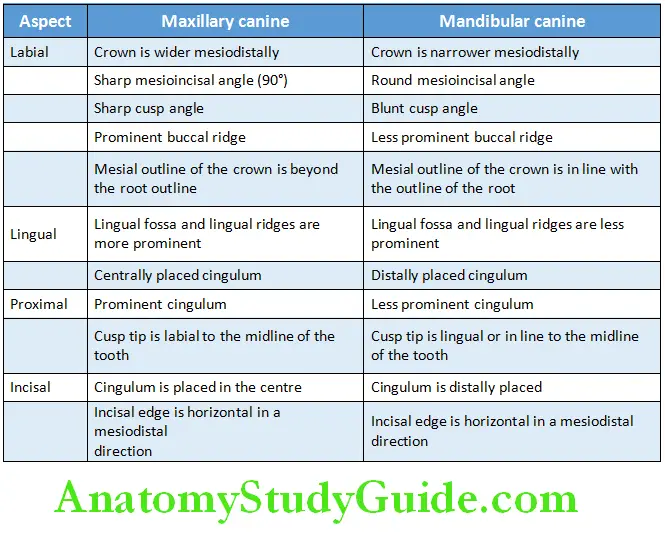
Differences Between The Maxillary And Mandibular Canine
Differences between maxillary and mandibular canine:
Leave a Reply