The Popliteal Fossa
Enumerate The Muscles Forming The Boundaries Of The Popliteal Fossa
Table of Contents
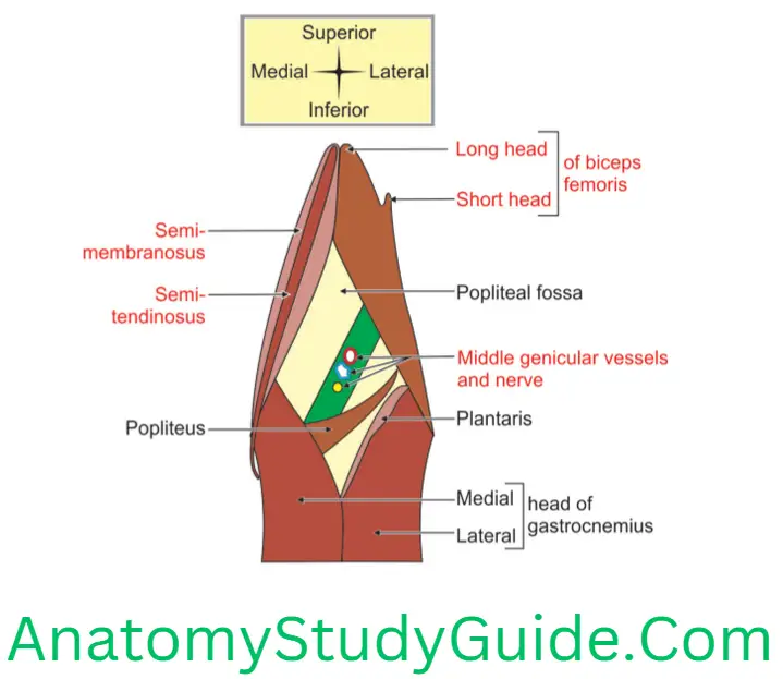
Boundaries Of The Popliteal Fossa : It is bounded by
1. Superomedially
- Semimembranosus medially, and
- Semitendinosus laterally
2. Superolaterally superficially: Tendon of biceps femoris.
3. Inferolaterally
- Lateral head of gastrocnemius, and
- Plantaris.
4. Inferomedially: Medial head of the gastrocnemius.
Read And Learn More: Anatomy Notes And Important Question And Answers
Enumerate The Structures Forming The Floor Of Popliteal Fossa
Floor Of Popliteal Fossa (anterior wall) is formed by following structures. They are from above downward.
1. Popliteal surface of femur. It is overlaid with fat,
2. Capsule of knee joint,
3. Oblique popliteal ligaments are the fibers of the posterior part of the fibrous capsule. They are parallel to the popliteus and, so, tend to save it from being overstretched,
4. Popliteal muscle, and
5. Fascia covering popliteus muscle: It is derived from the semimembranosus muscle. It is very thin laterally, and thick and strong medially.
Floor Of Popliteal Fossa is pierced by
1. Middle genicular vessels (branch/tributary of popliteal artery/vein),
2. Middle genicular nerve (branch/tributary), and
3. Genicular branch of the posterior division of obturator nerve.
Contents of the Popliteal Fossa
1. Main content of the popliteal fossa is fat.
2. The most Important Contents are
1. Tibial Nerve,
2. Common Peroneal Nerve,
3. Popliteal Artery and its branches. It gives
- Muscular branches to muscles of the popliteal fossa,
- Sural arteries to the gastrocnemius, and
- Genicular branches to the knee joint.
4. Popliteal vein and its muscular tributaries.
Describe Popliteal Fossa Under Following Heads
1. Popliteal Fossa Gross anatomy
2. Popliteal Fossa Boundaries,
3. Popliteal Fossa Roof,
4. Popliteal Fossa Floor,
5. Popliteal Fossa Contents,
6. Popliteal Fossa Relations, and
7. Popliteal Fossa Applied anatomy
Popliteal Fossa Introduction: It is a diamond-shaped fossa present on posterior aspect of knee joint. It is homologous with cubital fossa, present in front of the elbow joint.
1. Popliteal Fossa Gross anatomy
1. Shape of popliteal fossa in various positions
- Flexed knee: It is diamond-shaped. It is hollow area between the hamstring tendons.
- In the extended knee: Hamstring tendons lie against the femoral condyles. The fat of the popliteal space bulges the roof of the fossa.
2. Popliteal Fossa Communication: It communicates proximally to adductor canal and distally to posterior part of leg.
2. Popliteal Fossa Boundaries: It is bounded by
1. Superomedially
1. Superficially
- Semimembranosus medially, and
- Semitendinosus laterally.
- Supplemented by the Gracilis, Sartorius, and Adductor Magnus.
2. Deep: Medial supracondylar ridge of femur.
2. Superolaterally
- Superficially: Tendon of biceps femoris.
- Deep: Lateral supracondylar ridge of femur
3. Inferolaterally
- Lateral head of gastrocnemius, and
- Plantaris.
4. Inferomedially: Medial head of the gastrocnemius.
3. Popliteal Fossa Roof (posterior wall) is formed by following structures from superficial to deep.
1. Skin,
2. Superficial fascia, and
3. Fascia lata. It is strengthened by transverse fibres. It continues below fascia cruris. It is pierced by
- Sural nerve (cutaneous branch of tibial nerve),
- Sural communicating nerve, branch of common peroneal nerve,
- Posterior femoral cutaneous nerve of thigh,
- Short saphenous vein, and
- Lateral cutaneous nerve, branch of common peroneal nerve.
4. Popliteal Fossa Floor (anterior wall) is formed by the following structures. They are from above downward.
1. Popliteal surface of femur. It is overlaid with fat,
2. Capsule of knee joint,
3. Oblique popliteal ligaments are the fibres of the posterior part of the fibrous capsule. They are parallel to the popliteus and, so, tend to save it from being overstretched.
4. Popliteal muscle.
5. Fascia covering popliteus muscle: It is derived from the semimembranosus muscle. It is very thin laterally, thick and strong medially.
6. Floor is pierced by
- Middle genicular vessels (branch/tributary of popliteal artery/vein),
- Middle genicular nerve, and
- Genicular branch of posterior division of obturator nerve.
5. Popliteal Fossa Contents
1. Main content of popliteal fossa is fat.
2. The most important contents are
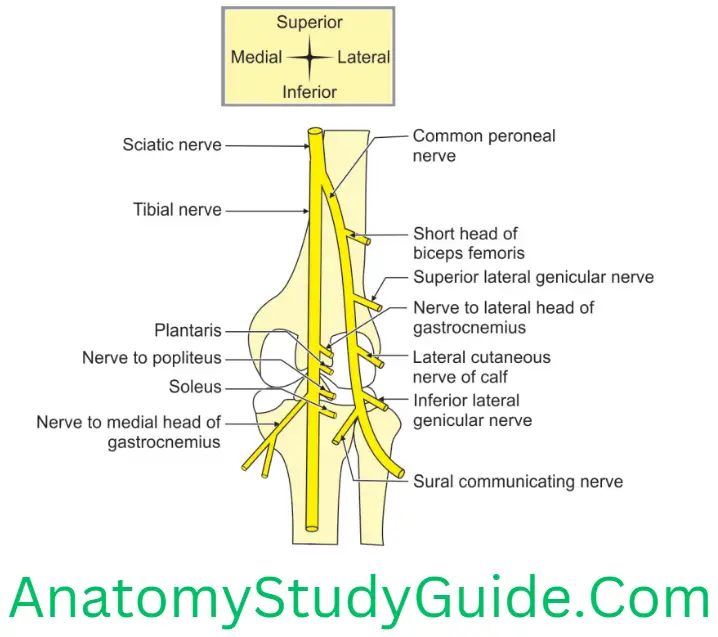
1. Tibial nerve, one of two terminal branches of sciatic nerve given at the upper part of popliteal fossa. In the fossa, this nerve gives branches to
- Both heads of the gastrocnemius,
- Plantaris,
- Popliteus,
- Soleus, a muscle of the leg lying deep to the gastrocnemius, and
- Genicular branches to the knee joint.
2. Common peroneal nerve: It is one of two terminal branches of sciatic nerve given at the upper part of popliteal fossa. It does not have any muscular branches in popliteal fossa except branch to short head of biceps. It gives two cutaneous and two articular branches in popliteal fossa. These are
- Sural communicating nerve (that joins the sural nerve),
- Lateral cutaneous nerve of the calf that supplies skin on the lateral side of the back of the leg, and
- Genicular branches to the knee joint.
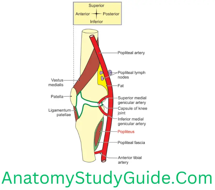
3. Popliteal artery: It gives
- Muscular branches to muscles of fossa,
- Sural arteries to gastrocnemius, and
- Genicular branches to knee joint.
4. Popliteal vein and its muscular tributaries.
3. The less important contents are
1. Descending genicular branch of posterior division of obturator nerve
2. Posterior cutaneous nerve of thigh
3. Popliteal lymph nodes
1. Number: They are 6 in number arranged in 3 sets (from superficial to deep),
- Superficial nodes,
- Intermediate nodes,
- Deep nodes,
2. Afferent lymphatics: They receive lymphatics from
- Superficial nodes, and
- Deep parts of the leg and foot.
3. Palpation of lymph nodes: They are palpated by the flexing of the knee which relaxes the deep fascia of the roof of the popliteal fossa.
4. Popliteal Fossa Pad of fat
6. Popliteal Fossa Relations: The relations of the contents are described at three different levels in

7. Popliteal Fossa Applied anatomy
Popliteal pulse is felt behind the knee, deep in the popliteal fossa with the knee flexed.
The tibial nerve is rarely injured in the popliteal fossa. Injury to tibial nerve in popliteal fossa is associated with motor paralysis of muscles supplied by it.
- Foot is dorsiflexed at ankle joint and held in eversion.
- There is a sensory loss on entire sole of foot.
All the popliteal swelling may not be from popliteal fossa. Hence, it is customary to examine the knee joint.
Following are the causes of swelling in the popliteal fossa.
- A pulsatile midline swelling in the popliteal fossa is due to aneurysm of popliteal artery. It demonstrates a bruit on auscultation. It is an audible rumbling made by turbulent blood flow.
- It compresses the popliteal vein and results in oedema of leg. It favours formation of thrombosis.
- Arterial adventitial cyst: It is a swelling arising from tunica adventitia of popliteal artery.
Features Of Popliteal Abscess
Popliteal Abscess Causes
- Infection of popliteal lymph nodes, and
- Acute suppurative osteomyelitis of the lower end of the femur.
Popliteal Abscess Features
- It is slow to heal when drained,
- It presses on the nerves and vessels of the popliteal fossa, and
- It is drained through an incision on the lateral aspect, anterior to the tendon of biceps femoris.
Popliteal swelling is unusually painful because of the
- Unyielding character of the walls of the fossa, and
- Fatty areolar tissue in which they are embedded.
A popliteal cyst (Baker’s cyst) is a synovial out pouching that arises from the posteromedial aspect of the knee joint. The synovial membrane of the knee joints out pouches between the medial head of gastrocnemius and the semimembranosus tendon to lie medially within the popliteal fossa. It disappears in flexion of knees.
Pes anserinus (like a goose): It is a bursa around the tendons of sartorius, gracilis, and semitendinosus.
Sebaceous cyst: A swelling of sebaceous gland in the skin.
Swelling of soft tissues: Lipoma, sarcoma.
Swelling of vein: Varicosities of the short saphenous vein in the roof of the fossa.
Swelling of knee joint-joint effusion.
Tumour of the lower end of femur or upper end of tibia.
Popliteal artery lies directly on the bone and can be damaged by supracondylar fractures.
Terminal Branches Of Popliteal Artery
Terminal branches Of Popliteal Artery : Two terminal branches are
- 1. Anterior tibial artery, and
- 2. Posterior tibial artery.
Describe Popliteal Artery Under Following Heads
1. Popliteal Artery Origin,
2. Popliteal Artery Termination,
3. Popliteal Artery Course,
4. Popliteal Artery Extent,
5. Popliteal Artery Relations,
6. Popliteal Artery Branches, and
7. Popliteal Artery Applied anatomy.
Popliteal Artery Introduction: Popliteal artery is deeply placed structure in the popliteal fossa.
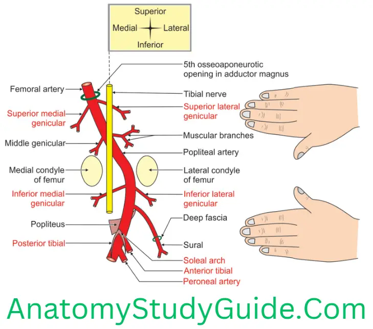
1. Popliteal Artery Origin: It is a continuation of the femoral artery at the 5th osseo-aponeurotic opening in adductor magnus.
2. Popliteal Artery Termination: It terminates at the lower border of popliteus by dividing into anterior and posterior tibial arteries.
3. Popliteal Artery Course: It runs downwards and laterally and passes in between two condyles. It ends at the lower border of popliteus. It terminates.
4. Popliteal Artery Extent: It extends a hand’s breadth above the knee to a hand’s breadth below the knee. It is about 8″ (20 cm) long.
5. Popliteal Artery Relations: For convenient purpose, it is divided into upper, middle and lower 1/3rd of popliteal fossa.
1. In the upper 1/3rd
1. Anterior or deep
- Popliteal surface of the femur and
- Fat.
2. Posterior or superficial: Semimembranosus.
3. Laterally
- Popliteal vein, and
- Tibial nerve.
2. Middle 1/3rd
- Anterior or deep: Capsule of knee joint
- Immediate posteriorly: Popliteal vein
- Most posterior or superficial: Tibial nerve
- Medially Medial condyle of femur and Medial head of gastrocnemius
5. Laterally
- Lateral head of gastrocnemius,
- Plantaris, and
- Lateral condyle of femur
3. Lower 1/3rd
1. Anteriorly or deep: Popliteus with its fascia.
2. Posterior or superficial
- Plantaris,
- Lateral head of gastrocnemius, and
- Nerve to lateral head of gastrocnemius.
- Posterior: Soleus muscle.
6. Popliteal Artery Branches
1. Muscular
1. Upper 2 to 3 supply the
- Adductor magnus, and
- Hamstrings.
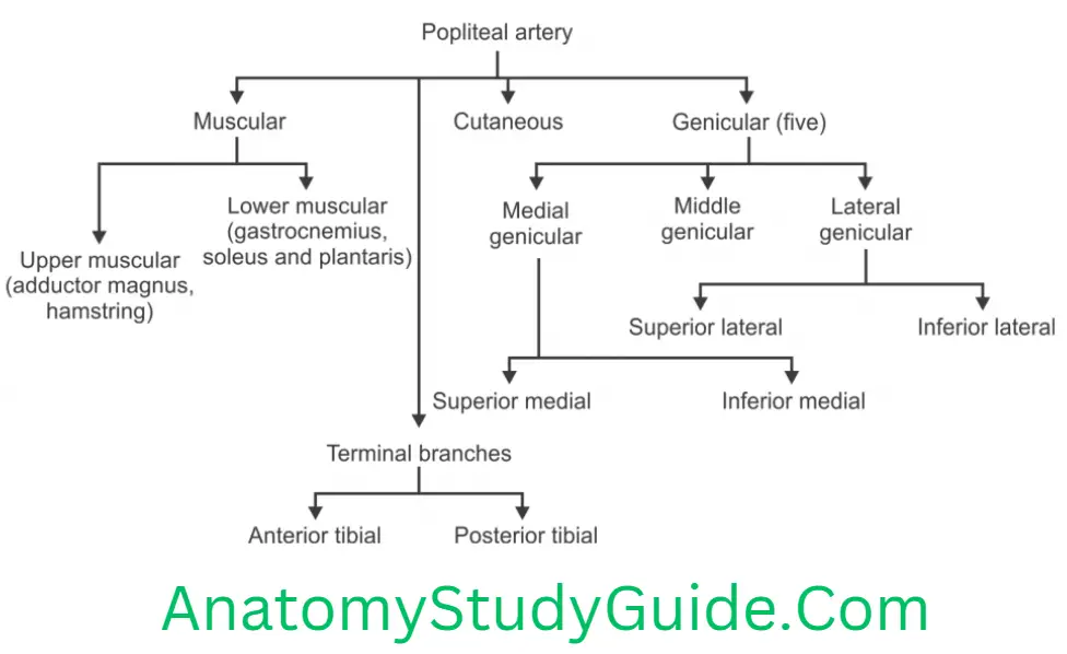
2. Lower muscular or sural branches supply
- Gastrocnemius,
- Soleus, and
- Plantaris.
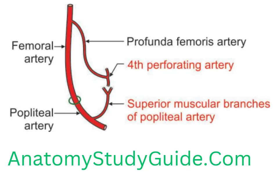
3. Upper muscular branches terminate by anastomosing with the 4th perforating artery.
2. Cutaneous branches arise either directly from the popliteal artery, or indirectly from its muscular branches. There are three superficial sural arteries. The central or median vessel is largest and most constant. It is sometimes named the sural cutaneous artery. It is given off close to the distal angle of the fossa. It passes distally in the groove marking the junction of the two heads of the gastrocnemius. It is a companion of the short saphenous vein.
3. Genicular branches are five in number
- Two superior,
- Two inferior, and
- Middle.
4. Terminal branches are two. They are
- Anterior tibial artery
- Posterior tibial artery
7. Popliteal Artery Applied anatomy
1. Popliteal artery is one of the arteries commonly used for peripheral pulsation.
2. Weakening or loss of the popliteal pulse is a sign of a femoral artery obstruction.
3. The blood pressure in the lower limb is recorded by the auscultation of the popliteal artery.
4. In atherosclerosis of popliteal artery, the graft can be tried from the lower part of popliteal artery as it is patent.
5. A common site of atheromatous occlusion is at the beginning of the popliteal artery near the adductor hiatus,
6. In coarctation of the aorta, the popliteal pressure is lower than the brachial pressure.
7. Abnormal localised dilation of an arterial wall is called aneurysm. The popliteal artery is more prone to aneurysm than any other arteries of the body. Pressure of the aneurysm on the
- Vein may cause venous thrombosis and peripheral oedema.
- Tibial nerve may cause severe pain in the leg. The artery lies deep to the tibial nerve; an aneurysm may stretch the nerve or compress its blood supply (vasa vasorum). The pain is referred to the skin overlying the medial aspect of the calf, ankle or foot.
8. True aneurysm means involvement of all the three constituents of the arterial wall, i.e. adventitia, media and the intima.
9. In operation of aneurysm of popliteal artery, it is ligated in the adductor canal.
10. The popliteal artery is exposed by
- Deep dissection in the midline within the popliteal fossa. Care is taken not to injure the more superficial vein and nerve.
Terminal Branches Of Tibial Nerve
1. Medial plantar nerve, and
2. Lateral plantar nerve.
Enumerate The Branches Of Tibial Nerve In Popliteal Fossa
Branches of tibial nerve in popliteal fossa are
1. Medial head of gastrocnemius,
2. Lateral head of gastrocnemius,
3. Plantaris,
4. Soleus, and
5. Popliteus.
Describe The Tibial Nerve Under Following Heads
1. Tibial Nerve Root value,
2. Tibial Nerve Course and relations,
3. Tibial Nerve Branches, and
4. Tibial Nerve Applied anatomy
Introduction: It is one of the two terminal branches of sciatic nerve given at the superior angle of popliteal fossa. It is a larger branch and supplies all the muscles of back of thigh, leg and foot.
1. Tibial Nerve Root value: Ventral divisions of ventral rami of
2. Tibial Nerve Course and relations
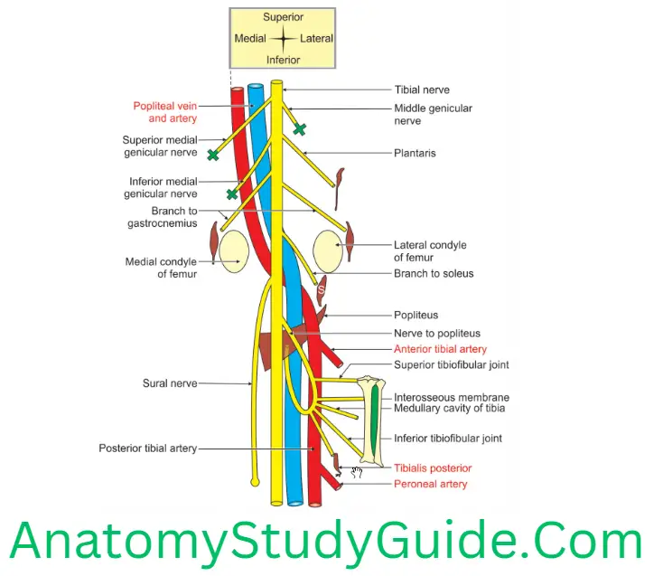
1. It lies superficial or posterior to the popliteal vessels.
2. It extends from the superior angle to the inferior angle of the popliteal fossa.
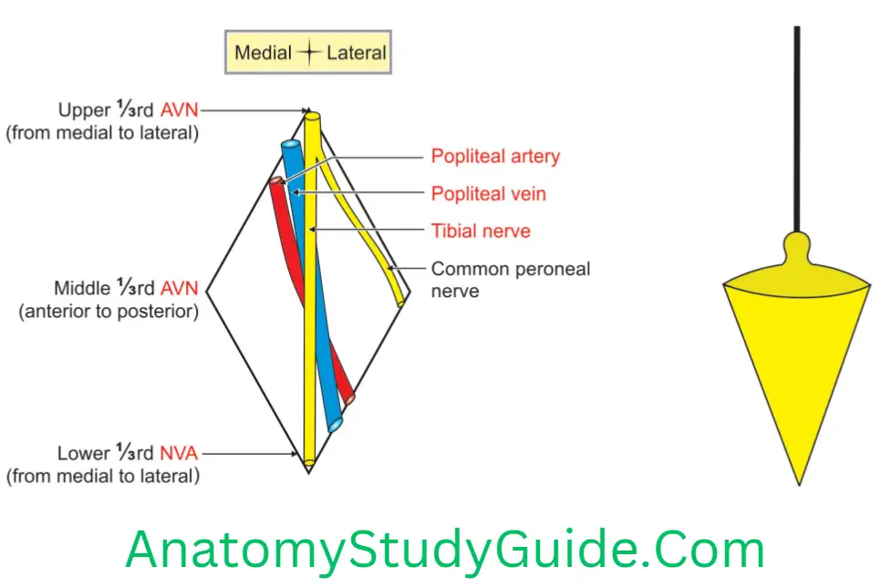
3. In the popliteal fossa, it drops like plumb from superior angle to inferior angle.
4. The relations of tibial nerve with popliteal vessels in the popliteal fossa.
- At the superior angle, tibial nerve is most lateral.
- In the middle, the nerve is most superficial.
- At the inferior angle, the nerve is most medial.
5. Distal to the popliteal fossa it passes anterior to the soleus.
6. It is the nerve of the posterior compartment of leg.
7. It descends with the posterior tibial vessels to lie between the heel and the medial malleolus.
8. It ends deep to the flexor retinaculum by dividing into the medial and lateral plantar nerves.
3. Tibial Nerve Branches
1. Collateral branches
1. Articular: Three genicular or articular branches arise in the upper part of the popliteal fossa. They are
- Superior medial genicular nerve. It lies above the medial condyle of femur, deep to the muscles.
- Middle genicular nerve. It pierces the posterior part of the capsule of the knee joint to supply structures in the intercondylar notch of femur.
- Inferior medial genicular nerve. It lies along the upper border of popliteus and reaches medial condyle of tibia.
2. Cutaneous nerve is called sural nerve. It originates in the middle of the fossa and leaves it at the inferior angle. It supplies the skin of
- Lower half of back of leg, and
- Lateral border of the foot till the tip of little toe.
3. Muscular branches arise in the distal part of the fossa for the
1. Lateral and medial heads of gastrocnemius,
2. Soleus,
3. Plantaris, and
4. Popliteus.
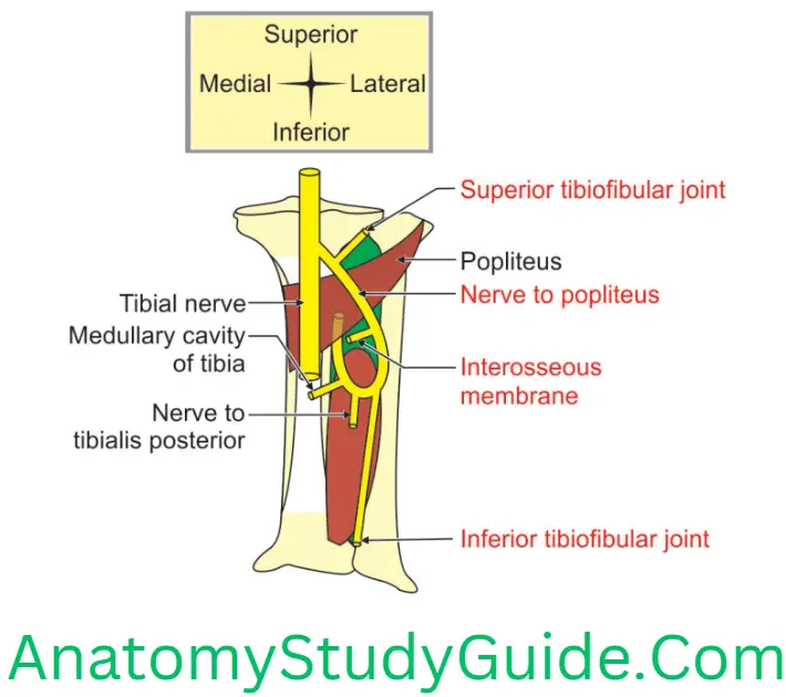
5. Nerve to the popliteus: Peculiarities of the nerve to popliteus:
- It crosses the popliteal vein and artery from medial to lateral.
- It runs downwards.
- It winds inferior border and runs its deep (anterior) surface.
- In addition to the popliteus, the nerve also supplies the
- Tibialis posterior,
- Superior tibiofibular joint,
- Medullary cavity of tibia,
- Interosseous membrane, and
- Inferior tibiofibular joint.
2. Terminal branches
1. Medial plantar, and
2. Lateral plantar
4. Tibial Nerve Applied anatomy
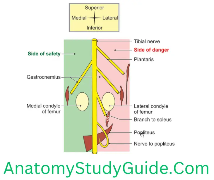
- Most of the muscular branches of tibial nerve arise from its lateral side except to the medial head of gastrocnemius. So, the medial side is called side of safety and lateral side is called side of danger. The tibial nerve is always approached from medial side to avoid the rupture to the muscular branches of the tibial nerve.
2. Damage to tibial nerve causes motor and sensory loss.
Muscles paralysed are
- Superficial and deep muscles of calf.
- Intrinsic muscles of sole.
Sensory loss
- Loss of sensation on whole of sole of foot.
- Plantar aspect of digits and nail beds on dorsum of foot.
3. Pressure of the aneurysm of popliteal artery on tibial nerve may cause severe pain in the leg.
4. Entrapment and compression of the tibial nerve (tarsal tunnel syndrome) occurs due to oedema and tightness in the ankle.
5. The pain in the heel may result from compression of tibial nerve by flexor retinaculum.
Branches Of Common Peroneal Nerve In The Fossa
Branches Of Common Peroneal Nerve In The Popliteal Fossa.
1. Common Peroneal Nerve In The Fossa Muscular: Short head of biceps femoris
2. Common Peroneal Nerve In The Fossa Cutaneous:
- Lateral cutaneous nerve of calf, and
- Sural communicating nerve.
3. Common Peroneal Nerve In The Fossa Articular
- Superior lateral genicular,
- Inferior lateral genicular, and
- Recurrent genicular.
4. Common Peroneal Nerve In The Fossa Terminal
- Deep peroneal, and
- Superficial peroneal.
Common Peroneal Nerve
Common Peroneal Nerve Introduction: It is one of the two terminal branches of sciatic nerve given at the superior angle of popliteal fossa. It is a smaller branch.
1. Root value: Dorsal division of ventral rami of segments of spinal cord.
2. Common Peroneal Nerve Course and relations
- It lies in the upper lateral part of popliteal fossa.
- It runs along the medial border of biceps femoris muscle.
- It winds around neck of fibula (postaxial bone).
- Then it lies in the substance of peroneus longus muscle.
3. Common Peroneal Nerve Branches
1. Muscular branch: Short head of biceps.
2. Cutaneous: Lateral cutaneous nerve of calf
3. Vascular: Sural communicating
4. Common Peroneal Nerve Articular
- Superior lateral genicular,
- Inferior lateral genicular, and
- Recurrent genicular.
5. Common Peroneal Nerve Terminal
1. Superficial branch supplies muscles of lateral compartment of leg. They include
- Peroneus longus
- Peroneus brevis
2. Deep branch supplies dorsiflexion (extensors) of the ankle joint. They include
- Tibialis anterior
- Extensor hallucis longus
- Extensor digitorum longus
- Peroneus tertius.
4. Common Peroneal Nerve Applied anatomy
1. It is the most commonly injured nerve.
2. It is injured due to
- Fracture of neck of fibula,
- ‘Lathi injury’ on the lateral side of knee joint, or
- Plaster on the leg: The nerve gets compressed between hard plaster and neck of fibula.
3. Effects of injury
Motor loss: Foot drop due to paralysis of dorsiflexors and evertors of foot.
Sensory loss
- Back of leg,
- Lateral side of leg, and
- Most of dorsum of foot.
Foot Drop
1. Foot Drop Definition: It is due to injury to common peroneal nerve. It leads to paralysis of dorsiflexion and eversion of foot resulting in “foot drop”.
2. Foot Drop Causes
- Fracture of neck of fibula,
- ‘Lathi injury’ on the lateral side of knee joint, or
- Plaster on the leg: The nerve gets compressed between hard plaster and neck of fibula.
3. Foot Drop Effects of injury
1. Motor loss: Foot drop due to paralysis of dorsiflexors and evertors of foot.
2. Sensory loss
- Back of leg,
- Lateral side of leg, and
- Most of dorsum of foot.
4. Foot Drop Paralysis of muscles
1. Tibialis anterior,
2. Extensor hallucis longus, and
3. Peroneus tertius.
5. Foot Drop Position of the foot
1. It is plantar flexed.
2. Inversion and plantar flexion are normal and ankle jerk is intact
Popliteus
(Pop—ham, posterior aspect of knee)
Popliteus Introduction: It is a flat, lar muscle present in the floor of lower part of popliteal fossa.
1. Popliteus Features
- It is deep muscle of posterior compartment of leg.
- It is key muscle of knee joint.
2. Popliteus Peculiarities
- It has tendinous proximal attachment and fleshy distal attachment.
- The proximal attachment is intracapsular and extra synovial.
- It forms the floor of lower part of popliteal fossa.
3. Popliteus Attachments
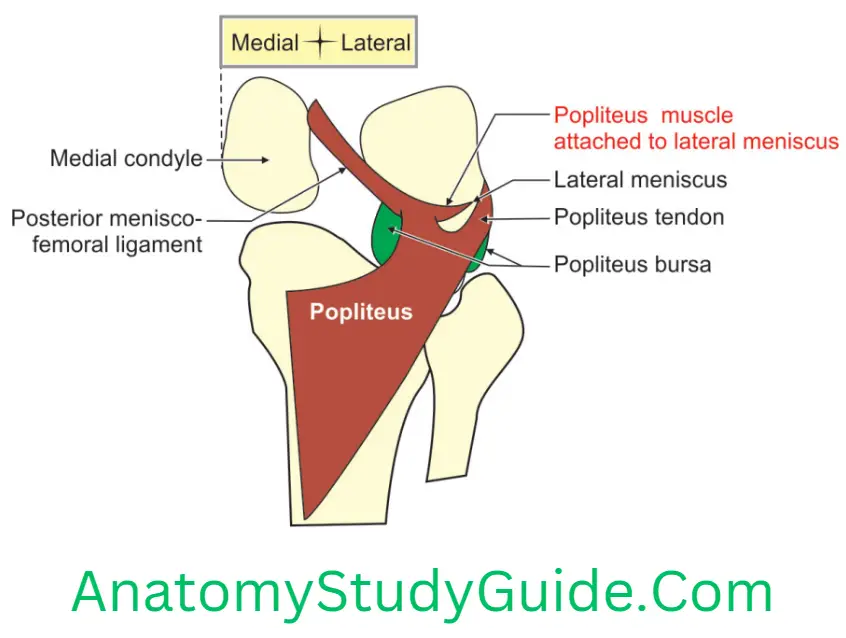
1. Proximal attachment: It arises from
- Popliteal groove present on the lateral surface of the lateral condyle of femur. Popliteal groove has anterior and posterior parts. The popliteus takes origin from the anterior part of the groove. The popliteus tendon occupies posterior part of the groove in full flexion of the knee. The popliteal tendon is about 1 length. Collagenous bands connect the tendon of popliteus to the arcuate popliteal ligament.
- Arcuate ligament
- The outer margin of lateral meniscus of the knee joint.
2. Distal attachment
- Posterior surface of tibia into medial 2/3rd of the triangular area above the soleal line.
- Fascia covering it.
4. Popliteus Nerve supply: It is supplied by a nerve to popliteus, a branch of tibial nerve.
5. Popliteus Relations
1. Anterior: Lateral condyle of tibia
2. Posterior
- Strong fascia, an extension of semimembranosus from the insertion.
- Popliteal vessels, and
- Tibial nerve.
6. Popliteus Actions
1. It unlocks the knee in initial phase of flexion, i.e. medial rotation of tibia on the fixed femur (when the foot is off the ground), or
- Lateral rotation of femur on fixed tibia (when the foot is on the ground).
- Flexes the knee joint.
2. PP lm PP Popliteus, Pulls, lateral meniscus Posteriorly, and Prevents it from being trapped at the beginning of the flexion.
3. In standing with the knee partly flexed, the popliteus contracts and helps the posterior cruciate ligament (PCL) to prevent anterior displacement of the femur.
7. Popliteus Nerve Applied anatomy: Injury to tibial nerve may result weakness of unlocking of knee joint.
Leave a Reply