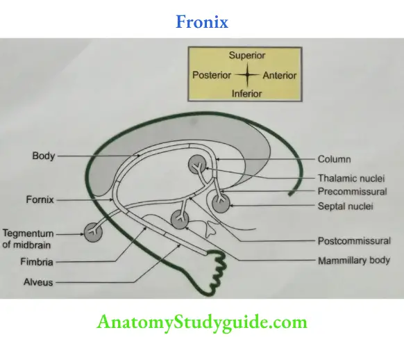
Fornix
- Fornix Definition: It is a bundle of white matter lying beneath the corpus callosum.
- Parts of fornix
- Alveus,
- Fimbria,
- Crus,
- Body, and
- Column.
Read And Learn More: Anatomy Important Question And Answers
Table of Contents
- Fornix Contents: It contains
- Association fibres arising from dentate gyrus.
- Commissural fibres which connect two hippocampi.
- Projection fibres which project to tegmentum of midbrain. It consists of
- Mammillotegmental tract.
- Mammilloreticular tract.
- Papez circuit: It is a neuronal circuit in the limbic system. It consists of
- Hippocampus.
- Fornix.
- Mammillary body (mammillothalamic tract).
- Anterior thalamic nuclei.
- Cingulate gyrus: Through cingulum again to hippocampus.
- Fornix Functions
- It is involved in experiencing the emotions.
- It also decides the response to the emotions.
Describe 3rd ventricle under following heads
- 3rd ventricle Gross anatomy,
- 3rd ventricle Boundaries,
- 3rd ventricle Recesses, and
- 3rd ventricle Applied anatomy.
- 3rd ventricle Gross anatomy
- 3rd ventricle Introduction: It is a narrow, midline cavity. It is present between two diencephalons of cerebral hemisphere.
- 3rd ventricle Extent: It extends from lamina terminalis to upper end of cerebral aqueduct.
- 3rd ventricle Communications
- Cranially, it communicates with both the lateral ventricles through interventricular foramen.
- Caudally, it communicates with 4th ventricle through cerebral aqueduct.
- 3rd ventricle Boundaries
- Roof is formed by
- Ependyma: It is a thin membrane lined by cuboidal or columnar epithelium. It lines the ventricle and stretches across the thalamus.
- Tela choroidea: It is a fold of pia mater projecting into ventricle.
- Body of fornix: It is a prominent bundle of fibres present on the medial aspect of cerebral hemisphere. It arises from hippocampus. It is suspended from corpus callosum by septum
- pellucidum.
- Floor is formed by following structures from anterior to posterior OT IMP Optic chiasma: Crossing of the optic nerves.
- Tuber cinereum (tuber-tubercle, cinereus-ash colour): It is ash-coloured elevation formed by grey matter present between mammillary body and optic chiasma.
- Infundibulum: It is a funnel
- shaped structure of the stalk of pituitary gland.
- Mammillary body: It is an important part of limbic system.
- Posterior perforated substance: It is a triangular interval between mammillary bodies and the midbrain which is pierced by numerous blood vessels.
- Tegmentum of midbrain: It is a part of cerebral peduncle of midbrain.
- Anterior wall: From above downwards
- Anterior column of fornix: The body of fornix anteriorly divides into anterior column of fornix. It contains important connections of limbic system. It correlates olfactory and visceral activities.
- Anterior commissure: It is a round bundle of white matter formed by
- Archipallial (archicortex): It belongs to rhinencephalon. It connects olfactory bulb and piriform area of both the sides (pear shape paleocortex present on the anterior part of parahippocampal gyrus).
- Neopallial: It is a larger commissure which connects two temporal lobes.
- Lamina terminalis: Thin plate derived from telencephalon (cranial end of neural tube) extending from rostrum of corpus callosum to dorsum of optic chiasma. It is encroached by anterior commissure in the upper part.
- Posterior wall
- Pineal body It is a small conical organ present between and above two superior colliculi.
- Posterior commissure: It connects two superior colliculi and also contains
- Corticotectal fibres, and
- Pretectal fibres.
- Cerebral aqueduct.
- Lateral wall
- Anteriorly: Anterior column of fornix.
- In upper part:
- Anterior two-thirds of thalamus and
- Interthalamic connexus.
- Lower part:
- Lower part of thalamus.
- Hypothalamic sulcus: Separates thalamus and hypothalamus and represents sulcus limitans of diencephalon.
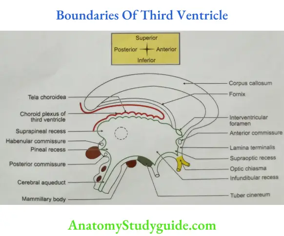
- Roof is formed by
- 3rd ventricle Recesses: These are prolongations of the 3rd ventricular cavity. These are described as
- Anteriorly:
- Anterior recess: It is present between anterior column of fornix and anterior commissure.
- Optic recess: It is present at the junction of anterior boundary and floor immediately above optic chiasma.
- In the floor: Infundibular recess: It is present in the stalk of infundibulum.
- Posteriorly:
- Pineal recess: In stalk of pineal body
- Suprapineal recess: It is present above the pineal body
- Anteriorly:
- 3rd ventricle Applied anatomy
- Ventriculography: It is a visualization of ventricles for determining the obstruction or dilatation of the 3rd ventricle.
- Obstruction of 3rd ventricle leads to raised intracranial pressure in adults and hydrocephalus in infants.
Enumerate the structures situated in the floor of the body of lateral ven- tricle.
Following are the structures situated in the floor of body of lateral ventricle.
- Body of caudate nucleus,
- Thalamostriate groove containing
- Stria terminalis, and
- Thalamostriate vein
- Lateral part of upper surface of thalamus,
- Fornix, and
- Choroid fissure.
Name the parts of the lateral ventricle.
- Anterior horn,
- Central part,
- Posterior horn, and
- Inferior horn.
Parts of Lateral Ventricle
Lateral Ventricle Introduction: It is a cavity of telencephalon, situated one in each cerebral hemisphere.
- Lateral Ventricle Communication: It communicates with the 3rd ventricle through interventricular foramen or foramen of Monro.
- Lateral Ventricle Situation: It is situated lateral to septum pellucidum and below corpus callosum
.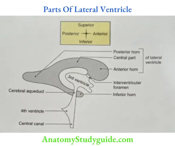
- Lateral Ventricle Contents: It contains
- Cerebrospinal fluid, and
- Choroid plexus in the central part and inferior horn of lateral ventricle.
- Lateral Ventricle Lining: It is lined by ependyma, a thin membrane.
- Lateral Ventricle Parts: It has
- Anterior horn,
- Central part,
- Posterior horn, and
- Inferior horn.
- Lateral Ventricle Development: It is developed from cavity of telencephalon of neural tube.
- Lateral Ventricle Applied anatomy: The blockage of interventricular foramina results into
- Hydrocephalus: Excessive accumulation of cerebrospinal fluid into lateral ventricle, and
- Increased intracranial pressure.
Enumerate the structures forming the floor of inferior horn of lateral ventricle.
Following are the structures forming the floor of the inferior horn of lateral ventricle. They are from medial to lateral.
- Choroid plexus,
- Fimbria (fringe),
- Hippocampus, and
- Collateral eminence.
Inferior Horn Of Lateral Ventricle
Inferior Horn Of Lateral Ventricle Introduction: It is the largest and longest of the three horns of lateral ventricle. It is direct continuation of the main ventricular cavity.
- Inferior Horn Of Lateral Ventricle Gross anatomy
- Situation: It is present in the temporal lobe.
- Direction: First it extends backwards and laterally around the pulvinar (pulvinar cushion) end of thalamus. It lies deep to the superior temporal sulcus and extends about an inch behind the temporal lobe.
- Inferior Horn Of Lateral Ventricle Boundaries: It consists of roof and floor .
- Inferior Horn Of Lateral Ventricle Roof: It is sloping and continues as lateral wall of inferior horn of lateral ventricle. It is formed by
- Lateral part of the tapetum of the corpus callosum. The fibres of the tapetum are formed by posterior fibres of trunk and anterior fibres of splenium.
- These fibres do not intersect corona radiata fibres.
- The tail of caudate nucleus: It is one of the important nuclei of basal nuclei. It is a part of neostriatum.
- Amygdaloid body: It is a part of limbic system and belongs to archistriatum.
- Stria terminalis: These are projection fibres of the amygdaloid nucleus. It connects amygdaloid nucleus to
- Septal nuclei,
- Hypothalamic nuclei, and
- Thalamic nuclei.
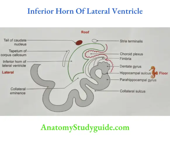
- Floor: Floor of the inferior horn presents following features (from medial to lateral).
- Choroid plexus: It is present most medially; it extends into the inferior horn through a choroid fissure between the fimbria (fringe) and the stria terminalis.
- Fimbria (fringe): The white matter of hippocampus is alveus. The fibres of the alveus (alveus-trough) converge backwards and medially to form fimbria. It is continuous with the crus of fornix below the splenium.
- Hippocampus: It is an elevation belonging to limbic system. It resembles seahorse hence called hippocampus. It lies medial to collateral eminence. It presents an enlarged anterior end with oblique grooves, which resembles paws of an animal. The fibres derived from the hippocampus form a thin sheath called alveus. It covers the ventricular surface of hippocampus.
- Collateral eminence: It is an elevation formed by projection of collateral sulcus. It forms a triangular area called collateral trigone. It is present at the junction of the floor of the posterior and inferior horn of lateral ventricle.
- Inferior Horn Of Lateral Ventricle Roof: It is sloping and continues as lateral wall of inferior horn of lateral ventricle. It is formed by
- Surgical approach of the inferior horn: A needle is introduced in the inferior horn through a hole at a point, 3 cm behind and above the centre of external acoustic meatus. The needle is passed in the direction of apex of the opposite auricle. It is about 5 cm from the surface.
Posterior Horn Of Lateral Ventricle
Posterior Horn Of Lateral Ventricle Introduction: It is a part of lateral ventricle which lies behind the splenium of corpus callosum.
- Posterior Horn Of Lateral Ventricle Gross anatomy
- Situation: It is present in the occipital lobe.
- Variation: It may be absent and may be of variable size.
- Direction: Backwards and medially.
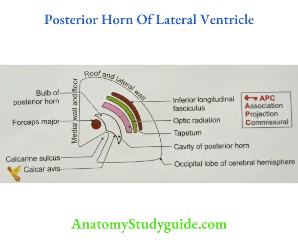
- Posterior Horn Of Lateral Ventricle Boundaries
- Medial wall and floor: It is formed by Bulb of the posterior horn: It is a raised area formed by forceps major. These are commissural fibres connecting two occipital lobes.
- Calcar avis (calcar-spur, avis-bird): It is a raised area formed by anterior part of calcarine sulcus.
- Roof and lateral wall are formed by
- Inferior longitudinal fasciculus (It is an example of association fibres.)
- Optic radiation:
- The fibres of the optic radiation arise from lateral geniculate body and terminate in the visual area of the occipital lobe (areas 17, 18 and 19).
- (It is an example of projection fibres.)
- Tapetum of the corpus callosum. (It is an example of commissural fibres.)
- Medial wall and floor: It is formed by Bulb of the posterior horn: It is a raised area formed by forceps major. These are commissural fibres connecting two occipital lobes.
Central Part Of Lateral Ventricle
- Extent: It extends from interventricular foramen (foramen of Monro) to splenium of corpus callosum.
- Roof: It is formed by inferior surface of body of corpus callosum.
- Floor: It is formed by
- Body of caudate nucleus.
- Thalamostriate groove which contains
- Stria terminalis: These are projection fibres of the amygdaloid body. It connects amygdaloid nucleus to
- Septal,
- Hypothalamic, and
- Thalamic nuclei.
- Thalamostriate vein.
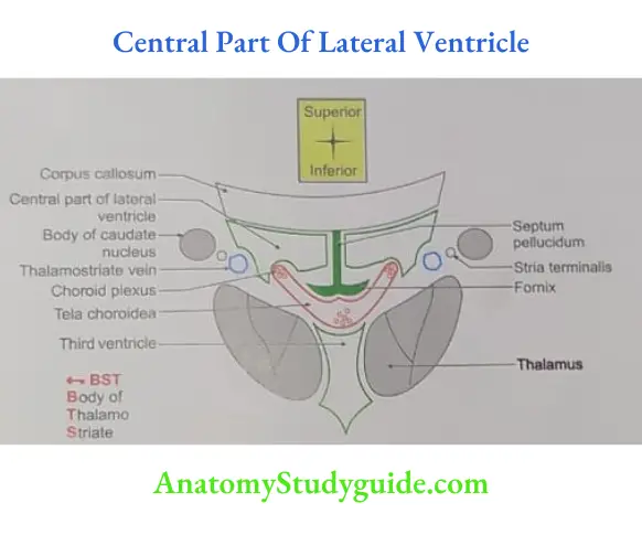
- Stria terminalis: These are projection fibres of the amygdaloid body. It connects amygdaloid nucleus to
- Lateral part of upper surface of thalamus.
- Fornix: It is a bundle of fibres connecting right and left hippocampus.
- Choroid fissure: It is a slit-like interval between the edge of the fornix and the upper surface of thalamus through which the choroid plexus enters the ventricles by invaginating the ependyma.
- Medial wall: It is formed by
- Posterior part of septum pellucidum. It is present anterosuperiorly.
- Body of fornix. It is present posteroinferiorly.
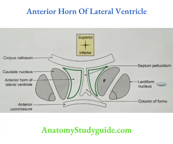
Anterior Horn Of Lateral Ventricle
- Situation: It is situated anterior to interventricular foramen.
- Extent: It extends forwards, laterally and downward in the frontal lobe of the brain. It is continuous with the central part posteriorly at the interventricular foramen. It presents a triangular outline in coronal section and possesses a roof, a floor, medial wall, lateral wall and anterior wall.
- Roof: It is formed by
- Anterior part of body of corpus callosum, and
- Posterior part of genu of corpus callosum.
- Floor: It is formed by
- Medially: Rostrum of corpus callosum.
- Laterally: Head of caudate nucleus.
- Medial wall: Anterior part of septum pellucidum.
- Anterior wall: Genu and rostrum of corpus callosum.
Hippocampus (seahorse)
- Hippocampus (seahorse) is part of limbic system.
- Mature neurons do divide in hippocampus. It is exception to other neurons.
- Hippocampus (seahorse) projects to the
- Nucleus accumbens through the fornix,and
- Orbitofrontal cortex (through the internal capsule)
- Papez (1937) described a closed circuit (the Papez circuit).
- It is connection between hippocampus and the cingulate cortex, via the mammillary bodies and anterior thalamus.
- He proposed that emotional expression is organized in the hippocampus.
- It is experienced in the cingulate gyrus and expressed via the mammillary bodies
- The Papez circuit is now widely accepted to be involved with cognitive processes, including mnemonic functions and spatial short-term memory.
- Definition: The hippocampus is a curved elevation of grey matter. It lies above the subiculum (to raise or lift) and medial parahippocampal gyrus.
- Length: 5 cm long.
- Morphology:
- Its anterior end is expanded.
- Its margin has 2 or 3 shallow grooves that give it a paw-like appearance, the pes hippocampi.
- The ventricular aspect is convex. It is covered by ependyma, beneath which fibres of the alveus converge medially on a longitudinal bundle of fibres, the fimbria of the fornix.
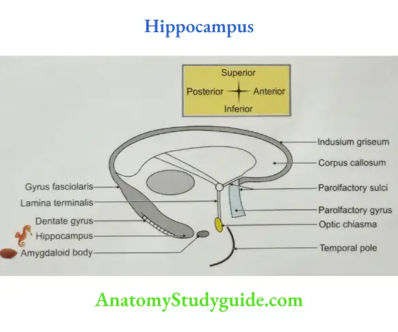
- Situation: Temporal lobe of the cerebral hemisphere.
- Extent: It extends throughout the length of the floor of the inferior horn of the lateral ventricle.
- Histology: The hippocampus is trilaminar archicortex. It consists of a single pyramidal cell layer, with plexiform layers above and below it. The thickness of the pyramidal cell layer varies from 10 to more than 30 cells.
- It can be divided into three distinct fields:
- CA1: Field CA1 is usually described as the most complex of the hippocampal subdivisions, and its appearance varies along its transverse and rostrocaudal axes.
- CA2
- The CA2 field has the most compact layer of pyramidal cells.
- It completely lacks a mossy fibre input from dentate granule cells and receives a major input from the supramammillary region of the hypothalamus.
- CA3
- Field CA3 pyramidal cells are the largest in the hippocampus, and the whole pyramidal cell layer in this field is about 10 cells thick.
- The most important feature of pyramidal cells in CA3 is that they receive the mossy fibre input from dentate granule cells on their proximal dendrites.
- It can be divided into three distinct fields:
- Functions
- It is responsible for correlating the new concepts learned for the first time with the pre-existing memory.
- It is considered to be like the telephone switchboard-correlating new facts with distant cortical areas.
- The conceived facts become stored in the cortex as memory. Later, different areas learn to ‘dial direct’, independent of the switchboard.
- Therefore, the significance of the hippocampus is to establish connection with the pre-existing concepts whenever a new concept is to be conceived.
- Hippocampus (seahorse) \ Blood supply: Anterior choroidal artery.
- Hippocampus (seahorse) Applied anatomy
- Bilateral ablation of hippocampus results in inability to build new memories and person suffers from loss of recent memory, i.e. amnesia of recent events. But there is no loss of memory of remote past events.
- The information which is not yet fully perceived needs consolidation by the hippocampus and therefore lost in its bilateral injury. The emotionally charged memories are affected more than non-emotional ones.
- In Alzheimer’s disease the changes of normal aging are more pronounced in the hippocampus.
- It is regarded as the cortical centre for autonomic reflexes. Hippocampal-amygdala complex is related to the memory of recent events.
- Lesions of this complex are associated with a loss of memory for recent events only. Patient is unable to commit any new facts to memory and does not remember recent events.
Leave a Reply