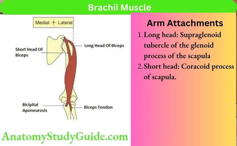
Arm Attachments And Actions Of Biceps Brachii
1. Arm Attachments
Table of Contents
- Long head: Supraglenoid tubercle of the glenoid process of the scapula
- Short head: Coracoid process of scapula.
Read And Learn More: Anatomy Notes And Important Question And Answers
2. Arm Attachments Actions
- Strong supinator of superior and inferior radioulnar joints,
- Flexor of the elbow joint, and
- Weak flexor of the shoulder joint.
Question-1:Describe Musculocutaneous Nerve Under Following Heads
1. Musculocutaneous Nerve Root value,
2. Musculocutaneous Nerve Course and relations,
3. Musculocutaneous Nerve Branches, and
4. Musculocutaneous Nerve Applied anatomy.
Answer:
Musculocutaneous Nerve Introduction:
It is a branch of the lateral cord of the brachial plexus. It is the motor nerve of the flexor compartment of the arm and the sensory nerve to the lateral skin of the forearm.
1. Musculocutaneous Nerve Root value
C5, C6 and C7.
2. Musculocutaneous Nerve Course and relations
It arises from the lateral cord of the brachial plexus.
1. The lateral cord of the brachial plexus lies lateral to the 2nd part of the axillary artery.
2. The nerve lies lateral to 3rd part of the axillary artery. It arises at the lower border of the pectoralis major.
In the axilla, it is related to the following structures.
- Anteriorly: Pectoralis major.
- Posteriorly: Subscapularis.
- Medially: Axillary artery.
- Laterally: Coracobrachialis.
It leaves the axilla, pierces coracobrachialis and enters the front of the arm.
In arm: It runs downwards and laterally.
- It passes between the biceps and brachialis.
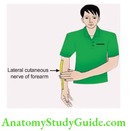
- It pierces deep fascia just below the elbow and continues as the lateral cutaneous nerve of the forearm.
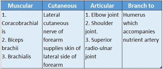
3. Musculocutaneous Nerve Branches and distribution
Distribution of musculocutaneous nerve
4. Musculocutaneous Nerve Applied anatomy
1. About the lesion of the musculocutaneous nerve.
- It is rare.
- It is due to a fracture of the neck of the humerus.
2. Lesion of the nerve causes:
- Loss of strong flexion and supination.
- Loss of biceps tendon reflex.
- Loss of sensation along the lateral aspect of the forearm.
- The pain and anaesthesia may be aggravated by the extension of the elbow.
3. Myotrophy: Marked weakness of flexion of the elbow is due to paralysis of the bicep brachii and coracobrachialis.
4. The nerve may be involved in Erb’s paralysis.
5. The musculocutaneous nerve in a cadaver is identified as
- First, identify the coracoid process.
- The medial muscle arising from the coracoid process is the coracobrachialis.
- The nerve piercing the coracobrachialis is the musculocutaneous nerve.
6. Biceps reflex: The integrity of the musculocutaneous nerve is tested by the biceps reflex. It is tested by tapping the tendon of the biceps brachii with the forearm pronated and partially extended at the elbow. The normal reflex is a brief jerk-like flexion of the elbow.
7. Surgical approach: The musculocutaneous nerve is exposed by opening up the deltopectoral groove. The nerve entering the coracobrachialis is identified as the musculocutaneous nerve. Here it lies below the lower border of teres major.
The muscles which are supplied by the musculocutaneous nerve can be remembered and recollected by putting the palmar surface of one band on the arm of the other side and saying “BBC” and by putting the hand on the lateral surface of the forearm and while saying it continues as “lateral cutaneous nerve of the forearm”.
The first letter “B” in “BBC” stands for “Biceps“.
The second letter “B” in “BBC” indicates “Brachialis“.
While the letter “C” in “BBC” represents” Coracobrachialis“.
Here I would like to draw your attention to the fact that the brachialis has a dual nerve supply. The musculocutaneous nerve supplies the medial 2/3rd of the brachialis and the lateral 1/3rd is supplied by the radial nerve.
Hence brachial is called MR brachialis. The letter “M” in “MR” indicates musculocutaneous and “R” stands for radial nerve.
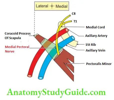
Medial Pectoral Nerve
- It arises from the medial cord, hence it is called the medial pectoral nerve.
- It arises from the anterior primary rami of the 8th cervical and 1st thoracic spinal nerve.
- It supplies pectoralis minor,
- It pierces the sternal fibres of the pectoralis major and supplies it.
- It has no cutaneous branch.
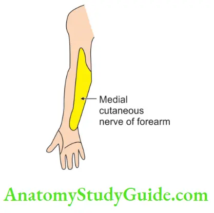
Medial Cutaneous Nerve Of Forearm
The medial cutaneous nerve of the forearm
- It arises from the anterior primary rami of C8 and T1.
- It is the smallest branch of the brachial plexus.
- It is much bigger than the medial cutaneous nerve of the arm.
- It runs between the axillary artery and vein and pierces the deep fascia at the middle of the arm.
- It supplies the skin over the biceps, almost to the elbow.
- It then divides into anterior and posterior branches.
- It supplies the skin along the ulnar border of the forearm up to the wrist.
- It is symmetrical with the lateral cutaneous nerve of the forearm.
- These cutaneous nerves meet along the anterior axillary line.
Question-2: Describe Brachial Artery Under The Following Heads
1. Brachial Artery Origin,
2. Brachial Artery Course and relations,
3. Brachial Artery Branches, and
4. Brachial Artery Applied anatomy
Answer:
1. Brachial Artery Origin
It is the continuation of the axillary artery distal to the lower border of teres major
2. Brachial Artery Course and relations
Peculiarities:
It is superficial throughout its course. It is accompanied by
1. Veins: Venae commitments (brachial veins)
Nerves accompanying the brachial artery and its branches
- The brachial artery accompanies the median nerve in the upper half of the arm.
- The superior ulnar collateral artery, a branch of the brachial artery, accompanies the median nerve in the lower half of the arm,
- Profunda brachii (a branch of the brachial artery) accompanies the radial nerve,
- Anterior descending artery (a branch of profunda brachial artery) accompanies the radial nerve, and
- The superior ulnar collateral artery (a branch of the brachial artery) accompanies the ulnar nerve.
Brachial Artery Relations
1. Anterior
- Skin,
- Superficial fascia,
- Deep fascia,
- Biceps brachii, and
- Bicipital aponeurosis at the bifurcation.
2. Posterior
- The long head of triceps,
- Medial head of
- Coracobrachialis, and
- Brachialis.
3. Lateral: Upper part median nerve.
4. Medial
- The medial cutaneous nerve of the arm, and
- The lower part of the median nerve.
Termination: The artery divides at the neck of the radius into radial and ulnar arteries.
3. Brachial Artery Branches
1. Cutaneous to the skin over the arm,
2. Muscular: Muscles of the arm (deltoid, biceps, brachialis, coracobrachialis),
3. Articular branches to
1. Shoulder joint,
2. Elbow joint by
- Superior ulnar collateral, and
- Inferior ulnar collateral artery
4. Nutrient branch to the humerus,
5. Anastomotic branch to the posterior circumflex humeral artery, and
6. Terminal branches: Radial and ulnar arteries.
4. Brachial Artery Applied Anatomy
- The brachial artery is ruptured in a supracondylar fracture of the humerus leading to Volkmann’s ischaemic contracture.
- The pulsations of the brachial artery are felt or auscultated in front of the elbow medial to the tendon of the biceps.
- Haemorrhage due to the brachial artery can be controlled by direct compression of the brachial artery. The compression should be in the middle of the arm on the tendon of the coracobrachialis and medial to the humerus.
- Blood pressure is recorded by auscultation of the pulsations of the brachial artery in the cubital fossa.
- The blood for blood gas analysis is collected from the brachial artery.
- The pulsations of the brachial artery are felt or auscultated in front of the elbow medial to the tendon of the biceps.
Branches Of Radial Nerve In Radial Groove
- Branch to the medial head of triceps,
- Branch to the lateral head of the triceps, and
- Anconeus.
Branches Of Radial Nerve In Axilla
- The long head of triceps, and
- The medial head of the triceps.
Branches Of Radial Nerve In Front Of Lower Part Of Arm
1. Muscular branch
- Brachioradialis,
- Extensor carpi radialis longus, and
2. Proprioceptive branch to brachialis.
Question-3: Describe Radial Nerve Under
1. Radial Nerve Root value,
2. Radial Nerve Course and relations,
3. Radial Nerve Branches, and
4. Radial Nerve Applied anatomy.
Answer:
Radial Nerve Introduction:
It is the nerve of the extensor compartment of the arm and forearm, supplying the skin over them and on the dorsum of the hand. It is the direct continuation of the posterior cord of the brachial plexus. It is the largest branch of the whole brachial plexus.
1. Radial Nerve Root value
It arises from the posterior cord of the brachial plexus carrying fibres from the ventral roots of spinal nerves C5, C6, C7, C8 and T1.
2. Radial Nerve Course and relations
It arises from the posterior cord of the brachial plexus. The posterior cord lies posterior to the 2nd part of the axillary artery and the radial nerve lies posterior to the 3rd part of the axillary artery. It lies medial to the axillary nerve.
1. Course of the radial nerve in the lower part of the axilla. The nerve passes downwards. It crosses the lower border of the posterior axillary wall. It includes crossing of the glistening tendon of latissimus dorsi, and subscapularis. Here it has the following relations.
- Anteriorly: Axillary artery and pectoralis major.
- Posteriorly: Subscapularis, teres major and latissimus dorsi.
- Medially: Axillary vein
- Laterally: Coracobrachialis muscle.
It passes through lower lar space and enters the radial groove along with the profunda brachial artery.
2. Course of the radial nerve in the arm: It enters the arm. It has the following relations. It is
- Posterior to the brachial artery,
- Medial to the humerus, and
- Anterior to the long head of the triceps.
3. Course of the radial nerve in the radial groove
- The nerve passes obliquely across the back of the humerus.
- The course is from medial to lateral in a shallow groove and between the long and medial heads of the triceps. It passes along with the profunda brachial artery.
- At the lower end of the radial groove, it pierces the lateral intermuscular septum and enters into the anterior or flexor compartment of the arm.
- In the flexor compartment of the lower arm, it descends in the intermuscular slit.
4. Course and relations of the radial nerve in the intermuscular slit
- In the upper part, it lies between the brachialis medially and brachioradialis laterally.
- In the lower part, it lies the brachialis medially and extensor carpi radialis longus laterally.
5. Course of the radial nerve in the forearm: At the level of the lateral epicondyle, it gives off two terminal branches. The branches are
- Superficial terminal branch, and
- Deep terminal branch. It is called the posterior interosseous nerve.
1. Superficial terminal branch. It is entirely cutaneous in its distribution. It supplies to the dorsum of the hand and fingers.
1. Course of the superficial terminal branch of radial nerve
In front of the forearm:
- It leaves the cubital fossa and enters the lateral side of the forearm.
- In the upper 2/3rd, it is accompanied by radial vessels.
- At the junction of the upper 2/3rd and lower 1/3rd, it runs laterally and reaches the posterolateral aspect of the forearm.
In hand
It reaches the wrist and travels in the roof of the anatomical snuffbox
2. Posterior interosseous branch.
1. Course of posterior interosseous nerve: This leaves the cubital fossa by piercing the supinator muscle. It enters the back of the forearm.
At the cubital fossa, it runs downwards on the surface of
- Supinator,
- Pronator teres, and
- Flexor digitorum superficialis.
At the forearm, it runs on the lateral side of the forearm under the cover of the brachioradialis.
In the middle 1/3rd of the forearm, it lies lateral to the radial artery.
In the extensor compartment of the lower part of the forearm.
It then leaves the flexor compartment of the forearm and winds around the lower end of the radius deep to the tendon of the brachioradialis. It crosses the structures forming an anatomical snuffbox. They are
- Abductor pollicis longus,
- Extensor pollicis brevis, and
- Extensor pollicis longus.
Note: On the posterior surface of the interosseous membrane, it enlarges into a pseudo-ganglion. It breaks into 2 or 3 branches which can be rolled on the surface of the tautened tendon of the extensor pollicis longus. They are distributed to the radial 2/3rd of the dorsum of the hand and proximal parts of the dorsal surfaces of the thumb and lateral 21/2 or 31/2 fingers.
3. Radial Nerve Branches of the radial nerve can be described as
- Collateral branches
- Terminal branches
1. Collateral branches
- Branches above the radial groove,
- In the radial groove, and
- Below the radial groove.
Branches above the groove are motor, sensory and articular.
1. Motor branches are to the
- Long, and
- The medial head of the triceps. It is called the ulnar collateral nerve.
2. Sensory branch is the posterior cutaneous nerve of the arm.
3. Articular branch supplies the elbow joint.
Branches in the groove are motor and sensory.
1. Motor branches are to
Lateral, and
The medial head of the triceps. The branch to the medial head of the triceps goes deep and supplies the anconeus.
2. There are two sensory branches given in the groove: To the skin of
- Arm, and
- Forearm.
1. The cutaneous branch given in the arm is called the lower lateral cutaneous nerve of the arm. It pierces the lateral head of the triceps to supply the skin over the lateral surface of the arm up to the elbow.
2. The cutaneous nerve of the forearm is called the posterior cutaneous nerve of the forearm. It runs straight behind the elbow to supply a strip of skin over the extensor surface of the forearm up to the wrist.
The branches given below the groove are
1. Motor to
- Brachioradialis and
- Extensor carpi radialis longus,
2. Proprioceptive to the lateral part of the brachialis.
2. Terminal branches: The radial nerve divides at the level of the lateral epicondyle into superficial and deep terminal branches (posterior interosseous nerve).
The superficial branch is the continuation of the main nerve. It is purely cutaneous and is distributed to the skin of
1. Lateral 1/2 of the dorsum of the hand
2. Proximal parts of the dorsal surface of the
- Thumb,
- Index fingers, and
- Lateral ½ of the middle finger.
A deep branch is motor and is called the posterior interosseous nerve. It supplies
1.Muscles in cubital fossa
Extensor carpi radialis brevis, and
Supinator
2. Muscles in the extensor compartment (7 muscles). They are conveniently divided into two groups for easy recollection.
1.1st group includes
Extensor muscles reaching wrist, index finger, digits and little finger. They are
- Extensor carpi ulnaris,
- Extensor indices,
- Extensor digitorum, and
- Extensor digiti minimi.
2.2nd group
The muscles form an anatomical snuffbox. They are
- Abductor pollicis longus,
- Extensor pollicis brevis, and
- Extensor pollicis longus
Note:
The symbol ” ” at the end of the nerve signifies the branch
The symbol ” ” at the end of the nerve signifies the cutaneous branch
Note:
- The radial nerve is also sensory to the wrist and carpal joints.
- The Dorsum of the hand proximal to 1st web space is an autonomous area for the radial nerve. The autonomous sensory area is part of a dermatome that has no overlap with adjacent nerves.
4. Applied anatomy of the radial nerve
The radial nerve is very commonly damaged in the region of the radial groove. The common causes are:
1. Saturday night palsy: When an intoxicated person falls asleep with his arm hanging over the edge of a chair. The prolonged pressure causes damage to the radial nerve as it is in the radial groove. As a result, in the morning, he finds that he is suffering from a wrist drop.
- It is characterized by flexion of the wrist and flexion of the metacarpophalangeal joint.
- The interphalangeal joints are not affected by wrist drop. They remain straight. This is because of the actions of interossei and lumbrical.
- Testing: This can be detected by testing elbow extension.
- It is purely a transient condition.
2. An interesting term, bridegroom’s paralysis, has been coined for the compression of the radial nerve in the radial grove. It results when the head of the spouse is lying on the arm of the other spouse.
3. The other causes of radial nerve injury include crutch paralysis and fracture of the shaft of the humerus.
4. All these conditions result in weakness and loss of power of extension of the wrist joint. This is called wrist drop.
5. There is minimum sensory loss which is confined to 1st dorsal interosseous. This is because there is an overlap of the nerve supply by the median and ulnar nerves. In addition to wrist drop, there is sensory loss over a narrow strip on the back of the forearm and the lateral side of the dorsum of the hand.
Injury to the superficial branch results in small sensory loss over the root of the thumb.
The integrity of the radial nerve can be confirmed by testing the sensations over the dorsum of the hand proximal to 1st web space.
Surgical approach
- Exposure of radial nerve.
- First, the radial nerve may be exposed from the back by developing an interval between the long and lateral head of the triceps. Here the nerve crosses the upper part of the medial head.
The superficial and deep branches of the radial nerve can be exposed by retracting the brachioradialis and extensor carpi radialis longus.
The deep branch is exposed by incising the deep part of the supinator muscle.
Distribution of radial nerve.
The keyword for the muscles supplied by the radial nerve is BEST.
The letter B represents Brachialis.
The letter E stands for Extensors of the forearm.
The letter S denotes Supinator.
The letter T represents Triceps.
Applied Anatomy Of Radial Nerve
Applied Anatomy:
Injury to the radial nerve at different levels.
Injury to the radial nerve at various sites and the effects of sensory and motor nerves
Wrist Drop
Wrist Drop Introduction:
It is loss of extension of the wrist due to paralysis of the extensor muscles of the wrist joint.
1. Wrist Drop Nerve Involved
It is produced by the lesion of the radial nerve and/or posterior interosseous branch of the radial nerve.
2. Wrist Drop Causes Of The Injury
In the axilla
- Saturday night palsy, and
- Crutch palsy.
In the middle of the arm
- Fracture of middle 1/3rd of the shaft of humerus, and
- Injection in the radial groove.
In the proximal part of the forearm: Fracture of proximal 1/3rd of radius
3. Wrist Drop Muscles paralysed
1. Extensor carpi radialis brevis
2. Supinator,
3. Muscles forming anatomical snuffbox.
- Abductor pollicis longus
- Extensor pollicis brevis
- Extensor pollicis longus
4. Other muscles
- Extensor indices,
- Extensor digitorum,
- Extensor digiti minimi, and
- Extensor carpi ulnaris.
4. Wrist Drop Clinical manifestations
1. Motor:
The patient is unable to extend the wrist which results in wrist drop.
2. Sensory:
A variable small area of anaesthesia is present over the dorsal surface of the hand and dorsal surface of the roots of the lateral 3½ fingers.
Profunda Brachii Artery
1. Profunda Brachii Artery Origin
It is the largest and deep (profound-deep) branch of the brachial artery.
2. Profunda Brachii Artery Course and relations
It accompanies the radial nerve in the lower lar space and radial groove. It pierces the lateral intramuscular septum and divides into branches.
3. Profunda Brachii Artery Branches
- Deltoid branch: It ascends between the long and lateral head of the triceps and anastomoses with the descending branch of the posterior circumflex humeral artery.
- Nutrient artery to the humerus.
- The radial collateral artery is one of the terminal branches and represents the continuation of the profunda artery. It ends by anastomosing with the radial recurrent artery.
- The middle collateral artery is the larger terminal branch. It ends by anastomosing with the interosseous recurrent artery.
Question-4: What Structures Pass Between The Medial And Lateral Head Of The Triceps
Answer:
- Radial nerve,
- Profunda brachial artery, and
- Profunda brachial vein.
Boundaries Of Cubital Fossa
- Medially by the lateral border of pronator teres, and
- Laterally by the medial border of brachioradialis.
Contents Of Cubital Fossa
1. Radial nerve and its terminal branches
- Superficial branch, and
- The posterior interosseous nerve.
2. Bicipital aponeurosis
3. Brachial artery and its terminal branches
Larger ulnar artery
1. Anterior ulnar recurrent,
2. Posterior ulnar recurrent,
3. Common interosseous,
- Anterior interosseous, and
- Posterior interosseous.
4. Smaller radial artery and its branch-radial recurrent
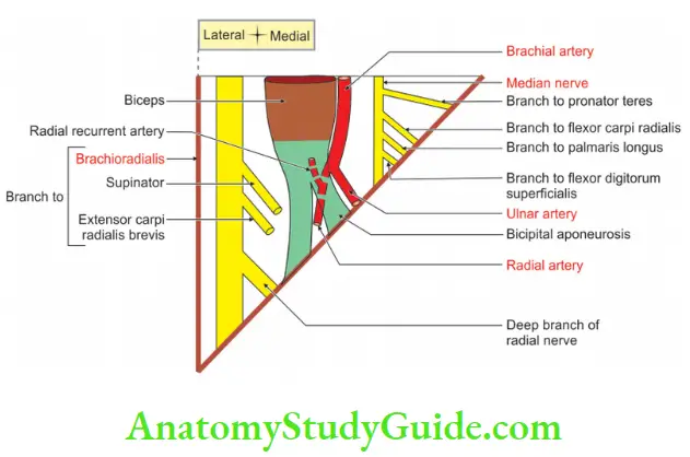
4. Brachial vein and its tributaries
5. Median nerve and its branches
Muscular artery
- Flexor carpi radialis,
- Palmaris longus,
- Flexor digitorum superficialis, and
- Flexor digitorum profundus (lateral half).
Applied Anatomy Of Cubital Fossa
1. Median cubital vein is the most fixed vein. Hence it is used for the withdrawal of blood for investigation purposes and for giving intravenous fluid.
2. The brachial artery is auscultated for recording blood pressure.
3. The brachial artery is selected for the withdrawal of arterial blood for blood gas analysis.
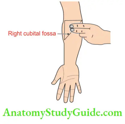
4. The cubital vein is used for the introduction of the cardiac catheter to secure blood samples from the great vessels and chambers of the heart.
5. The supracondylar fracture of the humerus results in the rupture of the brachial artery. It results in Volkmann’s ischaemic contracture.
Question-5: Describe Cubital Fossa Under the Following Heads
1. Boundaries,
2. Contents, and
3. Applied anatomy.
Answer:
Cubital Fossa Introduction:
It is a shallow lar fossa present in front of the elbow joint.
1. Cubital Fossa Boundaries
1. Medial boundary is formed by the lateral border of pronator teres.
2. Lateral boundary is formed by the medial border of the brachioradialis.
3. Cubital Fossa Roof: It is formed by
Skin and superficial fascia containing
1. Medial cutaneous nerve of forearm,
2. Lateral cutaneous nerve of forearm,
3. Medial cubital vein which is joined by
- Cephalic vein, and
- Basilic vein.
Deep fascia and bicipital aponeurosis.
4. Cubital Fossa Floor
- Upper part: Formed by the lower part of the brachialis.
- Lower part: Formed by the upper part of the supinator.
5. Base by an imaginary line joining the medial and lateral epicondyle.
6. Apex is formed by the meeting point of the medial border of brachioradialis and the lateral border of pronator teres.
2. Cubital Fossa Contents
1. Main contents: From lateral to medial are (Ravina Tandon ke Baton Me RT BM)
The Radial nerve divides into
Tendon of biceps
Brachial artery
Median nerve.
2. Minor contents are
1. Deep branch of the radial nerve: It continues as a posterior interosseous branch. It gives
2. Muscular branches to
- Supinator
- Extensor carpi radialis brevis
3. Terminal divisions of the branchial artery, namely
- The radial artery, and
- Ulnar artery
4. Musculocutaneous nerve. It lies at the lateral margin of the biceps tendon. Here it continues as the lateral cutaneous nerve of the forearm.
5. Muscular branches of median nerve in the cubital fossa are
- Pronator teres,
- Palmaris longus,
- Flexor carpi radialis,
- Flexor digitorum superficialis.
6. Anterior interosseous nerve, a branch of the median nerve.
7. Small isolated lymph nodes sometimes occur along radial, ulnar and interosseous arteries.
3. Cubital Fossa Applied anatomy
- The median cubital vein is the most fixed vein. Hence it is used for the withdrawal of blood for investigation purposes and for giving intravenous fluid.
- The brachial artery is auscultated for recording blood pressure.
- The brachial artery is selected for the withdrawal of arterial blood for blood gas analysis.
- The median cubital vein is used for the introduction of the cardiac catheter to secure blood samples from the great vessels and chambers of the heart.
- The supracondylar fracture of the humerus results in the rupture of the brachial artery and ends in a condition called Volkmann’s ischaemic contracture.
Leave a Reply