Venous And Lymphatic Drainage And Comparison Of Lower And Upper Limbs
Venous Perforators Of Lower Limb
Table of Contents
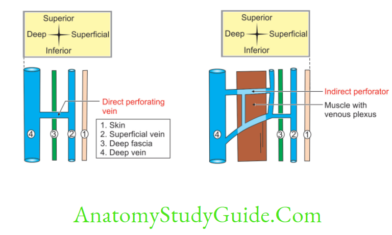
1. Lower Limb Definition: Venous perforators connect the superficial with the deep veins.
2. Lower Limb Number
- There are about 5 perforators along the great saphenous vein, and
- One perforator along the small saphenous vein.
Great Saphenous Vein
3. Lower Limb Features
- They are provided with valves.
- BThey permit flow of blood only from the superficial to the deep veins.
Read And Learn More: Anatomy Notes And Important Question And Answers
4. Lower Limb Applied anatomy: Failure of the valves gives rise to varicose veins.
Describe great Saphenous Vein under following heads
1. Saphenous Vein Formation,
2. Saphenous Vein Course and relations,
3. Saphenous Vein Peculiarities,
4. Perforating veins,
5. Saphenous Vein Termination,
6. Saphenous Vein Relations,
7. Saphenous Vein Tributaries,
8. Saphenous Vein Communicating veins, and
9. Saphenous Vein Applied anatomy.
Saphenous Vein Introduction: The great saphenous (Saphes—easily seen) vein is the longest vein of the body. It drains all the structures superficial to fascia lata in whole of the lower extremity except the medial side of leg between tendo calcaneus and tibia.
Great Saphenous Vein
1. Saphenous Vein Formation: It is formed by the
- Medial end of dorsal venous arch, and
- Medial marginal vein.
2. Saphenous Vein Course and relations: It begins at
Medial end of the dorsal venous arch
It is supplemented by medial marginal vein.
It runs in front of medial malleolus.
It crosses obliquely on medial surface of lower 1/3rd of tibia.
It ascends behind the medial border of tibia to reach knee.
It runs along medial side of thigh. It pierces the cribriform fascia over the saphenous opening. It drains into femoral vein.
3. Saphenous Vein Peculiarities: It contains about 10 to 15 valves, which prevent back flow of venous blood. One of the valves is always present at saphenofemoral junction.
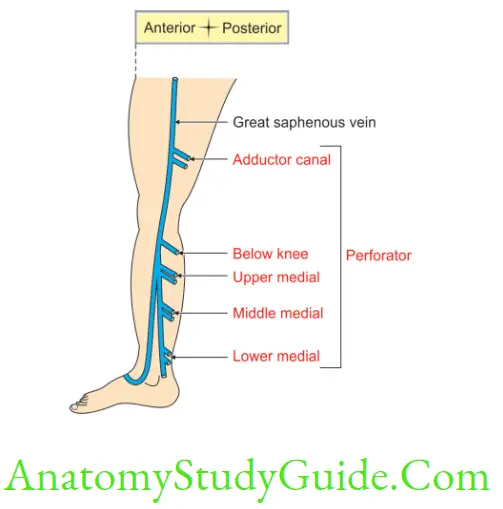
4. Saphenous Vein Perforating veins: They connect great saphenous vein to deep vein. They are three in number.
- Above the ankle,
- Below the knee, and
- Above the knee. It is present in the adductor canal. The perforating veins are provided with valves, which permit the flow of blood only from superficial to deep.
5. Saphenous Vein Termination: It terminates into upper part of femoral vein by piercing fascia lata.
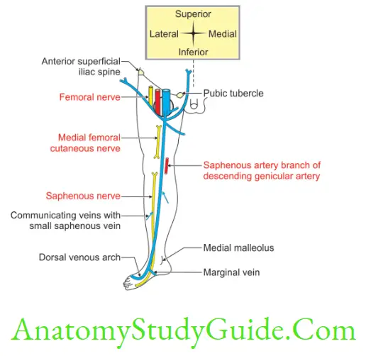
6. Saphenous Vein Relations: At ankle joint, the saphenous nerve is anterior to saphenous vein.
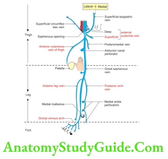
7. Saphenous Vein Tributaries
1. At the beginning: Medial marginal vein
2. Just below knee
Great Saphenous Vein
- Anterior vein of leg, and
- Posterior arch vein of calf.
3. In the thigh
- Posteromedial vein, and
- Anterior cutaneous vein of thigh.
4. Before piercing cribriform fascia
- Superficial epigastric vein,
- Superficial circumflex iliac vein, and
- Superficial external pudendal vein.
5. Before entering into femoral sheath: Deep external pudendal vein.
8. Saphenous Vein Communication vein: Small saphenous vein.
9. Saphenous Vein Applied anatomy
- Varicosity (torturous, dilated, enlarged and visible vein) of the veins is more common in people who are standing for long time (e.g. traffic police). The valves become incompetent and the flow of the blood is reversed. The defective veins become ‘high pressure leaks’. They produce varicose veins. The area of skin over the dependent part of vein becomes devitalized and results in varicose ulcers. It is one of non-healing ulcers.
- Great saphenous vein is used for arterial grafting. This is especially used in coronary artery bypass surgery.
- During venesection at the ankle joint, one should keep in mind the relation of saphenous nerve. Lesion of saphenous nerve results in loss of sensation on medial side of foot.
Venous Drainage of Lower Limb
1. There are three types of veins.
1. Superficial,
2. Deep, and
3. Perforating.
1. Features of Superficial Veins
- They lie superficial to deep fascia,
- They are thick-walled because of the presence of Smooth muscle, Fibrous, and Elastic tissues in their walls.
- They have valves in the lumen. The numbers of valves increase as they go to distal part. A large proportion of their blood is drained into the deep veins through the perforating veins.
2. Features of Deep Veins
- They have more valves than the valves in superficial veins.
- They are more efficient channels than the superficial veins.
2. The Superficial Veins are
1. Great or long saphenous vein.
2. Small Saphenous Vein.
1. Small Saphenous Vein Formation: The vein is formed on the dorsum of the foot by the union of the
- Lateral end of the dorsal venous arch with the
- Lateral marginal vein.
Great Saphenous Vein
2. Small Saphenous Vein Course
- It passes behind the lateral malleolus.
- It ascends on the back of leg.
- It lies lateral to the tendo calcaneus.
- It continues to ascend along the middle line of the calf.
- It ends at the lower part of the popliteal fossa.
- It pierces the deep fascia and opens into the popliteal vein.
3. Small Saphenous Vein Drainage area: It drains the
- Lateral border of the foot,
- Heel, and
- Back of the leg.
4. Small Saphenous Vein Connected to the
- Great saphenous, and
- Deep veins.
5. Small Saphenous Vein Accompanying structure: Sural nerve.
3. Deep Veins: They drain the entire structures deep to deep fascia (fascia lata). The deep veins are
1. Deep Veins Medial plantar vein
2. Deep Veins Lateral plantar
- Dorsalis pedis,
- Anterior tibial,
- Posterior tibial,
- Peroneal,
- Popliteal, and
- Femoral veins and their tributaries.
4. Deep Veins Perforating veins: They connect the superficial with the deep veins. Their valves permit flow of blood in only one direction. The flow is from the superficial to the deep veins. They are three in number.
1. Above the ankle,
2. Below the knee, and
3. Above the knee.
It is present in the adductor canal.
Leave a Reply