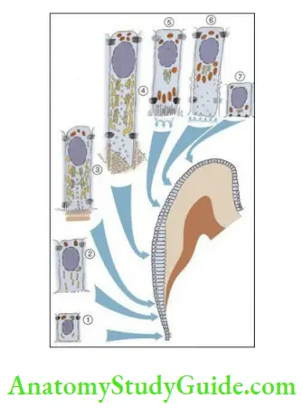
Ameloblast Life Cycle
The life cycle of an Ameloblast can be divided into the following six stages based on the function:
- Morphogenic
- Organizing
- Formative
- Maturative
- Protective
- Desmolytic
All or a few of the above-mentioned stages of the developing ameloblasts can be observed in a tooth germ because the differentiation of ameloblasts is most advanced in the incisal edge or the cusp tip and least in the area of the cervical loop.
Read And Learn More: Oral Histology Notes
Morphogenic stage:
- The cells of the IEE interact with the adjacent cells of the mesenchyme and determine the shape of the crown and the DEJ before the formation of enamel.
- Before the deposition of dentin, the early differentiating ameloblasts have a large oval nucleus, numerous mitochondria and small Golgi apparatus towards the stratum intermedium end. Rough endoplasmic reticulum, free ribosomes and pinocytic invaginations are present towards the dental papilla end.
- The cells of the IEE are linked by terminal bars at the proximal and distal ends. The stratum intermedium end is called the proximal end and the dental papilla end is called the distal end. The terminal bars are the thickenings of the opposing cell membranes and associated with underlying cytoplasmic condensation.

- The cells of the IEE in this stage are short and columnar in this stage.
- The basal lamina that separates the IEE and the dental papilla is the location of the future DEJ.
Organizing stage:
- After morphological changes begin in the IEE, concomitant changes begin in the adjacent cells of the dental papilla that differentiate into odontoblasts. The differentiation of the odontoblasts is controlled by the release of growth factors by the IEE.
- The cells of the IEE now become elongated and the nucleus-free distal end becomes as lengthy as the proximal part containing nuclei. Reversal of functional polarity occurs as the centrioles and Golgi migrate from the proximal to the distal ends. Concentration of mitochondria is seen in the proximal ends of the cell. The elongation of the epithelial cells towards the dental papilla leads to the disappearance of the cell-free zone between the IEE and dental papilla. The epithelial cells thus come in contact with the connective tissue of the pulp and differentiate into odontoblasts.
- Dentin formation begins at the end of the organizing stage, which is critical to the life cycle of the cells of the IEE.
- The IEE receives nutrition from the blood vessels of the dental papilla. The formation of dentin cuts off the supply of nutrition. They are now supplied by proliferation of capillaries from the dental sac that surround them and may penetrate the OEE. The reversal of the source of nutrition is also accompanied by the decrease and gradual disappearance of the stellate reticulum and thus shortening the distance between the capillaries and stratum intermedium and the ameloblast layer.
Formative stage:
- Once the first layer of dentin is laid down, the ameloblasts enter the formative stage. Dentin formation is necessary for the enamel matrix formation to begin.
- The ameloblasts retain the same length and arrangement during the formation of the enamel matrix. With the initiation of secretion of enamel matrix, there are changes in the number and organization of the organelle and inclusions of the cytoplasm.
- Blunt processes develop on the surface of the ameloblasts that enter the predentin through the basal lamina.
- Once the entire thickness of enamel is formed, the formative phase ends and the Tomes’ process retracts and the distal end of the ameloblast becomes flat.
Maturative stage:
- The maturation or complete mineralization of enamel occurs after the entire thickness of enamel is laid down in the incisal and occlusal area, whereas enamel matrix formation continues in the cervical parts.
- The ameloblasts decrease in length and display microvilli and cytoplasmic
vacuoles containing material resembling enamel matrix at the distal end during enamel maturation.
Protective stage:
- Once the enamel is matured, the ameloblasts become flat. The primary enamel cuticle, which is a thin layer of proteins, separates these cells from the enamel. The flattened ameloblasts merge with the remnants of the enamel organ and form the reduced enamel epithelium. The primary enamel cuticle and the reduced enamel epithelium together form the Nasmyth’s membrane, which protects the surface of the enamel during eruption.
Desmolytic stage:
- The reduced enamel epithelium proliferates and induces atrophy of the connective tissue that separates it from the oral epithelium to allow for the fusion of the two epithelial cells in the future. The epithelial cells may release enzymes to degrade the connective tissue by desmolysis.
- Early degeneration of the reduced enamel epithelium might prevent eruption of a tooth.
Leave a Reply