Bony Pelvis, Pelvic Muscles, And Vessels
Bony Pelvis
Question 1. What is the pelvis? Give its formation and subdivisions.
Answer:
Table of Contents
Pelvis Definition and Formation:
- The pelvis is a basin-shaped complex of bones at the junction of the trunk and legs (Latin pelvis = basin).
- It contains the intestines, urinary bladder, and internal male and female sex organs.
- It is formed by 4 bones: Two hip bones – one on either side and sacrum and coccyx behind.
- These bones are joined by 4 joints, two sacroiliac joints (plane type of synovial joints), a sacrococcygeal joint, (secondary cartilaginous joint), and a pubic symphysis (secondary cartilaginous joint).
Read And Learn More: Anatomy Question And Answers
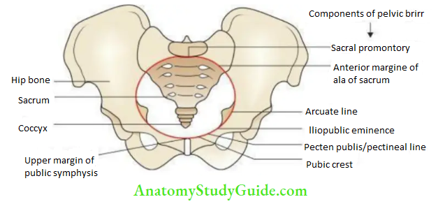
Subdivisions and pelvis:
- The pelvis is divided into two parts: A shallow upper part called the greater pelvis and a deep lower part called the lesser pelvis by pelvic brim (also called the pelvic inlet)
- The pelvic brim is formed by the sacral promontory posteriorly, the upper margin of pubic symphysis anteriorly, and iliopectineal line (arcuate line + pectinal line) on either side.
Pelvis Bones
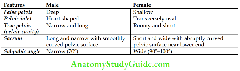
Question 2. What are the differences between male and female pelves?
Answer:
The differences between male and female pelves are given in the Table.
Sex Differences Between Male and Female Pelves:
Pelvic Muscles
Question 3. Enumerate muscles of the pelvis.
Answer:
Muscles Of The Pelvis:-
These are 4 pairs of pelvic muscles:
- Levator ani
- Coccygeus
- Piriformis
- Obturator externus
Question 4. Describe the pelvic diaphragm under the following headings:
- Introduction
- Formation
- Openings
- Relations
- Functions and
- Applied anatomy.
Answer:
1. Pelvic Introduction: The pelvic diaphragm is a gutter-shaped muscular partition between the pelvis and perineum.
2. Pelvic Formation: It is formed by 4 muscles, two from each side:
- Levator ani
- Coccyges
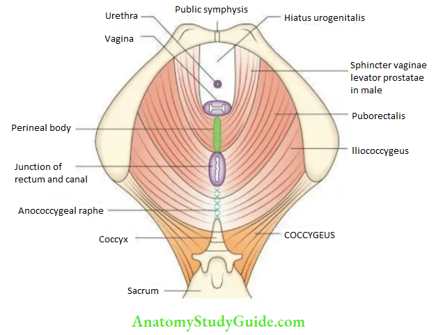
The two levator ani form the major part of the pelvic diaphragm.
1. Levator ani:
It consists of two parts: the anterior part called pubococcygeus and the posterior part called iliococcygeus.
- Origin:
- Pubococcygeus arises from the pelvic surface of the body of the pubis and the anterior half of the tendinous arch/white line of pelvic fascia.
- Iliococcygeus arises from the posterior half of the tendinous arch/white line of pelvic fascia and the pelvic surface of the ischial spine.
- Insertion: The fibers run backward, downward, and medially with different degrees of obliquity and are inserted as follows:
- Anteriormost fibers (puboprostate or pubovaginalis): These fibers pass by the sides of the prostate in males (levator prostate) or vagina in females (sphincter vaginae) to insert into the perineal body.
- Middle fibres (puborectalis): These fibres wind around the posterior aspect of the anorectal junction and continue with the similar fibres of the opposite muscle forming a U-shaped loop termed puborectal sling.
- Posterior most fibers (iliococcygeus): These fibers pass downwards and medially and are inserted into the anococcygeal raphe and tip of the coccyx
Nerve supply: the Anterior half of the levator ani is supplied from the perineal surface via the perineal branch of the pudendal nerve (S2 and S3), and the posterior half is supplied from the pelvic surface by the 4th sacral nerve.
Pelvis Bones
2. Coccygeus (ischiococcygeus):
It is a triangular muscle situated behind the levator ani and makes a small contribution in the formation of the pelvic diaphragm.
- Origin: Ischial spine and sacrospinous ligament.
- Insertion: Sides of the upper two pieces of the coccyx and the last piece of the sacrum.
- Nerve supply: By ventral rami of the 4th and 5th sacral nerves.
3. Openings of the pelvic diaphragm:
- Hiatus urogenital: It is a triangular gap between the anterior fibers of the two levator ani muscles. It provides passage to the urethra in males and the urethra and vagina in females. This gap is closed from below by the urogenital diaphragm.
- Hiatus rectal: It is a round opening between the perineal body and the anococcygeal raphe. It provides a passage to the anorectal junction.
Note:
Hiatus of Schwabe:
- Sometimes there is an abnormal opening present in the pelvic diaphragm when levator ani fails to arise from the obturator fascia.
- It is called as hiatus of Schwabe.
- Through, this gap between the obturator fascia and the tendinous arch of the obturator fascia, the pelvic viscera may herniate into the ischiorectal fossa of the corresponding side.
4. Relations of the pelvic diaphragm:
- Superior/pelvic surface
- Pelvic fascia
- Urinary bladder
- Prostate
- Rectum
- Inferior/perineal surface
- Anal fascia
5. Functions of the pelvic diaphragm:
- Supports the pelvic viscera
- Constriction of vagina
- Elevation of prostate
- Facilitates defecation and parturition
- Aids in micturition
6. Pelvic Applied anatomy: The pelvic diaphragm may be injured during parturition. This may cause uterine or rectal prolapse.
Pelvis Bones
Arteries of the pelvis:
Question 5. Write a short note on the internal iliac artery.
Answer:
1. Internal iliac artery Origin: It is one of the two terminal branches of the common iliac artery.
2. Internal iliac artery Termination: It terminates by dividing into anterior and posterior divisions at the upper margin of the greater sciatic foramen.
3. Internal iliac artery branches:
- Anterior divisions: It gives off 6 branches in the male/female
- Superior vesical artery
- Obturator artery
- Middle rectal artery
- Inferior vesical artery (replaced by the vaginal artery in females)
- Internal pudendal artery
- Inferior gluteal artery
- Uterine artery (in females only)
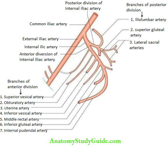
Note: All the branches from the anterior division are visceral branches except inferior gluteal and obturator arteries, which are parietal branches.
Posterior Divisions:
- It gives off 3 branches:
- Iliolumbar
- Lateral sacral (usually 2 in number)
- Superior gluteal artery
Note: All the branches of the posterior division are parietal branches.
Pelvis Bones
Question 6. Describe the obturator artery in brief. Write a note on ‘corona mortise and abnormal obturator artery.
Answer:
Obturator Artery:-
The obturator artery arises from the anterior division of the internal iliac artery. It passes forward along the lateral wall of the pelvis and enters the obturator canal to reach the front of the thigh.
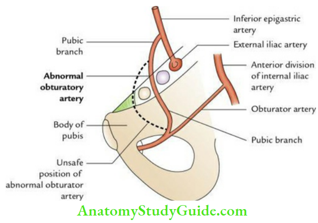
Corona Mortis:
- The important pubic branch of the obturator artery runs upward and medially to anastomose with the pubic branch of the inferior epigastric artery on the pelvic surface of the superior pubic ramus.
- This anastomosis establishes collateral circulation between external and internal iliac arteries and is called ‘corona mortis’. The variant vessel if cut can lead to a fatal haemorrhage. It is called because it means death (L., corona mortis = crown of death).
Abnormal Obturator Artery:
- Occasionally the anastomosis between a pubic branch of the obturator artery and the pubic branch of an inferior epigastric artery is so large that the obturator artery appears to be a branch of the inferior epigastric artery, and then it is termed an abnormal obturator artery.
- If it lies along the lateral margin of the femoral canal, it is called a safe position.
- If it lies along the medial margin of the femoral canal, i.e. on the free margin of the lacunar ligament, it is called an unsafe position as it is likely to be cut during enlargement of the femoral ring to relieve the strangulated femoral hernia.
Leave a Reply