• Skip to main content
  • Skip to secondary menu
  • Skip to primary sidebar
  • Skip to footer
  • Anatomy
    • Anatomy Question And Answers
    • Face Anatomy
    • Neck Anatomy
    • Head Anatomy
    • Oral Anatomy
    • Lower Limb
    • Upper Limb
  • Endodontics
    • Paediatric Dentistry
  • General Histology
    • Oral Histology
    • Genetics
  • Pediatric Clinical Methods
  • Complete Dentures
    • Pharmacology for Dentistry
  • Medical Physiology
    • Body Fluids
    • Muscle Physiology
    • Digestive System
    • Renal Physiology
    • Endocrinology
    • Nervous System
    • Respiratory System
    • Cardiovascular System
    • Reproductive System
    • Oral Physiology
  • General Medicine
  • General Pathology
    • Systemic Pathology
    • Oral Pathology
    • Neoplasia
    • Homeostasis
    • Infectious Diseases
    • Infammation
    • Amyloidosis Notes
  • Periodontology
  • General Surgery
    • Basic Principles Of Surgery
    • General Surgery

Anatomy Study Guide

Anatomy Study Guide

  • About Us
  • Contact Us
  • Privacy Policy
  • Terms of Use
  • Disclaimer
  • Sitemap
Home » Choledochal Cyst Classification, Clinical Features and Investigations

Choledochal Cyst Classification, Clinical Features and Investigations

October 21, 2023 by Sainavle Leave a Comment

Choledochal Cyst

It is a congenital cyst occurring in the CBD due to partial or complete weakness of the wall of the CBD. Majority of cases manifest by 1-2 years of age.

Table of Contents

  • Choledochal Cyst
  • Caroli’s Disease

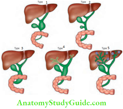
Choledochal Cyst Classification- Recent advances: Type 6—cystic dilatation of cystic duct.

Gallbladder And Pancreas Types of choledochal cysts

Choledochal Cyst Clinical Features

  • Age: Majority of cases manifest in children within 1-2 years of age. It can also present in adults.
  • More common in females—4:1.
  • Abdominal distension can be due to a large cyst. The cyst can be palpated per abdomen in the right hypochondrium.
  • Slow progressive jaundice, recurrent attacks with abdominal pain and pyrexia.

Choledochal Cyst Investigations

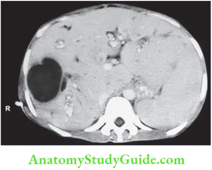
  1. USG will confirm presence of abnormal cyst. It is usually unilocular cyst.
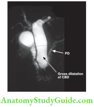
  2. MRC: It will define the relation between lower end of the bile duct and pancreatic duct to know basic anatomy.
  3. CT is also useful to know intrahepatic and extra- hepatic dilatation.
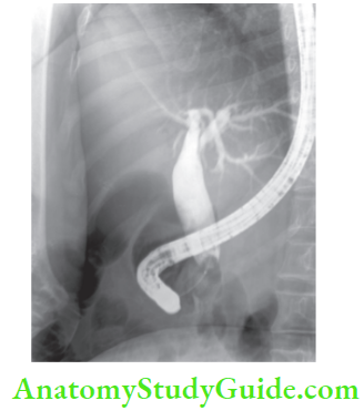
  4. ERCP may be done but it will not give any more information than MRC.

Gallbladder And Pancreas MRC showing choledochal cyst

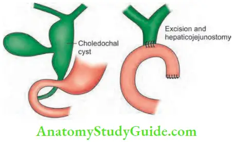
Choledochal Cyst Treatment: This anomaly is premalignant. Change to carcinoma is a well-recognised complication and it carries poor prognosis. Hence, excision of the cyst and reconstruction is the treatment of choice

  1. Type 1: Excision of the cyst followed by Roux-en-Y hepaticojejunostomy.
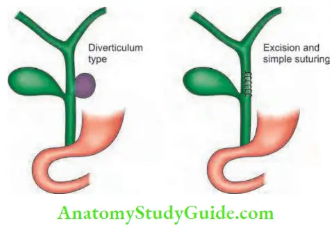
  2. Type 2: Excision of the diverticulum with suturing of CBD.
  3. Type 3: Endoscopic sphincterotomy is adequate (choledochocele).
  4. Type 4: They are difficult to treat. Due to recurrent cholangitis, if total excision is not possible due to adhesions between the cyst and portal vein, posterior wall of the cyst can be left behind, after removal of mucosa. This is described as Lilly’s technique.
  5. Type 5 will be described later.

Gallbladder And Pancreas ERCP showing choledochal cyst type 3 treated by sphincterotomy

Gallbladder And Pancreas Type 1 Cyst

Gallbladder And Pancreas Type 2 excision and suturing

Choledochal Cyst Complications

  1. Recurrent cholangitis with high-grade fever, resulting in biliary cirrhosis.
  2. Rupture of the cyst resulting in biliary peritonitis.
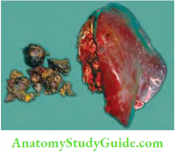
  3. CBD stones
  4. Carcinoma in the cyst (25-30% of cases). It is a cholangiocarcinoma common in types 1 and 5.

Caroli’s Disease

It is a hereditary condition wherein there is dilatation of intrahepatic ducts with stenotic segments in between. Multiple, irregular saccular dilatations are characteristic. It is also an example of type 5 choledochal cyst. Two types:

  1. Simple: Presents later with abdominal pain and sepsis.
  2. Periportal: It occurs in childhood, presents as recurrent cholangitis.
  • Diagnosed by ultrasound and CT scan. MRI and ERCP are other investigations.
  • Associated lesions are given.
    • Congenital hepatic fibrosis
    • Medullary sponge kidney
    • Polycystic liver

Gallbladder And Pancreas Caroli’s disease showing large cyst and intrahepaticdilation

Caroli’s Disease Complications

  • Cholangitis: It occurs due to constant obstruction.
  • Stones: Obstruction and stasis precipitate stone formation.
  • Biliary cirrhosis
  • Cholangiocarcinoma

Caroli’s Disease Treatment: Hepatectomy, liver transplantation.

Gallbladder And Pancreas Left lobectomy with extraction of stone Caroli’s disease

Filed Under: Gastrointestinal Surgery

Reader Interactions

Leave a Reply Cancel reply

Your email address will not be published. Required fields are marked *

Primary Sidebar

Recent Posts

  • Esophagus Anatomy
  • Lacrimal Apparatus: Anatomy, Parts & Function
  • Scalp Temple And Face Question and Answers
  • Orbicularis Oculi Muscle Anatomy
  • Extraocular Muscles Anatomy
  • Ciliary Ganglion Anatomy
  • Femoral sheath Anatomy
  • Femoral Artery – Location and Anatomy
  • Adductor Canal: Anatomy And Function
  • Ankle Joint: Anatomy, Bones, Ligaments And Movements
  • Risk Factors For Breast Cancer
  • Cervical Tuberculous Lymphadenitis Notes
  • Carbuncles: Causes, Symptoms, and Treatments
  • Sinuses And Fistulas Notes
  • Cellulitis: Treatments, Causes, Symptoms
  • Pyogenic Liver Abscess: Causes, Symptoms, and Diagnosis
  • Acid Base Balance Multiple Choice Questions
  • General Surgery Multiple Choice Questions
  • Hypertrophic Scarring Keloids Multiple Choice Questions
  • Surgical Site Infection Multiple Choice Questions
  • Facebook
  • Pinterest
  • Tumblr
  • Twitter

Footer

Anatomy Study Guide

AnatomyStudyGuide.com is a student-centric educational online service that offers high-quality test papers and study resources to students studying for Medical Exams or attempting to get admission to different universities.

Recent

  • Esophagus Anatomy
  • Lacrimal Apparatus: Anatomy, Parts & Function
  • Scalp Temple And Face Question and Answers
  • Orbicularis Oculi Muscle Anatomy
  • Extraocular Muscles Anatomy

Search

Copyright © 2024 · Magazine Pro on Genesis Framework · WordPress · Log in