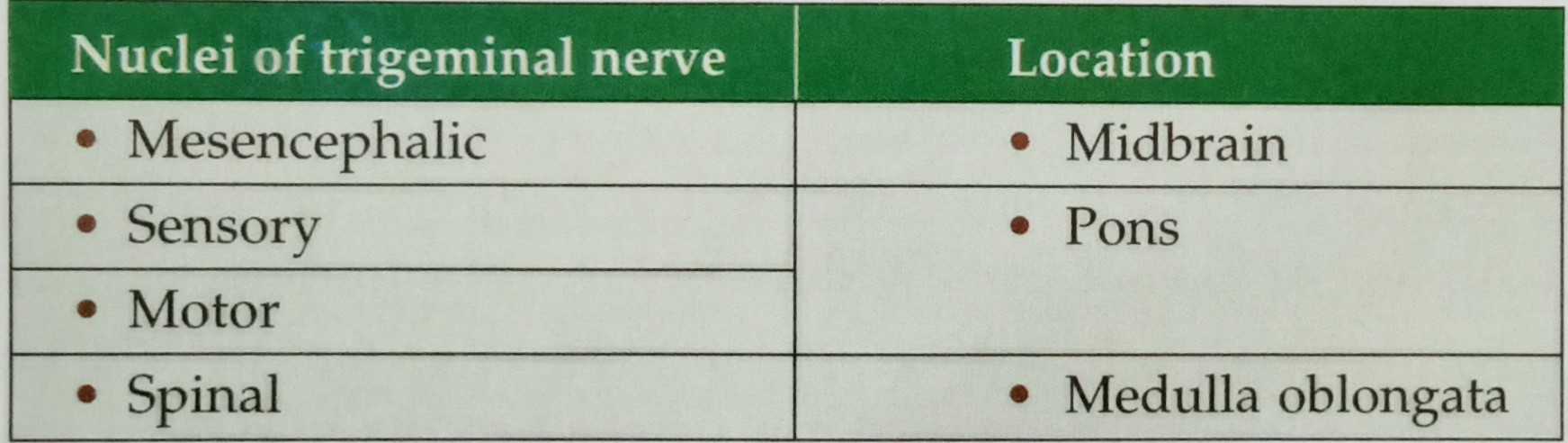
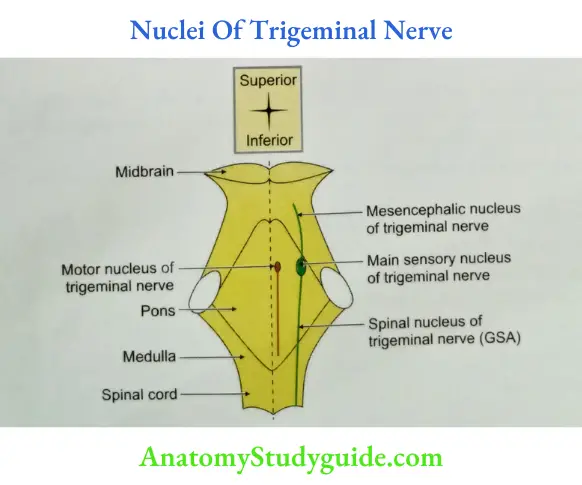
Question 1 Enumerate the nuclei associated with 5th cranial nerve.
Answer:
Table of Contents
Read And Learn More: Anatomy Important Question And Answers
Medial Longitudinal Bundle (fasciculus)
Medial Longitudinal Bundle Introduction: It is vertical running bundle of axons, present in the midline of brainstem.
- Medial Longitudinal Bundle Medial Longitudinal Bundle Extent: It extends from upper border of midbrain to the upper cervical part of spinal cord.
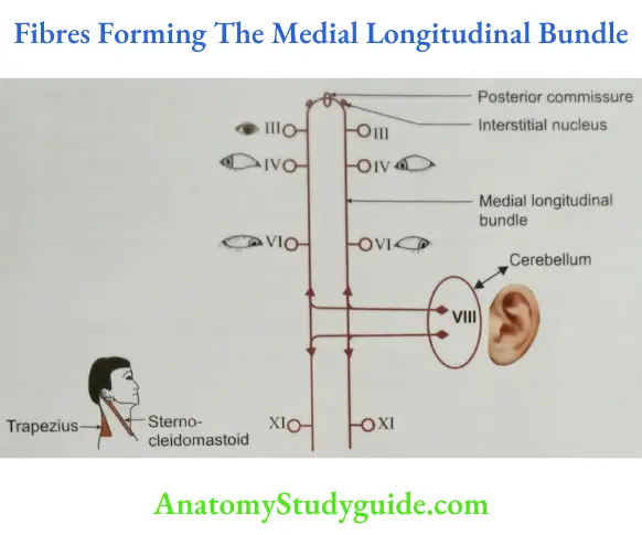
- Medial Longitudinal Bundle Medial Longitudinal Bundle Formation: It connects the nuclei of the following cranial nerves (Fig. 4.2) A. Vestibular-VIIIth cranial nerve.
- Oculomotor-IIIrd cranial nerve.
- Trochlear-IVth cranial nerve.
- Abducent-VIth cranial nerve.
- Spinal accessory-XIth cranial nerve.
- Reticular nuclei.
- Medial Longitudinal Bundle Functions
- Coordination of conjugate movements of eyeball.
- The movements of head and neck in response to audio and visual reflexes.
- Medial Longitudinal Bundle Location: Just in front of (central canal/aqueduct of 4th ventricle) cavity.
- Medial Longitudinal Bundle Applied anatomy: Lesion of intralaminar nuclei of medial longitudinal bundle results in loss of coordination of conjugate movements of eyeball.
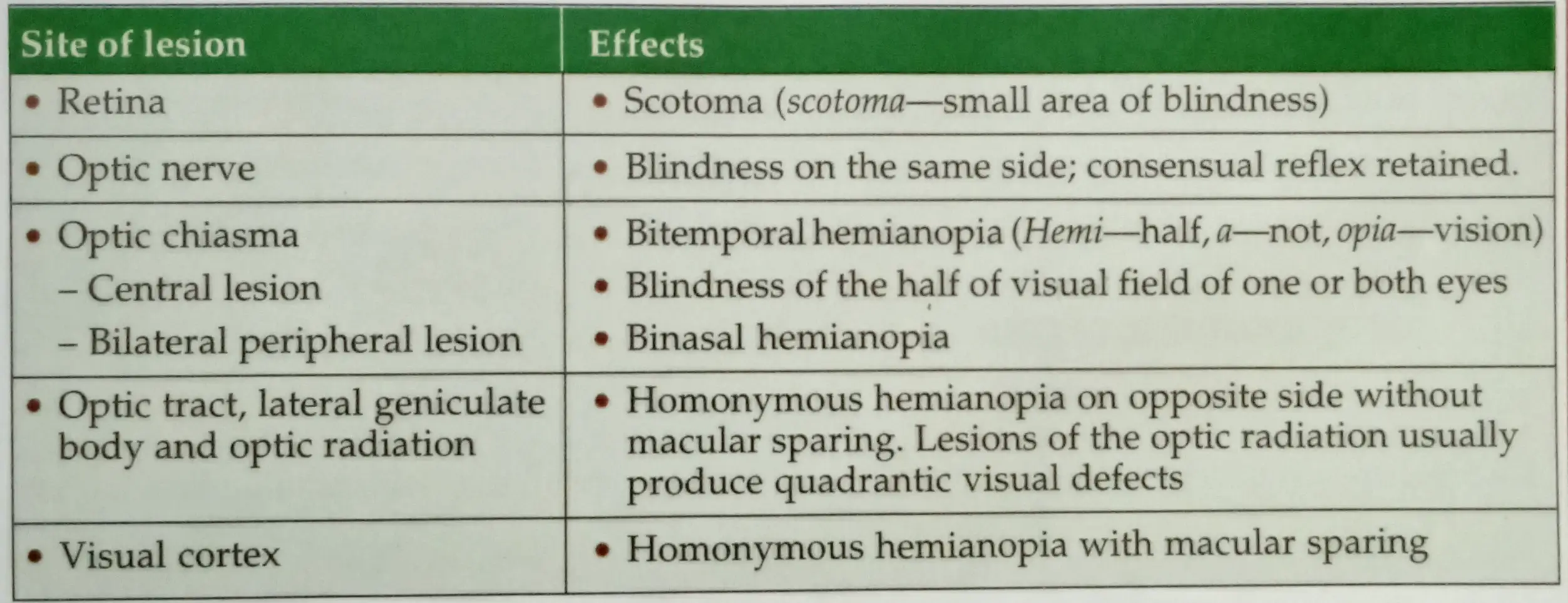
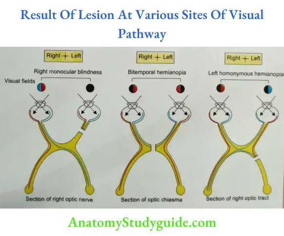
Describe visual (optic) pathway and the effects of lesion of different parts of visual pathways.
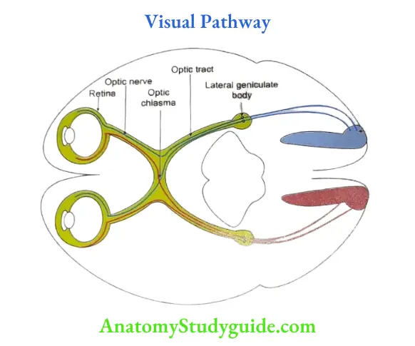
The visual pathway includes structures which are concerned with the reception, transmission and perception of visual impulses.
Structures in visual pathway
- Retina: It is a thin, delicate inner layer of eyeball. It contains rods and cones which are receptors for the visual pathway. It contains bipolar cells and ganglion cells. The axons of the ganglion cells form optic nerve.
- Peculiarities of optic nerve
- In a strict sense, the optic nerve is not a cranial nerve, but a prolongation of the white matter of the brain, because it is developed from the stalk of optic vesicle.
- It is the only cranial nerve covered by three meninges of the brain; hence the nerve undergoes atrophy in prolonged increase of cerebrospinal fluid pressure.
- The myelination of the optic nerve is derived from the oligodendroglia, but not from the Schwann cells. Hence, it does not have endoneurium and if damaged cannot regenerate.
- It has rich blood supply.
- It is a tract and not a nerve. (The nerve is usually first order neuron.)
- Optic chiasma: In the chiasma, the nasal fibres of each optic nerve decussate. They pass into the optic tract of the opposite side. The temporal fibres from each retina pass on to their own side.
- Optic tract: Each optic tract winds round the cerebral peduncle of the midbrain. It divides near the lateral geniculate body into two roots.
- Lateral root: It is thick and terminates in the lateral geniculate body. A few of its fibres pass to the superior colliculus, the pretectal nucleus and the hypothalamus.
- Medial roots: It is believed to contain the fibres of Gudden’s commissure. (The fibre bundles situated dorsal to the optic chiasma.) Each optic tract contains temporal fibres of the same side and nasal fibres of the opposite side.
- Lateral geniculate body: The larger fibres of the optic tract synapse in the lateral geniculate body. These are visual fibres.
- Optic radiation (geniculocalcarine tract): It begins from the lateral geniculate body. It passes through the retrolentiform part of the internal capsule. It ends in the visual cortex.
- Visual cortex: The optic radiation ends in the striate area (area 17) where the colour, size, shape, motion, illumination and transparency are appreciated separately. Objects are identified by integration of these perceptions with past experience stored in the parastriate and peristriate areas (areas 18, 19). The area of the visual cortex that receives impulses from the macula is relatively much larger than the part related to the rest of the retina.
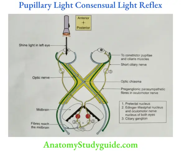
Pathway Of Light Reflex
Light reflexes are mediated through the
- Retina,
- Optic nerve,
- Optic chiasma,
- Optic tract,
- Lateral geniculate body,
- Pretectal nucleus,
- Edinger-Westphal nucleus of the 3rd nerve,
- 3rd cranial nerve,
- Ciliary ganglion,
- Short ciliary nerves, and
- Constrictor pupillae.
- Throwing light on one eye produces constriction of the pupil in both eyes (consensual reflex).
- This is due to bilateral connections of the pretectal nucleus with the Edinger-Westphal nuclei.
Pathway Of Accommodation Reflex
Constriction of the pupil also takes place when looking at a near object. This is mediated through the
- Retina,
- Optic nerve,
- Optic chiasma and tract,
- Lateral geniculate body,
- Optic radiation,
- Visual area of the cortex,
- Superior longitudinal bundle,
- Frontal eye field,
- 3rd nerve nucleus, Corticonuclear fibers
- 3rd cranial nerve,
- Ciliary ganglion, and
- Ciliary and sphincter pupillae muscles.
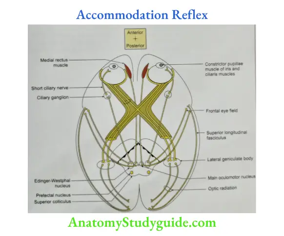
Note that the pretectal nucleus is not involved in the accommodation reflex.
Accommodation Reflex Applied anatomy:
- In lesions of the pretectal nucleus, the light reflex is lost, but the pupil contracts on accommodation.
- This is called the Argyll-Robertson pupil (accommodation reflex present-ARP).
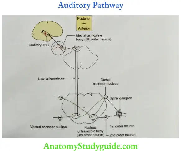
Pathway Of Hearing (auditory pathway)
- The 1st order neurons are located in the spiral ganglion. They are bipolar cells. The peripheral processes innervate the organ of Corti, while the central processes terminate in the dorsal and ventral cochlear nuclei.
- The 2nd order neurons lie in the dorsal and ventral cochlear nuclei. Most of the axons arising in these nuclei cross to the opposite side (in the trapezoid body and terminate in the superior olivary nucleus. (Many fibres end in the nucleus of the trapezoid body or of the lateral lemniscus). Some fibres are uncrossed.
- The 3rd order neurons lie in the superior olivary nucleus present in the pons. The axons form the lateral lemniscus and reach the inferior colliculus.
- The 4th order neurons lie in the inferior colliculus. Their axons pass through the inferior brachium to reach the medial geniculate body. (Some fibers of the lateral lemniscus reach the medial geniculate body without relay in the inferior colliculus.)
- The 5th-order neurons lie in the medial geniculate body. The axons form the auditory radiation, which passes through the sublentiform part of the internal capsule to reach the auditory area of the temporal lobe.
Leave a Reply