Development Of Teeth
Have you anytime thought how the hardest tissue of the body called enamel and the softest tissue called the pulp are formed together in an organ called the tooth? The story of the formation of the tooth, known as odontogenesis, is an interesting one. Odontogenesis is studied in two parts:
Table of Contents
- Formation of the crown, which includes the formation of tissues such as enamel, dentin and pulp.
- Formation of the root, which includes formation of the cementum, root pulp, root dentin, and the supporting structures of the tooth including the periodontal ligament, alveolar bone.
Read And Learn More: Oral Histology Notes
Tooth Germ Formation
Stomatodeum:
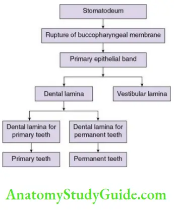
- The primitive oral cavity is called the stomatodeum. It is formed around the 3rd week after gestation and is covered by the epithelial cells which are derived from the ectoderm.
- When the embryo is 4 weeks old, the rupture of buccopharyngeal membrane takes place and the communication between the ectoderm and endoderm is established, which means communication between the future oral cavity derived from the ectoderm and the foregut derived from the endoderm is established.
Primary epithelial band:
When the embryo is around 5 weeks old, the ectodermal cells show condensation to form a band-like structure called the primary epithelial band. The connective tissue component of the teeth and the supporting structures are derived from the neural crest cells. These cells migrate and reach the primitive oral cavity. Thus, the connective tissue here is called ectomesenchyme.
Dental and vestibular lamina:
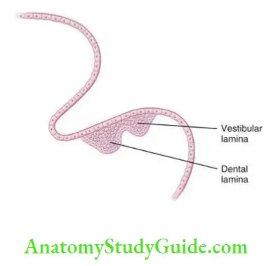
There are two extensions arising in the epithelial band.
- One on the buccal side
- It shows initial proliferation and then degeneration of the central epithelial cells.
- This leads to the formation of a sulcus or depressed area called the lip furrow, which is the future vestibule.
- This extension of the epithelial cells is known as the vestibular lamina or the lip furrow band.
- One on the lingual side
- It shows condensation of the epithelial cells in an arch shape, on the lower jaw and on the upper jaw.
- These correspond to the future tooth-bearing area called gum pads at birth.
- Furthermore, condensation of the epithelial cells in certain areas on these arch-shaped structures are seen as bulges, which correspond to the future teeth.
Dental lamina:
Histologically, there are epithelial strands which later give rise to the tooth germ, i.e. the tooth forming structures arising from these bulges. This looks like the stalk of the bud. These structures are called dental lamina.
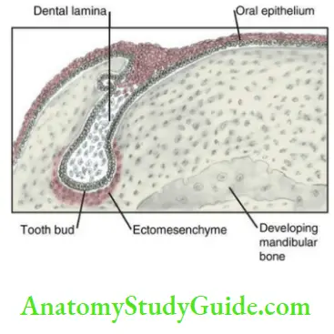
Salient Features Of Dental Lamina:
- Active from the 5th week of intrauterine period to 5 years of age
- Connects the tooth germ with the oral ectoderm
- Derived from the primitive epithelial band
- Activity starts at the midline of the arch and proceeds from anterior to posterior
- Dental lamina for deciduous teeth, successional lamina, dental lamina for the permanent molars is present.
- After the function the dental lamina ruptures and degenerates
- The persistent, inactive cells are the cell rests of Serres.
From each stalk, a future tooth will form.
- Humans have 32 permanent and 20 deciduous teeth in their lifetime, but all the 52 teeth do not form simultaneously.
- The sequence of formation of the teeth follows that of the pattern of the eruption of these teeth into the oral cavity, i.e. the deciduous teeth will form ahead of the permanent teeth.
- At birth, the evidence of formation of all the deciduous teeth and that of the permanent first molars is available. The remaining of the permanent teeth form in the coming years.
Successional lamina:
The dental lamina for the successor permanent teeth (incisors, canines, premolars) gets formed by the lingual extension of the dental lamina of the deciduous teeth. This dental lamina is called the successional lamina.
Accessional lamina:
The distal extension of the primary epithelial band provides the dental lamina for the permanent molars, which is called accessional lamina.
- Dental lamina is active from the 5th week of the intrauterine period to 5 years of age.
- After its function, the dental lamina breaks and degenerates. If the cells persist in the gingiva, they are called the cell rests ofSerres.
Parts Of The Tooth Germ:
Odontogenesis is mainly studied histologically
Stages Of Tooth Development:
There are various changes observed during the course of the tooth formation. To identify these changes, the researchers have first focused on the shape of the primitive structure which gives rise to the enamel called the enamel organ.
Stages of tooth development:
There are various changes observed during the course of the tooth formation.
To identify these changes, the researchers have first focused on the shape of the primitive structure which gives rise to the enamel called the enamel organ.
Stages Of Tooth Development
The stages of tooth development based on the shape of the enamel organ
- Bud stage
- Cap stage
- Early bell stage
- Advanced bell stage
Bud stage
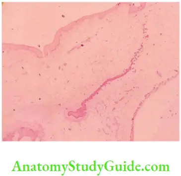
At about the 8th week of the embryonic life, the dental lamina shows proliferation of the epithelial cells at certain areas. Furthermore, the cells also condense to give a structure which resembles a bud. Thus, this stage is recognized as a bud stage.
- In this mass of cells, the peripheral cells are low columnar in shape and the central cells are polygonal in shape. The epithelial cells are rich in RNA and oxidative enzymes and instruct the initiation of tooth formation.
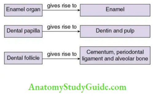
- Surrounding the enamel organ are the ectomesenchymal cells. These cells also show proliferation and condensation just beneath the enamel organ. These cells are called the dental papilla.
- The enamel organ and dental papilla are surrounded by condensation of
ectomesenchymal cells called the dental follicle or dental sac The epithelial cells are separated from the adjacent dental papilla and the dental follicle by a basement membrane.
The continuous mitotic activity and further differentiation of the cells leads to a capshaped enamel organ and thus called the cap stage.
Cap stage:
The cap stage can be identified from the 11th to the 13th week of intrauterine life.
During this period, the tooth germ will assume the shape which marks the stage of morphogenesis.
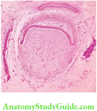
- The enamel organ shows unequal rate of cell division at different parts. The cells at the periphery divide more actively than the centre, as a result a cap-shaped enamel organ is formed
- The cells present at different parts of the cap assume different shapes and thus perform different functions.
- Outer enamel epithelium: The convex, outer part of the cap is lined by short cuboidal cells.
- Inner enamel epithelium: The concave, inner part of the cap is lined by tall, columnar cells.
Stellate reticulum:
The cells in between the outer enamel epithelium and inner enamel epithelium are polygonal in shape and show some changes leading to star-shaped cells forming the layer called stellate reticulum.
- These cells synthesize glycosaminoglycans and secrete the same to the
extracellular compartment. The glycosaminoglycans are hydrophilic in nature, thus they absorb water from the surrounding ectomesenchyme. Due to this fluid accumulation, the polygonal cells are pushed apart but the cells are connected to each other with desmosomes. They get pulled by each other with the junctions and assume a star shape and form a network called the stellate reticulum. - The fluid in between these cells is rich in albumin. This acts as a shock absorber by acting like a cushion to protect the inner enamel epithelial cells which later form the enamel. Thus, enamel pulp is the other name for the stellate reticulum.
Membrane preformativa:
Immediately below the concavity of the cap, the dental papilla cells show proliferation and condensation. These cells are separated from the inner enamel epithelial cells by the basement membrane.
- This membrane shows the folding representing the future cusp tips.
- The enamel and the dentin matrix are later deposited along this membrane and the dentinoenamel junction is formed.
- This structure decides the shape of the crown, so it is called membrane
preformativa, which is the blue print of the crown. - Similarly, the dentinocemental junction decides the shape of the root. This marks the stage of morphogenesis.
Genetic aspect:
A group of genes called the Homeobox genes get expressed at different sites of the forming jaw, thus the form or the pattern (such as the incisor, canine, premolar, molar) of the tooth is determined by these site-specific Homeobox genes. This is called the field model hypothesis.
Transient structures in the enamel organ:
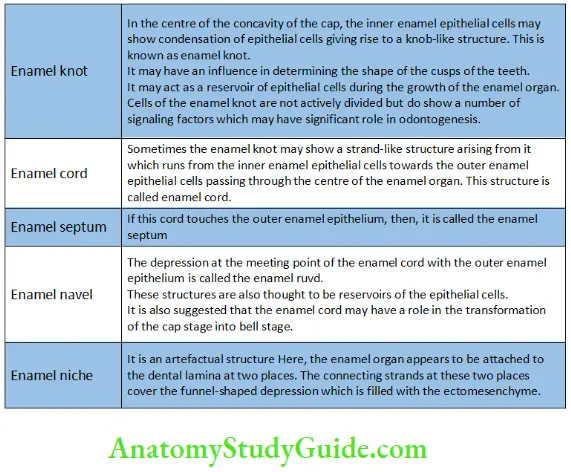
There are few histologic features which may appear in the enamel organ in the cap and early bell stage. These are temporary or transitory structures which play some role in odontogenesis
Transient Structures in the Enamel Organ
Dental follicle:
Both enamel organ and dental papilla are surrounded by proliferation and condensation of the ectomesenchymal cells called the dental follicle.
Bell stage:
This can be studied under the early and advanced bell stages.
Early bell stage:
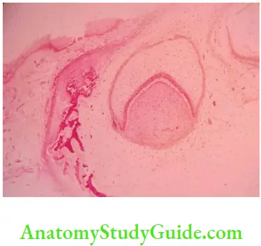
At about 14th to 16th week of intrauterine period, the division of the cells at the margins continues profusely to further change the shape of the enamel organ. The concavity in the centre of the cap deepens and the enamel organ assumes the shape of a bell.
Cells Of The Early B Ell Stage:
The cell layers of the enamel organ at bell stage
- Inner enamel epithelium
- Stratum intermedium
- Stellate Reticulum
- Outer enamel epithelium
Mnemonic: ISRO
At the bell stage, the enamel organ shows the following four distinct types of cell layers:
- Inner enamel epithelium
- Stratum intermedium
- Stellate reticulum
- Outer enamel epithelium
Inner enamel epithelium:
The cells of the inner enamel epithelium reside on the basement membrane.
They form a single row of cells.
- The inner enamel epithelial cells induce the dental papilla cells to differentiate into odontoblasts.
- The odontoblasts secrete dentin and in turn influence the inner enamel epithelial cells to change the morphology.
- The inner enamel epithelial cells change their shape to acquire the specialized function of secreting the enamel matrix. They become tall, columnar with the nucleus away from the basement membrane, and accumulate rich number of cellular organelles suggesting secretory function. These are called the ameloblasts, the enamel forming cells. This mechanism is known as reciprocal induction.
Stratum intermedium
Few layers of cells just above the inner enamel epithelium constitute the stratum intermedium, which means the layer in between the inner enamel epithelium and earlier formed stellate reticulum.
- The cells have squamous shape and show good number of cellular organelles such as Golgi apparatus, ribosomes, mitochondria, etc. This suggests that these cells do have an important role in the supply of nutrition to the ameloblasts, synthesis and transport of ions required for enamel formation.
- They show rich amount of alkaline phosphatase enzyme required in the mineralization of the enamel.
Stellate reticulum
This is a network of star-shaped cells as seen in the cap stage. At the bell stage, in addition to protection of the enamel forming cells, they play another role.
- When the first layer of dentin is formed by the odontoblasts, the nutritional supply reaching the inner enamel epithelium through the dental papilla is cut off. For a short time, the stratum intermedium supplies the nutrition.
- Meanwhile, the stellate reticulum begins to collapse, first in the deepest invagination part, later on the cervical part. By this action, the capillaries in the dental follicle come closer to the inner enamel epithelial cells. This supply of nutrition allows them to further function as ameloblasts.
Once the stellate reticulum collapses, it is difficult to distinguish these cells from the cells of the stratum intermedium.
Outer enamel epithelium
- This layer shows the cuboidal cells.
- No dramatic changes are seen in this layer.
However, after the collapse of the stellate reticulum, this layer shows the infoldings which bring the capillaries of the dental follicle closer to the ameloblasts.
Dental papilla
The cells of the dental papilla, which lie condensed beneath the concavity of the cap, start moving apart and show morphological changes.
- The basal lamina of the inner enamel epithelial cells separates them from the dental papilla cells.
- There is a small acellular zone filled with fibrils. The organizing influence of the inner enamel epithelial cells changes the stellate-shaped cell to tall columnar. As a result, the acellular zone becomes occupied with the cells which are called the odontoblasts. They secrete the dentin matrix at the basal lamina end.
- This zone which demarcates the enamel forming and the dentin forming cells is the membrane preformativa which marks the future dentinoenamel junction.
- The deposition of enamel and dentin takes place in the opposite direction.
- Enamel is formed from the dentinoenamel junction towards outer surface.
- Dentin formation is also from the dentinoenamel junction but towards inner side to the pulp.
Dental follicle:
There are some changes taking place in the zone surrounding the enamel organ and the dental papilla.
- More number of fibrils are formed in this zone. These fibrils become fibres and show arrangement into bundles. They start orienting themselves in relation to the enamel organ and the dental papilla.
Breaking of dental lamina:
Till this stage, the tooth germ is connected with the oral ectoderm through the dental lamina. The dental lamina now begins to fragment and the cells undergo degeneration.
- Sometimes the cell groups may remain inactive. These are known as cell rests of Serres. (Any disturbance can make them make active and they may produce odontogenic lesions later in life.)
Growth centres:
The division and differentiation of the inner enamel epithelial cells does not take place uniformly throughout the enamel organ.
- It begins at the dentinoenamel junction of the incisal edge and the cusp tips, which can be recognized as the growth centre.
- It then follows the slopes and proceeds to the cervical area.
Advanced bell stage
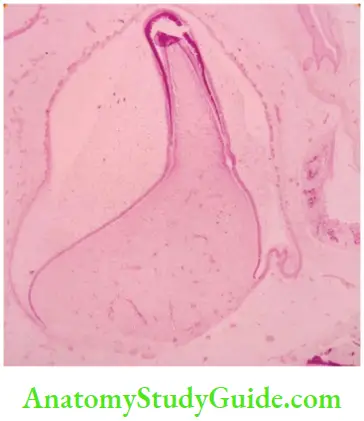
This stage can be identified between the 18th and 20th week of the embryo.
- This stage is characterized by the incremental deposition of dentin and enamel which gets mineralized immediately.
- The rhythmic process continues till the entire crown formation is complete.
- The apposition and mineralization processes continue to form the crown of the tooth.
- Following the crown formation, the root formation begins at the cervical area.
At about the 20th week of the intrauterine period, the formation of the permanent tooth germs can be identified. This will continue in the same manner as described for the primary dentition so far. At the age of 5 years, the tooth buds for the third molar teeth starts forming. Thus, the dental lamina is active till this 5 years of age.
Root formation
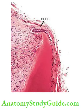
Cervical loop
The inner enamel epithelium and the outer enamel epithelium join at the neck of the forming tooth. Here the stratum intermedium and the stellate reticulum are absent. This structure is called the cervical loop or zone ofreflexion.
Hertwig’s epithelial root sheath
The root formation begins at the cervical portion from the cervical loop. The cells of this loop start proliferating to form a structure called Hertwig’s epithelial root sheath (HERS).
- The inner enamel epithelial cells are resting on a basement membrane which separates it from the dental papilla. The outer enamel epithelial cells are in proximity with the dental follicle cells.
- Similar to the crown formation, in the root formation also there is induction of the dental papilla cells by the inner enamel epithelial cells to differentiate into the odontoblasts. These cells secrete dentinal matrix of the root dentin.
- Once this dentin is formed, HERS undergoes fragmentation and the cells
degenerate. This breaking up of the epithelial cells makes the dental follicle cells to come in direct contact with the root dentin - As a result of this, the dental follicle cells differentiate into cementoblasts and start secreting the cementum on top of the root dentin. Thus, the cementodentinal junction is formed. Furthermore, the dental follicle cells differentiate into fibroblasts to form the periodontal ligament and some of the cells differentiate into osteoblasts to form the alveolar bone proper – the socket of the tooth.
- The number, shape and size of the roots are determined by HERS.
- Once it ruptures, the cells degenerate. If they persist, they are seen as inactive cell groups in the periodontal ligament. They are called the cell rests ofMalssez.
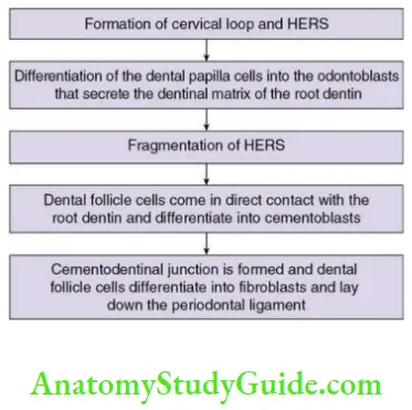
Root formation, periodontal ligament formation, alveolar bone proper formation and the eruptive movements of the teeth – all these work in a synchronous fashion. Although HERS will decide the number of the roots in a tooth, there are some differences in the formation of root of single-rooted tooth and a multirooted tooth.
Root formation in a single-rooted tooth:
HERS is composed of outer and inner epithelial cell layers present at the cervical portion of the tooth.
- This will show a horizontal bend called the epithelial diaphragm. This structure remains fixed during root formation.
- Root formation happens from the cervical portion towards the coronal side. There is no lengthening of HERS towards the apex of the tooth, as one easily assumes. Only proliferation of the cells of the epithelial diaphragm takes place.
- As the root dentin gets formed, the tooth starts moving coronally. The dental follicle cells form the periodontal ligament fibres which get oriented in different directions and help to pull the tooth along its path.
- HERS is not seen as a full-length sheath, instead, the cells proliferate and rupture rapidly. Once the full length of the root is formed the root dentin and the cementum formation overtake, that of the proliferation of epithelial diaphragm. This results in the narrowing of the cervical opening resulting in the conical shape of the root and formation of the apical foramina.
Formation of multiple roots in multirooted tooth:
The epithelial diaphragm shows a tongue-like extension. The number of this extension depends on the number of roots required to form.
- The mandibular molars will have two extensions.
- The maxillary molars will have three extensions.
These extensions from different sides grow and get fused, thereby leading to multiple diaphragms. Then each diaphragm grows into a root similar to the single root formation.
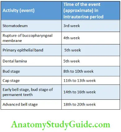
As multiple roots are formed, multiple foramen are also formed. Table 1.2 shows the various events and their approximate time during tooth development.
Timeline of Events in Tooth Development
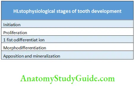
Histophysiological Events
Histophysiological events or processes can be identified during various stages of the tooth development. There may be overlapping of these events in various stages of the tooth development based on the shape of the enamel organ.
Histophysiological Events in Tooth Development
Initiation:
Odontogenesis is a complex phenomenon highly characterized by epithelial–mesenchymal interactions.
- The epithelium of the first pharyngeal arch has the potential for odontogenesis in the presence of the first arch mesenchyme derived from the neural crest cells called the ectomesenchyme. This epithelium stimulates the initiation of odontogenesis, and the tooth germ begins to form. After this stimulation from the epithelium, the ectomesenchyme attains the potential to form tissues of the tooth.
- In this event of initiation, the stimuli to form the tooth bud is communicated to the surrounding mesenchyme. In the absence of this initiation, no tooth would form and this condition is called anodontia.
Proliferation:
This is characterized by the division of the cells. As a result, the tooth bud is formed which grows into the cap stage.
Histodifferentiation:
Histodifferentiation stage can be recognized in the bell stage.
- The inner enamel epithelial cells differentiate into ameloblasts and the dental papilla cells differentiate into odontoblasts. The ameloblasts secrete enamel and odontoblasts lay down dentin.
- To prepare the cell for the secretory function, the cells assume tall columnar shape and other characteristics. These changes mark the histodifferentiation stage.
Morphodifferentiation:
- The ‘nonuniform’ growth of the enamel organ leads to the various forms and shapes of the tooth.
- The membrane preformativa which is the future dentinoenamel junction decides the shape and size of the crown. Thus, it is considered as the blue print of the crown of the tooth.
These events take place at the cap stage. It is morphogenesis. As the changes continue to take place in the bell stage to determine the final shape and size of the tooth, it is called the morphodifferentiation stage.
Apposition and mineralization:
In the late bell stage, the ameloblasts and odontoblasts secrete enamel and dentin matrix. This secretion takes place in an incremental pattern called apposition. This phenomenon is regular, rhythmic with alternate periods of activity and rest.
The organic matrix of enamel and dentin will become mineralized by the deposition of calcium and phosphate ions. This formation of the hard tissue follows the predetermined shape and size of the tooth.
Correlation between the histomorphological and the histophysiological stages of tooth development
- Initiation: Dental lamina
- Proliferation: Bud, cap and bell stage
- Histodifferentiation: Cap and bell stage
- Morphodifferentiation: Cap, bell, advanced bell stage
- Apposition: Formation of enamel and dentin
Development And Growth Of Teeth Clinical Considerations:
Any disturbance in these stages will lead to clinical problems. One can identify these problems and possibly assess the event and time that was faulty during odontogenesis.
Change in the number of teeth
- The disturbance in the initiation will result in
- Increase in number, decrease in number or absence of the teeth
- The congenital absence of the tooth is called anodontia.
- If few teeth are present, it is oligodontia. Commonly, maxillary lateral incisor, third molar and mandibular second premolar are the ones which may be absent.
- If the number is increased, the extra tooth is called the supernumerary tooth. Mesiodens and paramolars are examples of this.
Change in the size of the tooth
- The disturbance in the proliferation and morphodifferentiation of the tooth forming cells may result in increase or decrease in its size.
- Macrodont is an abnormally large-sized tooth and microdont is an
abnormally small-sized tooth.
- Macrodont is an abnormally large-sized tooth and microdont is an
Change in the shape of the tooth
- The disturbance in the morphodifferentiation will result in altered shape of the tooth.
- Dilaceration, which is a sharp bend or curve in the root, is an example.
Change in the structure of the teeth
- Stages of histodifferentiation, apposition and mineralization will lead to an improper quality of enamel and dentin.
- Vitamin A deficiency leads to failure of ameloblasts to influence the
dental papilla. As a result, the odontoblasts are not properly differentiated. The dentin formed here is also defective, called osteodentin. The defective enamel matrix formation by the ameloblasts in various situations may lead to enamel hypoplasia. The improper calcification of enamel leads to hypomineralization of the tooth where weak enamel leads to easy breaking of the tooth.
- Vitamin A deficiency leads to failure of ameloblasts to influence the
Abnormal roots
- Disturbance in HERS will result in abnormalities of the root.
Enamel pearls
- If HERS does not rupture and degenerate on time, the cells of the inner enamel epithelium may differentiate into ameloblasts and secrete a globule of enamel, usually seen on the furcation area of the multiple root. It is called the enamel pearl.
Accessory root canal
- If HERS breaks before the formation of the radicular dentin, it results in an accessory root canal.
Development And Growth Of Teeth Synopsis:
- Odontogenesis is a complex process which involves induction and interactions between the ectoderm and the ectomesenchyme, derived from the neural crest cells.
- The stomatodeum gives rise to primary epithelial band, from which the dental lamina and vestibular lamina arise. The dental lamina gives rise to the tooth germ and connects it to the oral ectoderm.
- The tooth germ shows three parts – enamel organ, which is ectodermally derived and gives rise to the enamel; the dental papilla derived from the ectomesenchyme gives rise to the dentin and the pulp; and the dental follicle derived also from the ectomesenchyme gives rise to the cementum, periodontal ligament and the alveolar bone.
- Based on the morphology of the enamel organ, the stages of the tooth
development are bud, cap, early bell and the advanced bell stages. - Based on the physiological changes, the stages are initiation, proliferation, histodifferentiation, morphodifferentiation, apposition and mineralization. There are histological changes marked in these stages.
- The enamel organ shows inner enamel epithelium, stratum intermedium, stellate reticulum, outer enamel epithelium. There are dental papilla and the dental follicle cells surrounding the enamel organ which get induced by the epithelial cells to secrete the respective tissues.
- Dentin is the first formed hard tissue of the tooth. The dentinoenamel junction decides the shape of the tooth.
- After the crown formation, the root formation begins at the neck of the tooth from the cervical loop. This structure gives rise to Hertwig’s epithelial root sheath, which acts as the blue print of the root. The root formation continues along with the eruptive movements of the tooth.
- The dental lamina and HERS rupture after their function, if the cells remain they are called cell rests of Serres and Malssez, respectively.
- Any disturbance in the various stages of the tooth development will result in the change in the number, size, shape, structure, etc. The roots can also show the variations due to the disturbances.
Leave a Reply