Epithelial Tissue: Structure with Diagram, Function, Types and Location
Simple Squamous Epithelium
Introduction: The cells of the simple squamous epithelium are flat. They have length, and breadth but no thickness.
Table of Contents

1. Epithelial Tissue Features: Cells of simple squamous epithelium have the following features—“ABC”. They
- Are Avascular
- Rest on a Basement membrane.
- Are Compactly arranged. Cells are separated by the minimal amount of intercellular substance.
| Body Fluids | Muscle Physiology | Digestive System |
| Endocrinology | Face Anatomy | Neck Anatomy |
| Lower Limb | Upper Limb | Nervous System |
Squamous Epithelial Tissue
2. Epithelial Tissue Morphology
1. Cells are arranged in single layer. They are irregular which are thin and platelike.
2. The nuclei are round and centrally situated.
Read And Learn More: Anatomy Notes And Important Question And Answers
3. The cell junction is marked by zonulae occludentes.
4. Surface view
- Cells look polygonal, or mosaic.
- Nuclei are bulging.
3. Epithelial Tissue Functions
1. It helps in transport of material across the cell.
2. The tight junctions between the cells prevent entry of various substances through them.
3. It facilitates movement of viscera in serous cavities.
Squamous Epithelial Tissue
4. Sites or examples of simple squamous epithelium:
1. Serous membranes like
- Pleura (covering of lungs),
- Pericardium (covering of heart), and
- Peritoneum (covering and the abdominal organs and abdominal cavity).
2. Alveoli of lung.
3. Loop of Henle—descending and ascending loop of Henle in kidney.
4. Endothelium of blood vessel.
5. Bowman’s capsule.
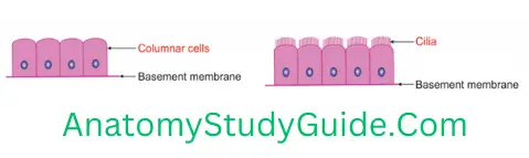
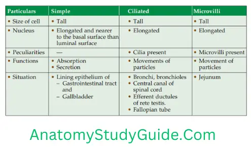
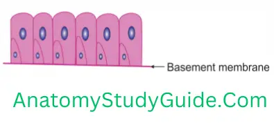
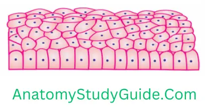
Simple Columnar Epithelium
Simple Columnar Epithelium Introduction: The cells are column-like or rectangular (on vertical section).
Simple Columnar Epithelium Function
1. Simple Columnar Epithelium Features
- The cells of the simple columnar epithelium are arranged in single layer.
- All the cells touch basal lamina (basement membrane).
- The height of the cells is more than width.
- The nuclei are vertically elongated.
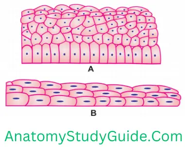
Pseudostratified Epithelium
(Pseudo—false)
Introduction: The simple columnar epithelium giving appearance of stratified (multilayered) epithelium is called pseudostratified epithelium.
Pseudostratified Columnar Epithelium
1. Pseudostratified Epithelium Features
- The cells are of variable height. Some cells are short and some cells are tall.
- All cells lie on the basement membrane.
- Short cells do not reach lumen.
- The nuclei are situated in the centre of each cell irrespective of the length of the cell.
- The nuclei are arranged in more than one layer which gives impression of many layered cells.
Pseudostratified Epithelium Classification: The pseudostratified epithelium is divided into following types
1. Pseudostratified non-ciliated: The luminal surface of the cells does not bearcilia, e.g. epithelium of
- Male urethra, and
- Vas deferens.
Pseudostratified Columnar Epithelium
2. Pseudostratified ciliated columnar: The luminal surface of the cells bears cilia, e.g. epithelium of
- Upper respiratory tract,
- Trachea, and
- Larger bronchi.
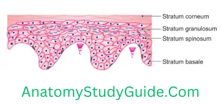
Stratified Squamous Epithelium
Stratified Squamous Epithelium Introduction: The multilayered epithelium having squamous (flat) cells on the top.
1. Stratified Squamous Epithelium Morphology
- The deepest cells are columnar.
- The middle cells are cuboidal or polyhedral.
- The superficial cells are flat or squamous.
2. Stratified Squamous Epithelium Classification: The stratified squamous epithelium is divided into following types depending upon the presence and absence of keratin.

Transitional Epithelium (Urothelium)
Transitional Epithelium (Urothelium) Introduction: The cells of the transitional epithelium are multilayered, having a capacity for distension.
1. Transitional Epithelium (Urothelium) Arrangement of cell: The cells of
- Deepest rows are columnar.
- Middle rows are pear-shaped.
- Superficial layers are umbrella-shaped.
2. Transitional Epithelium (Urothelium) Peculiarities
- It shows mitosis.
- The cells are multilayered (stratified). The top cells have the capability of stretching.
- The cells are embedded in lipids which prevents the entry of toxins.
3. Transitional Epithelium (Urothelium) Sites
1. Pelvis of the kidney,
2. Ureter, and
3. Urinary bladder.
What is brown fat?
1. Brown Fat Definition: The adipose tissue showing many vacuoles is called multilocular adipose tissue. It is brown in colour. Hence, it is called brown fat.
2. Brown Fat Characters
- It is highly vascular.
- It is smaller in size as compared to unilocular.
- It has centrally placed spherical nucleus.
- It contains many mitochondria.
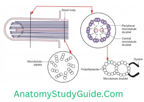
Draw and label a section of a cilium
Classify compound epithelium with examples
The compound epithelium is subdivided into
1. Stratified squamous
- Stratified squamous keratinised, e.g. skin
- Stratified squamous non-keratinised, e.g. oesophagus
2. Stratified columnar, e.g. moderate-sized ducts of salivary gland
3. Stratified cuboidal, e.g. secondary ovarian follicles
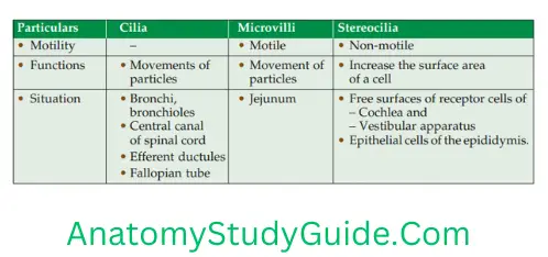
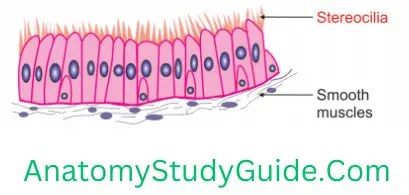
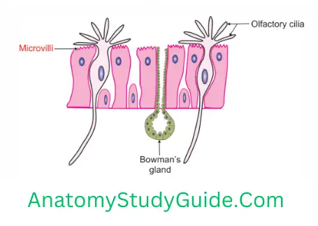
What Are The Differences Between Microvilli, Cilia And Stereocilia? Explain With The Help Of Diagrams
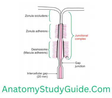
Junctional Complexes
The structures present on the lateral and the basal surfaces of epithelium are called junctional complexes. The epithelial cells are tightly attached to each other or to the extracellular matrix by specialised junctions.
1. There are several types of cell adhesion molecules (CAMs). They are membrane proteins, responsible for the specialised junctions.
2. The proteins consist of three domains
1. Intracellular binding protein: It interacts with the cytoskeleton and acts as an anchor within the cell.
2. Transmembrane protein: It protrudes through the plasma membrane.
3. Extracellular link protein: It binds with
- Other CAMs of the same type (homophilic binding) or with
- Different types of CAMs or extracellular matrix (heterophilic binding).
3. They are classified into two groups
1. Calcium-dependent
- Cadherins,
- Integrins, and
- Selectins.
2. Calcium independent: There are of two groups
- NCAMs (neural cell adhesion molecules): Expressed by nerve cells
- ICAMs (intercellular cell adhesion molecules): Expressed by leucocytes
C. Types of junctions: Epithelial cells are tightly attached to each other by specialised junctions. These junctions are of three types
- Occluding,
- Anchoring, and
- Gap junctions.
4. Functions
- They bind the adjacent cells with each other.
- They act as a barrier.
Leave a Reply