Introduction and Osteology
Spina bifida
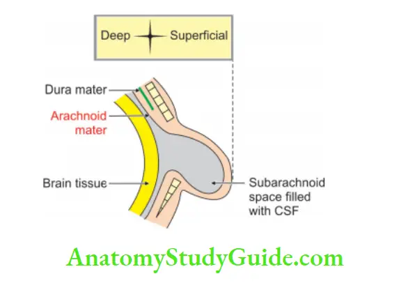
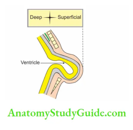
It is a gap present on the back in the midline. It is due to the failure of the fusion of two neural arches. The meninges and spinal cord may herniate through this gap. Depending upon the contents of the spina bifida, they are named as:
Table of Contents
1. Meningocele: It is a protrusion of only meninges. It is in the form of cystic swelling. It is filled with cerebrospinal fluid.

| Body Fluids | Muscle Physiology | Digestive System |
| Endocrinology | Face Anatomy | Neck Anatomy |
| Lower Limb | Upper Limb | Nervous System |
Read And Learn More: General Histology Questions and Answers
2. Meningomyelocele: It is a protrusion of meninges and spinal cord.
3. Syringomyelocele: It is a protrusion of the meninges and spinal cord with a dilated central canal.
4. Myelocele: It is a protrusion of the substance of the spinal cord through a defect in the vertebral canal.
5. Spina bifida occulta: There is a failure of fusion of two neural arches, but there is no protrusion. There is no swelling on the surface. This is usually associated with talipes equinovarus (or club foot). In this condition, the foot is inverted, adducted and plantar flexed.

Sacral hiatus
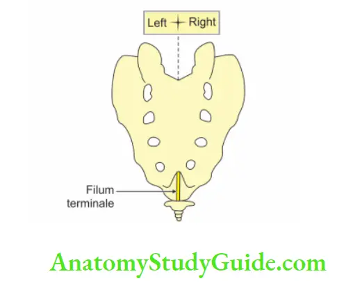
1. It is the opening present at the lower end of the sacral canal. It is formed by the failure of the fusion of laminae of the 5th sacral vertebra. It is covered by the dorsal sacrococcygeal ligament.
The structures passing through are: (CSF)
- Coccygeal nerve (C)
- 5th Sacral nerves, and (S)
- Filum terminal. (F)

2. Applied anatomy
The sacral hiatus can be entered by a needle that pierces
- Skin
- Fascia, and
- Posterior sacrococcygeal ligament.
Caudal anaesthesia:
- Anaesthetic solution is injected into the sacral canal through the sacral hiatus. It affects spinal roots emerging from the dural sheath.
- The solutions act on the spinal roots of the 2nd, 3rd, 4th, and 5th sacral and coccygeal nerves.
- It is used in obstetrics to anaesthetize the perineum. It blocks fibres of pain arising from the cervix of the uterus.
3. Surface anatomy: The sacral hiatus lies about 5 cm above the tip of the coccyx and beneath the skin of the cleft between the buttocks.
Leave a Reply