Maxillary Sinus
The maxillary sinus (also known as the antrum of Highmore) is a pneumatic space that is lodged in the body of the maxilla. This sinus communicates with the environment by way of the middle nasal meatus and the nasal vestibule. It is one of the paranasal sinuses of the head and neck region.
Table of Contents
Paranasal Sinuses:
- Maxillary sinus
- Frontal sinus
- Ethmoid sinus
- Sphenoid sinus
Read And Learn More: Oral Histology Notes
Maxillary Sinus Development
The development of the maxillary sinus begins when the embryo is at about 32 mm crown-rump length.
- It expands vertically into the primordium of the maxillary body and reaches a diameter of 1 mm when the crown-rump length is 50 mm.
- The sinus gradually increases and reaches a size of 7.5 mm when the embryo is of 250 mm crown-rump length.
Development of the Maxillary Sinus
Maxillary Sinus Structure
The maxillary sinus is described as a four-sided pyramid with the base towards the nasal cavity and apex at the body of the zygomatic bone.
The boundaries of the maxillary sinus are
- Anterior – facial surface of the maxilla
- Posterior – infratemporal surface
- Superior – floor of the orbit
- Inferior – alveolar and the zygomatic surface
- Medial – nasal cavity
- Lateral – body of the zygomatic bone
The base of the sinus is the thinnest and presents with a perforation. This perforation is known as the ostium and is at the level of the middle nasal meatus. Occasionally, in addition to the main ostium, two or more accessory ostia are seen that connect the sinus with the middle nasal meatus.
Location of the Ostia:

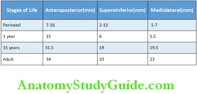
Maxillary Sinus Anatomical Description
The anatomical description of the maxillary sinus is given in Table
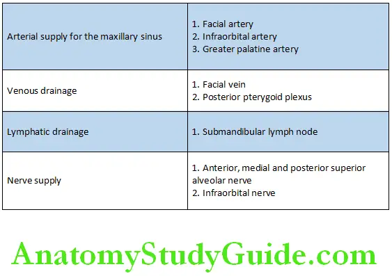
Vessels, Lymph and Nerve Supply of the Maxillary Sinus:
Maxillary Sinus Recesses
Some of the processes of maxilla are consequently filled with air. These areas are called recesses. Recesses are seen in the alveolar process, zygomatic process, frontal process and palatine process.
The floor of the maxillary sinus is in close proximity to the roots of the maxillary posterior teeth in the following order:
- Roots of first molar
- Roots of second premolar
- Roots of second molar
Histology Of The Lining Of The Maxillary Sinus
Three microscopically distinct layers line the pneumatic space of the maxillary sinus:
- An epithelial layer
- The basal lamina
- The subepithelial layer
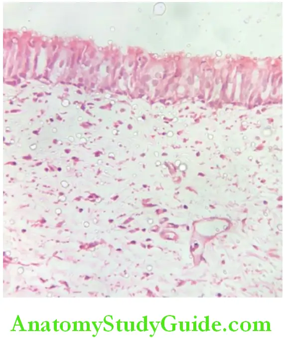
Epithelial layer:
The epithelial lining of the maxillary sinus is derived from the olfactory epithelium of the middle nasal meatus and is therefore pseudostratified columnar ciliated epithelium.
The lining epithelium comprises of
- Columnar ciliated cells
- Nonciliated columnar cells
- Mucous producing goblet cells
- Basal cells
Columnar ciliated cell:
This cell contains a nucleus and an electron-lucent cytoplasm. The cytoplasm contains numerous mitochondria and enzyme-containing organelles. The cell contains numerous basal bodies that serve as attachment of the ciliary microtubules.
The Cilia:
Each columnar cell comprises numerous cilia. Each cilium is composed of a free part or the shaft that is outside the cell and a part that extends into the superficial part of the cell. The free part is covered with a cell membrane.
A cross-section of the cilium shows:
- A central pair of microtubules surrounded with nine pairs of microtubules (9+1 configurations) arrangement is seen. The ring of nine pairs of microtubules is situated half way between the centre and the cell membrane. The cilium extends into the cell for a short distance.
Goblet cells:
The goblet cell is a secretory cell with a goblet-shaped morphology.
- The cell has a stem-like narrow base and bowl-shaped cell body.
- The nucleus is present in the basal segment of the cell. The basal segment also contains mitochondria, rough endoplasmic reticulum and Golgi apparatus.
- The cell body is packed with membranous vesicles filled with mucus. Golgi apparatus are also present and they are located at the periphery of the cell.
- The protein component is formed in the rough surfaced endoplasmic reticulum and the carbohydrate component is added in the Golgi apparatus.
- From the Golgi apparatus, the membrane-bound zymogenic granules bud off into the cell body.
- When these granules are pushed towards the apex of the cells, they coalesce weith each other and finally release their contents onto the epithelium by exocytosis.
- A few microvilli are present on the surface of the cell membrane.
- The mucous secreted by the goblet cell accumulates on the surface of the epithelium. Foreign particles and microorganism accumulate on this mucosal blanket.
- By means of the ciliary beating the mucous blanket along with the deposits moves from the sinus interior toward the nasal cavity.
The basal lamina:
The ducts of the subepithelial glands pierce through the basal lamina to drain their secretion on the surface of the epithelium.
The subepithelial layer:
The subepithelial layer contains glandular tissue. The glands contain a varying proportion of serous and mucous acini.
- The serous cells are stained with ninhydrin–Schiff and Sudan Black B and contain electron-dense homogenous secretary material. The secretory material consists primarily of water with small amounts of neutral nonspecific lipids, proteins and carbohydrates.
- The mucous cells are stained with Alcian blue 8GX (this stains acid sialomucins or sulphomucins or both). The mucous cell produces an electron-lucent, heterogeneous material. This material consists of compound glycoproteins of mucopolysaccharides or both.
Myoepithelial cells surround the acini composed of either both the types of secretory cells or purely serous or mucous acini.
The subepithelial layer of the sinus lining consists of:
- Blood vessels
- Numerous nonmyelinated axons
- Few myelinated axons
- Fibroblasts, fibrocytes, collagen bundles and connective tissue elements
The maxillary nerve complex supplies the autonomic axons and the general sensory components to the maxillary sinus. Both the divisions of the autonomic nervous system control the secretions from the subepithelial glands.
Functions Of The Maxillary Sinus
Functions of the maxillary sinus:
- Lightens the weight of the skull
- Adds resonance to the voice
- Humidifies and warms the inspired air
- Protects the brain and internal structures against exposure to cold air
- Enhances craniofacial resistance to mechanical shock
- Produces bactericidal lysozymal activity in the nasal cavity
Maxillary Sinus Clinical Significance
Developmental anomalies:
- Agenesis – there is complete absence of the maxillary sinus.
- Aplasia – defective development of the maxillary sinus
- Hypoplasia – underdeveloped maxillary sinus
- Supernumerary maxillary sinus – two completely separate sinuses
- Pituitary gigantism – sinus assumes a much larger volume than in healthy
individuals - Congenital syphilis – the sinus is suppressed
Dental Association:
- The maxillary sinus and orodental complex are topographically related. A pathological condition may be transferred from the maxillary sinus to the orodental apparatus and vice versa.
- Surgical manipulation of the maxillary first molar or the maxillary second molar frequently causes this tooth to break through the partitioning bony lamina and thus establish an oro-antral fistula. If the fistula is left untreated, the lumen of the fistula may epithelialize and permanently connect the maxillary space with the oral cavity.
Treatment: Surgical closure
- This can also occur when a molar or a premolar tooth is associated with a periapical granuloma, cyst or an abscess.
- Extraction of a tooth with hypercementosis can also result in perforation.
Note: It is advisable to evaluate the relationship between the premolar and the molar tooth with the floor of the maxillary sinus radiographically before any surgical intervention.
Infection:
- The chronic infection of the mucoperiosteal layer of the sinus might involve the superior alveolar nerves that are close to the maxillary sinus.
- The neuralgia that ensues might mimic pain of dental origin. A careful inspection of all the upper teeth as well as the maxillary sinus is necessary to identify the cause of this condition.
- There is a pathogenic association of the sinus with the oro-dental apparatus due to the topographic relationship and extensive vascular connection between the two regions by the superior alveolar vessels. Nonspecific bacterial sinusitis may be followed by some oral manifestation. The infection caused by streptococci, staphylococci, pneumococci or virus of the common cold are likely to spread from either of the two regions to involve the other.
Malignant neoplasms:
Conditions such as adenocarcinoma, squamous cell carcinoma, osteosarcoma, fibrosarcoma, lymphosarcoma of the maxillary sinus can produce their primary manifestation in the maxillary teeth. This may cause pain, loosening of teeth, supra eruption of teeth bleeding of the gingival tissues.
Maxillary Sinus Synopsis
- The maxillary sinus (also known as the antrum of Highmore) is a pneumatic space that is lodged in the body of the maxilla.
- This sinus communicates with the environment by way of the middle nasal meatus and the nasal vestibule. It is one of the paranasal sinuses of the head and neck region.
- The maxillary sinus is described as a four sided pyramid with the base towards the nasal cavity and apex at the body of the zygomatic bone.
- The base of the sinus is the thinnest and presents with a perforation. This perforation is known as the ostium and is at the level of the middle nasal meatus.
- Some of the processes of maxilla are consequently filled with air. These areas are called recesses.
- Recesses are seen in the alveolar process, zygomatic process, frontal process and palatine process.
- Three microscopically distinct layers line the pneumatic space of the maxillary sinus which include an epithelial layer, the basal lamina and the subepithelial layer.
- The epithelial lining of the maxillary sinus is derived from the olfactory epithelium of the middle nasal meatus and is therefore pseudostratified columnar ciliated.
- The lining epithelium comprises columnar ciliated cells, nonciliated columnar cells, mucous producing goblet cells, basal cells.
- Columnar ciliated cells contain a nucleus and an electron-lucent cytoplasm.
- Goblet cell is a secretory cell with goblet-shaped morphology.
- The basal lamina has ducts of the subepithelial glands piercing through it.
- The subepithelial layer contains glandular tissue. The glands contain a varying proportion of serous and mucous acini.
- The functions of the maxillary sinus include lightening the weight of the skull, resonance to the voice, humidifying and warming the inspired air, protecting the brain and internal structures against exposure to cold air, enhancement of craniofacial resistance to mechanical shock and production of bactericidal lysozyme to the nasal cavity.
Leave a Reply