Lungs
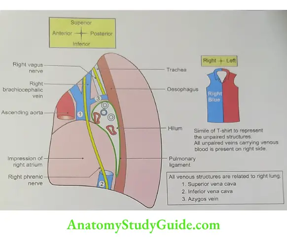
OLA-6 Visceral relations of right lung
1. Chambers of heart
- Right atrium,
- Right auricle, and
- Right ventricle-small part.
2. Superior vena cava and its tributaries
- Right brachiocephalic vein, and
- Azygos vein.
3. Inferior vena cava,
4. Oesophagus,
5. Trachea,
6. Right vagus nerve, and
7. Right phrenic nerve.
Read And Learn More: Anatomy Important Question And Answers
OLA-7 Visceral relations of left lung
1. Chambers of heart
- Left auricle,
- Left ventricle-small part, and
- Right ventricle-adjoining part.
2. Arch of aorta and its branch: Left subclavian artery.
3. Descending thoracic aorta,
4. Thoracic duct,
5. Oesophagus,
6. Trachea,
7. Left vagus nerve and its branch-left recurrent laryngeal nerve.
8. Left phrenic nerve.
OLA-8 Difference between right and left lungs
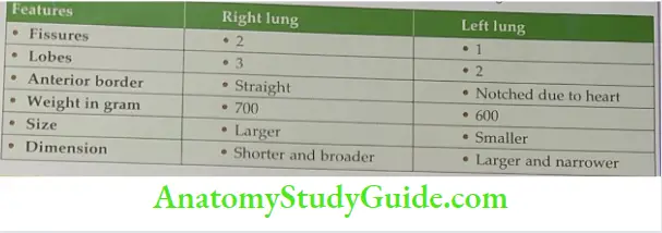
Mediastinal surfaces of the right and left lungs
1. Right side (venous)
Cardiac impressions and chambers
- Right atrium and auricle
- Right ventricle (small part)
Veins
- Superior vena cava,
- Inferior vena cava, and
- Brachicephalic vein.
- Hilum is arched by azygos vein.
Nerves
- Vagus nerve, and
- Phrenic nerve.
Trachea
Oesophagus.
2. Left side (arterial)
Cardiac impressions and chambers
- Left ventricle
- Left auricle
- Left atrium
- Right ventricle
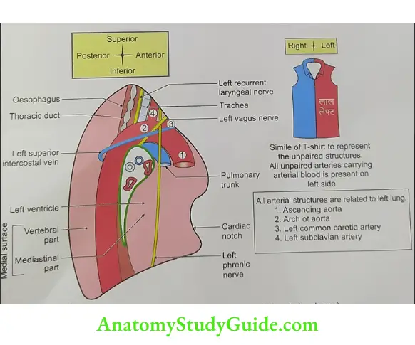
Arteries
- Hilum is arched by arch of aorta
- Descending thoracic aorta
- Subclavian artery
Veins
- Brachiocephalic vein
- Pulmonary trunk.
Nerves
- Vagus nerve
- Recurrent laryngeal nerve
- Phrenic nerve
Thoracic duct
Oesophagus.
Draw and label structure of the roots of the right and left lungs.

Plane 1:
- Superior right pulmonary Vein.
- Inferior right pulmonary Vein
Plane 2: Pulmonary Artery
Plane 3:
- Eparterial Bronchus
- Hyparterial Bronchus
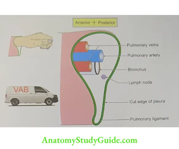
Plane 1:
- Superior left pulmonary Vein.
- Inferior right pulmonary Vein
Plane 2: Pulmonary Artery
Plane 3: Bronchus
Azygos lobe
(A—not; zygos-paired)
1. Types: Accessory or supernumerary lobes of lungs are called azygos lobes. They are of three types
- Upper azygos lobe,
- Lower azygos lobe, and
- Lobe of azygos vein.
The formal two types have little practical importance. And these are described in reference to hilum of the lung.
2. Incidence: 1% of population.
3. It affects the upper lobe of the right lung. The apex of the lung splits into medial and lateral parts of the fissure. The bottom of the lung contains the arch of the azygos vein suspended by a pleural septum, the mesoazygos. The medial part of the split apex forms the lobe of the azygos vein.
4. Development: It is because of the upward development of apical bronchus. It develops medial to the arch of azygos vein instead lateral to it.

5. Applied anatomy
- The plane X-ray of the chest shows a small dense shadow close to the right sternal angle.
- It is one of the differential diagnoses of enlarged lymph node in the chest.
Blood supply of lung
1. Arterial supply
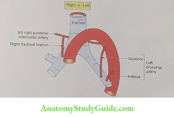
The nutrition of the lungs is by bronchial arteries.
- These are small arteries varying in origin, size and number.
- On the right side, there is 1 bronchial artery. It is a branch of third posterior intercostal artery or from the upper left bronchial artery.thoracic aorta.
- On the left side, there are two bronchial arteries. They arise from descending
Deoxygenated blood to the lungs is brought by the pulmonary arteries.
Oxygenated blood is returned to the heart by pulmonary veins.
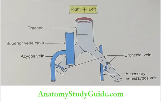
2. Venous drainage: They are divided into
- Superficial bronchial veins
- Deep bronchial veins
- Superficial bronchial veins
Deep bronchial vein
- Draining areas
- Intrapulmonary bronchial tree
- Parenchyma of lung
- Drain into pulmonary vein.
Movements of respiration
Introduction: The lungs expand during inspiration and retract during expiration. These movements are governed by the following two factors.
- Air is inspired during inspiration.
- Air is expelled during expiration.

Principles of movements
- Along with the up and down movements of the 2nd to 6th ribs, the body of the sternum also moves up and down called pump-handle movement. This results in formation of sternal angle. The middle of the shaft of the rib lies at a lower level than the plane passing through the two ends. Therefore, during elevation of the rib, the shaft also moves outwards. This causes increase in the transverse diameter bucket-handle movement. of the thorax. Such movements occur in the vertebrochondral ribs and are called
- The thorax resembles a cone, tapering upwards. As a result, each rib is longer than the next higher rib. On elevation, the larger lower rib comes to occupy the the thorax. position of the smaller upper rib. This also increases the transverse diameter of diaphragm. C.
- Vertical diameter is increased by the “piston movements” of the thoracoabdominal
Suprapleural membrane (Sibson’s fascia)
Introduction: It is the dome thoracic cavity.
1. Formation: It has shaped musculofacial membrane which roofs
- Muscular part: It is formed occasionally by scalenus (uneven) minimus muscle.
- Fascial part: It is formed from endothoracic fascia.
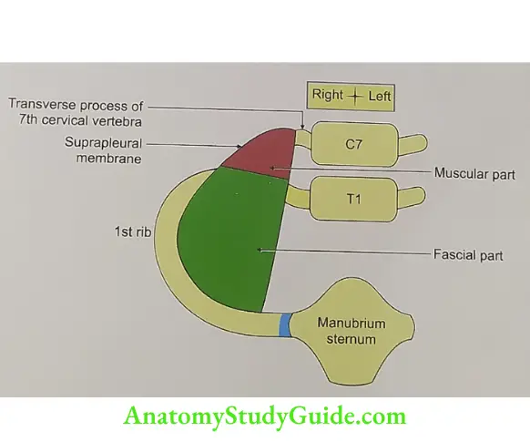
2. Attachments
- Anteriorly: Inner border of 1st rib.
- Posteriorly: Tip of the transverse process of 7th cervical vertebra.
- Medially: Continuous with the pretracheal fascia.
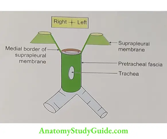
3. Functions: Protects the apex of the lung and cervical pleura during respiratory movements.
4. Applied anatomy: Rupture of suprapleural membrane results into herniation of cervical pleura.
LAQ-3 Draw and describe bronchopulmonary segment under following heads
1. Gross anatomy, and
2. Applied anatomy.
Definition: It is structurally separate and functionally independent respiratory, surgical segment or unit of lung aerated by tertiary or segmental bronchus. It includes tertiary bronchus plus segment of lung aerated by tertiary lung. It is not bronchovascular segment.
1. Gross anatomy
- Shape: Pyramidal
- Apex: Directed towards the root of lung
- Base: Towards the surface of lung.
- Segment:
- Each segment is aerated by tertiary bronchus.
- Each segment is separated by intersegmental alveolar septum.
- The septum is occupied by tributaries of pulmonary vein.
- But the pulmonary vein is intersegmental.
- Each segment contains
- Segmental bronchi,
- Pulmonary artery,
- Bronchial artery, and
- Bronchial vein.
- There are 10 segments in each lung . They are described below

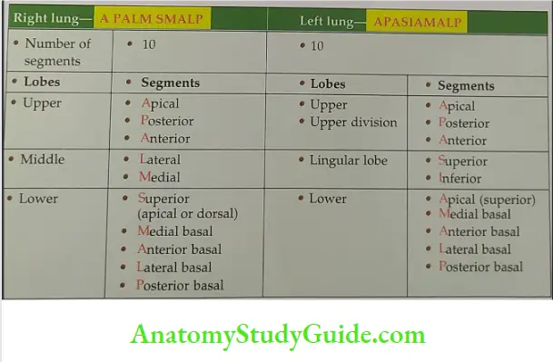
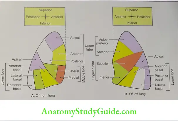
Intersegmental plane: It is the connective tissue septa between two adjacent lobes. It is continuous on the surface with pulmonary subpleural connective tissue.
Relations:
- Pulmonary arteries lie dorsolateral to bronchus
- Important relations of bronchopulmonary segment.

- Pulmonary vein is intersegmental in position.
- One pulmonary vein drains more than one bronchopulmonary segment.
- Each segment is drained by more than one pulmonary vein. Pulmonary veins do not accompany the bronchial or pulmonary arteries. Near the hilum of the lung, the pulmonary veins lie ventromedial to the bronchus.
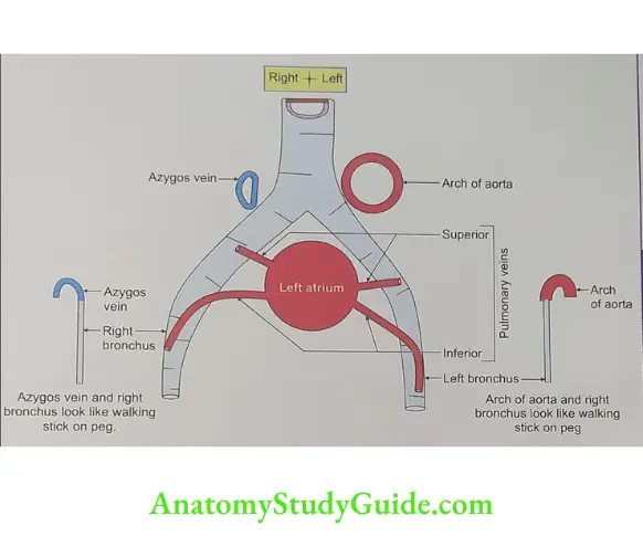

Venous drainage: Intersegmental pulmonary vein.
Lymphatic drainage: There are two sets of lymphatics both of which drain into bronchopulmonary nodes.
- Superficial lymphatics drain into peripheral lung tissue. It lies beneath the pulmonary pleura. The vessels pass round the borders of lung and margins of fissure. They reach the hilum.
- Deep lymphatics drain into
- Bronchial tree,
- Pulmonary vessels, and
- Connective tissue septa.
- They run towards the hilum and drain into bronchopulmonary nodes.
2. Applied anatomy
- Bronchography: It is the study of bronchopulmonary segments. It is done radiologically by instillation of radio-opaque dye. Study of bronchopulmonary segments helps to localize the affected segment of the lung and helps in postural drainage.
- Foreign body in the respiratory passage is one of the important causes of lung abscess. (Mendelson’s syndrome: This disorder is produced, as a complication of anaesthesia. It is by inhalation of gastric content with pH of less than 2.5. It is common in posterior segment of upper lobe and apical segment of lower lobe on the right side. Because these segments are most dependent in recumbent positions.)
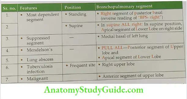
- Each bronchopulmonary segment acts as an independent unit; hence infection is restricted only to respective segment, except in tuberculosis.
- The benign neoplasm is restricted to each segment. However, malignant growths are not restricted to the respective segment.
- Tertiary bronchus guides the surgeon for segmental resection. After inflating the lung, the collapse segment of lung fails to inflate. It helps the surgeon to identify the diseased segment of lung.
- The knowledge of bronchopulmonary segment helps for draining the secretion o various segments. Various positions are adapted for drainage of various lobes. Righ lateral position is adapted to drain the abscess of left lung and vice versa.
Leave a Reply