• Skip to main content
  • Skip to secondary menu
  • Skip to primary sidebar
  • Skip to footer
  • Anatomy
    • Anatomy Question And Answers
    • Face Anatomy
    • Neck Anatomy
    • Head Anatomy
    • Oral Anatomy
    • Lower Limb
    • Upper Limb
  • Endodontics
    • Paediatric Dentistry
  • General Histology
    • Oral Histology
    • Genetics
  • Pediatric Clinical Methods
  • Complete Dentures
    • Pharmacology for Dentistry
  • Medical Physiology
    • Body Fluids
    • Muscle Physiology
    • Digestive System
    • Renal Physiology
    • Endocrinology
    • Nervous System
    • Respiratory System
    • Cardiovascular System
    • Reproductive System
    • Oral Physiology
  • General Medicine
  • General Pathology
    • Systemic Pathology
    • Oral Pathology
    • Neoplasia
    • Homeostasis
    • Infectious Diseases
    • Infammation
    • Amyloidosis Notes
  • Periodontology
  • General Surgery
    • Basic Principles Of Surgery
    • General Surgery

Anatomy Study Guide

Anatomy Study Guide

  • About Us
  • Contact Us
  • Privacy Policy
  • Terms of Use
  • Disclaimer
  • Sitemap
Home » Mandibular Central Incisors Notes

Mandibular Central Incisors Notes

November 20, 2023 by Joankessler parkland Leave a Comment

Carving Of The Permanent Incisors

The steps in carving of the permanent maxillary central incisor will be described in detail. The permanent maxillary lateral incisor and permanent mandibular incisors can be carved following the same steps but with variation in features.

Table of Contents

  • Carving Of The Permanent Incisors
  • Carving Of The Permanent Maxillary Right Central Incisor
  • Carving Of The Permanent Maxillary Lateral Incisor
  • Carving Of The Permanent Mandibular Central Incisor
  • Carving Of The Permanent Mandibular Lateral Incisor

Note: Measurements to be noted down from the respective chapters

Read And Learn More: Oral Physiology Notes

Carving Of The Permanent Maxillary Right Central Incisor

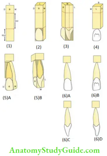
1. Mark the cervicoincisal dimension of the crown and the root on the wax block. The portion of the wax block beyond the root is called the base. Label the Surfaces at the bottom of the block as B, L, M, D indicating buccal, lingual, mesial and distal.

Note: B is used instead of L (for labial) to avoid confusion

2. Mark the mesio-distal dimension on the buccal surface of the crown at the cervical line and at the contact area. Draw the outline of the labial aspect on the crown. Carve the wax outside the outline (shaded portion) and thus create the mesial and distal surface of the crown. Make the disto-incisal line angle more rounded than the mesio-incisal line angle in order to differentiate between the permanent right and left central incisor.

3. Mark the midline of the block on the proximal aspect. Mark the labio-lingual dimension of the crown at the cervical line, at the crest of curvature and the incisal edge (2 mm). The incisal edge has to pass through the midline of the block. Carve the wax outside the outline and thus create the labial and the lingual surface of the crown with the lingual surface being narrower(lingual convergence) than the labial surface.

4. Carve the W shaped lingual fossa. The lingual fossa can be carefully carved with the discoid portion of the instrument.

5. Mark the shape of the root on the buccal and proximal side of the wax block and carve the wax outside the outline of the root (shaded portion). Make the root lingually converging.

6. Check for the dimension and complete carving the anatomy of the tooth in detail. Carve the cervical line and polish the tooth.  The steps in carving the permanent maxillary lateral incisor and the mandibular incisors are the same.

The exceptions are mentioned below:

Carving Of The Permanent Maxillary Lateral Incisor

  1. Dimensions of the crown and the root.
  2. Outlines.
  3. Shape of the lingual fossa.
  4. Outline of the incisal edge

Carving Of The Permanent Mandibular Central Incisor

  1. Dimensions of the crown and the root.
  2. Outlines.
  3. The incisal edge is lingual to the midline.
  4. Shape of the lingual fossa.

Carving Of The Permanent Mandibular Lateral Incisor

  • Dimensions of the crown and the root.
  • Outlines.
  • The incisal edge is lingual to the midline.
  • The crown appears to be twisted on the root base.
  • Shape of the lingual fossa.

Carving Of The Permanent Incisors Carving of the permanent mandibular central inciso

Filed Under: Oral Physiology

Reader Interactions

Leave a Reply Cancel reply

Your email address will not be published. Required fields are marked *

Primary Sidebar

Recent Posts

  • Esophagus Anatomy
  • Lacrimal Apparatus: Anatomy, Parts & Function
  • Scalp Temple And Face Question and Answers
  • Orbicularis Oculi Muscle Anatomy
  • Extraocular Muscles Anatomy
  • Ciliary Ganglion Anatomy
  • Femoral sheath Anatomy
  • Femoral Artery – Location and Anatomy
  • Adductor Canal: Anatomy And Function
  • Ankle Joint: Anatomy, Bones, Ligaments And Movements
  • Risk Factors For Breast Cancer
  • Cervical Tuberculous Lymphadenitis Notes
  • Carbuncles: Causes, Symptoms, and Treatments
  • Sinuses And Fistulas Notes
  • Cellulitis: Treatments, Causes, Symptoms
  • Pyogenic Liver Abscess: Causes, Symptoms, and Diagnosis
  • Acid Base Balance Multiple Choice Questions
  • General Surgery Multiple Choice Questions
  • Hypertrophic Scarring Keloids Multiple Choice Questions
  • Surgical Site Infection Multiple Choice Questions
  • Facebook
  • Pinterest
  • Tumblr
  • Twitter

Footer

Anatomy Study Guide

AnatomyStudyGuide.com is a student-centric educational online service that offers high-quality test papers and study resources to students studying for Medical Exams or attempting to get admission to different universities.

Recent

  • Esophagus Anatomy
  • Lacrimal Apparatus: Anatomy, Parts & Function
  • Scalp Temple And Face Question and Answers
  • Orbicularis Oculi Muscle Anatomy
  • Extraocular Muscles Anatomy

Search

Copyright © 2024 · Magazine Pro on Genesis Framework · WordPress · Log in