Vitamin-B12 Deficiency (Megaloblastic Anaemia)
Case 1: Degenerative Changes In The Spinal Cord
A patient came to the hospital with neurological symptoms; his MRI shows degenerative changes in the spinal cord. Peripheral smear shows megaloblastic RBCs and serum methyl malonate level was raised.
Table of Contents
- Diagnosis – Megaloblastic anaemia due to Vitamin B12 deficiency.
- Deficient vitamin – Cobalamin (Vitamin B12).
- Active form – adenosylcobalamin and methylcobalamin. Sources – food of animal origin – liver, meat, milk, also synthesized by intestinal bacteria.
Read And Learn More: Biochemistry Clinical Case Studies With Answers
Important Reactions
- Methyl malonyl CoA mutase.
- Methyl malonyl CoA ————-B12—> Succinyl CoA.
- Methionine synthase.
- Homocysteine ———–B12——-> Methionine.
Deficiency Manifestations
- Megaloblastic anaemia.
- Subacute combined degeneration of the spinal cord.
- Methyl malonate excretion in urine.
Mnemonic to remember Cobamine Mms Scam
- Corrin ring with central cobalt.
- B12– two active forms.
- Adenosylcobalamin.
- Methylcobalamin.
- An intrinsic factor of the castle is required for absorption.
- Neurological manifestations in deficiency.
- Extrinsic factor (other names).
- Methyl malonyl CoA mutase.
- Methionine synthase.
- Subacute combined degeneration of the spinal cord.
- Anaemia.
- Methyl malonate excretion in urine.
Folate Trap

- THF (Tetrahydrofolate) gets trapped as methyl THF in vitamin B12 deficiency. There is a functional deficiency of folate. Folate is available as methyl THF but the body can’t utilize methyl THF.
- So in B12 deficiency, there is a functional folate deficiency known as folate trap. Folate is responsible for the synthesis of TMP (Thymidylate) and Purines.
- Decreased nucleotide synthesis leads to decreased DNA and RNA synthesis, hence cell division arrested in the S phase of the cell cycle. The cell grows continuously without a cell division and macrocytosis occurs.
Vitamin A Deficiency
Case 1: Diminished Vision During The Night
25 years old man came to OPD with a complaint of diminished vision during the night. On examination– triangular opaque spot in the conjunctiva of the right eye. Diagnosis– Night blindness due to Vitamin A deficiency.
- RDA– 1000 Retinol equivalent in males and 800 RE in females.
- Sources– Cod or shark liver oil, meat, eggs, milk, carrots, papaya, mango, green vegetables, etc.
Vitamin A Deficiency Manifestation
World Health Organization (WHO) classification of vitamin A deficiency

Vitamin A Treatment
- Vitamin A-rich diet.
- Daily oral vitamin A supplements.
- In severe deficiency– 200000 IU is given.
Vitamin A Functions
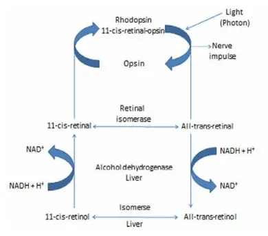
- Vision– Involved in Wald’s visual cycle.
- Help to maintain germinal epithelial cell integrity.
- Antioxidant– reduces the risk of cancer and prevents heart attack.
- Growth and development.
- Glycoprotein synthesis and mucopolysaccharide sulfation.
- Bone and teeth formation.
Three Vitamers
- Retinol
- Retinal
- Retinoic acid
Megaloblastic Anaemia (Folate Deficiency)
Case 1: Macrocytic RBCs And Reticulocytosis
A 30-year-old pregnant lady came to the hospital with complaints of breathlessness, weakness, loss of weight, and smoothness of tongue. Laboratory investigations showed macrocytic RBCs and reticulocytosis. Hb was 7 gm%, and urine was positive for FIGLU. USG shows neural tube defects in the fetus.
- Diagnosis– Megaloblastic anemia due to Folate deficiency.
- Deficient vitamin– Folic acid.
- Sources– Green leafy vegetables, cereals, pulses, etc.
- Active form– THF.
Biochemical Functions
Actively involved in one-carbon metabolism and synthesis of many compounds
- C2 and C8 of purine nucleotides.
- dTMP- pyrimidine nucleotide synthesis.
- Glycine, serine, choline, ethanolamine synthesis.
- Initiation of protein synthesis.
Reason For Megaloblastic Anaemia
Folate is responsible for the synthesis of TMP and Purines. Decreased nucleotide synthesis leads to decreased DNA and RNA synthesis, hence cell division is arrested in the S phase of the cell cycle. The cell grows continuously without a cell division and macrocytosis occurs (megaloblastic RBCs).
Folate Trap

THF gets trapped as methyl THF in vitamin B12 deficiency. There is a functional deficiency of folate. Folate is available as methyl THF but the body can’t utilize methyl THF. So in B12 deficiency, there is a functional folate deficiency known as folate trap.
Folate Antagonists
- Aminopterin and Amethopterine (methotrexate)
- Inhibit dihydrofolate reductase and inhibit folate synthesis, decreases purines and pyrimidine synthesis hence resulting in impaired DNA synthesis and inhibition of cell division. These drugs are used in the treatment of leukemia.
- Trimethoprim– antibiotic.
- Sulfonamide– an antibiotic-structural analog of PABA, inhibits dihydropteroate synthase and inhibits folic acid synthesis.
Vitamin K Deficiency (Hemolytic Disease of Newborn)
Preterm infants are more susceptible to Vitamin K deficiency because of- a sterile intestine, poor placental transfer, low stores of Vitamin K, and an immature liver system. Clinical symptoms– prolonged bleeding, increased prothrombin time (PT).
Role of Vitamin K
- It is required for post-translational modification of clotting factors– II, VII, IX, X, osteocalcin, protein C and S.
- It causes gamma-carboxylation of glutamate residues in clotting factors- II, VII, IX, X. This negatively charged Glutamic acid binds with positively charged calcium ions.
- The Caprothrombin complex then binds with the phospholipid on the platelets and causes the conversion of prothrombin to thrombin. Thrombin converts fibrinogen to fibrin clots. Laboratory investigation– increased prothrombin time.
RDA-70-140 μg/day
Vitamin K Sources
- Vitamin K1– Phylloquinone– Spinach and green leafy vegetables.
- Vitamin K2– Menaquinone– Synthesized by intestinal bacteria.
- Vitamin K3– Menadione– Synthetic form.
The antagonist of Vitamin K
- Dicoumarol and Warfarin.
- These drugs act as anticoagulants by inhibiting vitamin K epoxide reductase.
Leave a Reply