Forearm And Hand
Branches Of Radial Artery In Forearm
Branches in
Table of Contents
1. Forearm
- Radial recurrent artery
- Muscular branches
2. Hand
Branches of the deep palmar arch
- Branch to superficial palmar branch,
- Princeps pollicis artery, and
- Radialis indices artery.
- Palmar metacarpal branch
Dorsum of hand: Dorsal carpal branch
Anterior Forearm Muscles
Read And Learn More: Anatomy Notes And Important Question And Answers
Question-1: Describe Ulnar Artery Under The Following Heads
1. Ulnar Artery Origin,
2. Ulnar Artery Course and relations,
3. Ulnar Artery Branches, and
4. Ulnar Artery Applied anatomy.
Answer:
Ulnar Artery Introduction:
It is the artery of the medial side of the forearm.
1. Ulnar Artery Origin:
This larger terminal branch of the brachial artery arises 1 cm below the bend of the elbow.
2. Ulnar Artery Course and relations:
1. Peculiarities: It is accompanied by
- Venae commitantes, and
- The ulnar nerve in the distal 2/3rd of the forearm.
2. Extent: It extends from the elbow to the wrist joint.
Anterior Forearm Muscles
Ulnar Artery Termination: Ulnar artery forms the superficial palmar arch in the hand.
Ulnar Artery Course: It passes downward, medially.
- It leaves the cubital fossa deep to the ulnar head of the pronator teres.
- It lies between two nerves, laterally the median and medially the ulnar nerve.
Anterior Forearm Muscles
Relations
Anterior
1. Upper half
Muscles
- Pronator teres,
- Flexor carpi radialis,
- Flexor digitorum superficialis, and
- Palmaris longus
Nerve: Median nerve
Lower half
- Skin
- Superficial fascia
2. Posterior
Anterior relations of the ulnar artery in the upper and lower 1/2 of the forearm
1. Flexor digitorum profundus, and
2. Pronator quadratus.
Lateral: Flexor digitorum superficialis.
Medial: Flexor carpi ulnaris and ulnar nerve.
Anterior Forearm Muscles
1. Branches :
1. Muscular branch to all the superficial group of muscles on the medial side of forearm and hand
2. Cutaneous
3. Articular branch to
- Elbow, and
- Wrist joint.
4. Branches to nerves
- Ulnar, and
- Median.
5. Nutrient arteries to
- Radius, and
- Ulna.
6. Communicating branches to
- Anterior,
- Posterior carpal arch, and c. Superficial palmar arch.
7. Terminates as
- Superficial palmar arch, and
- Branch to the synovial sheath.
Muscles Of Anterior Forearm
2. Ulnar Artery Applied anatomy:
It is felt lateral to the flexor carpi ulnaris and above the pisiform bone.
Note: For easy recalling remember the digit ‘2′
1. One of 2 terminal branches of the brachial artery.
2. Extends between 2 joints, namely elbow and wrist joint, i.e. neck (of radius) to head (of ulna).
3. Passes between 2 nerves:
- Median, and
- Ulnar.
Muscles Of Anterior Forearm
4. Relations with 2 groups of muscles
- Superficial, and
- Deep.
5. Lies on 2 muscles
- Brachialis, and
- Flexor digitorum profundus.
6. Branches: Mnemonic for branches of the ulnar artery is.(“BRANCHES“)
Branches to
2 Bones: Radius, and ulna.
2 Recurrent: Anterior ulnar recurrent, and posterior ulnar recurrent.
2 Articular to: Elbow joint, and wrist joint.
2 Nerves: Ulnar, and median.
2 Carpal bones: Anterior, and posterior carpal.
2 Branches to Hand: Superficial palmar arch and deep palmar arch.
2 Interosseous membrane: Anterior interosseous and posterior interosseous artery
2 groups of muscles: Superficial, and deep.
Muscles Of Anterior Forearm
Question-2: Name The Boundaries And Contents Of Anatomical Snuffbox
Answer:
Boundaries and contents:
Boundaries, floor, and contents are formed by “3” structures each.
1. 3 structures form boundaries, 3 structures form the floor and 3 structures form the contents.
2. Laterally: Tendon of
- Abductor pollicis longus, and
- Extensor pollicis brevis.
Medially: Extensor pollicis longus.
2. Floor: From proximal to distal 3 bones
- Scaphoid,
- Trapezium, and
- The base of 1st metacarpal bone.
3. Contents are 3:
From superficial to deep
- Cephalic vein (over the roof),
- The superficial branch of the radial nerve, and
- Radial artery.
Anterior Forearm Superficial Muscles
Anatomical Snuffbox
Anatomical Snuffbox Introduction:
It is a depressed lar area present on the lateral side of the wrist and becomes prominent when the thumb is fully extended.
1. Boundaries and contents:
Boundaries, floor, and contents are formed by “3” structures each.
3 structures form boundaries, 3 structures form the floor and 3 structures form the contents. (“3”)
Laterally: Tendon of abductor pollicis longus and tendon of extensor pollicis brevis.
Medially: Extensor pollicis longus.
2. Anatomical Snuffbox Floor:
From proximal to distal 3 bones
- Scaphoid,
- Trapezium, and
- The base of 1st metacarpal bone.
3. Anatomical Snuffbox Contents:
From superficial to deep
- Cephalic vein (over the roof),
- The superficial branch of the radial nerve, and
- Radial artery.
4. Anatomical Snuffbox Applied anatomy:
A person having a fracture of the scaphoid complains of pain in the wrist. There is no impairment in function
Anterior Forearm Superficial Muscles
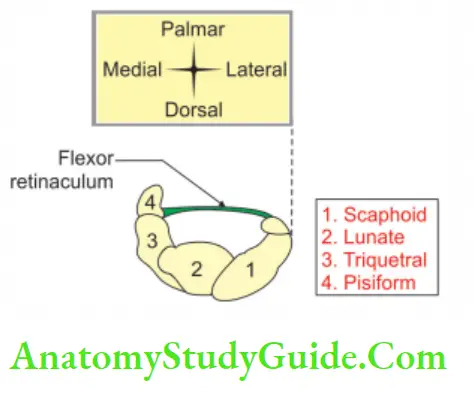
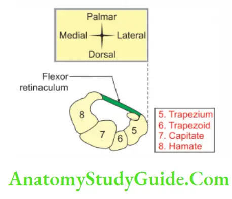
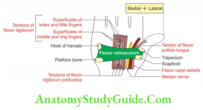
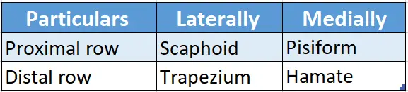
Question-3: What Are The Attachments Of The Flexor Retinaculum?
Answer:
1. Attachments Of The Flexor Retinaculum Proximally and
- Laterally to the tubercle of the scaphoid, and
- Medially to hook the pisiform.
2. Attachments Of The Flexor Retinaculum Distally and
- Laterally to the crest of the trapezium, and
- Medially to hook the hamate.
3. Attachments Of The Flexor Retinaculum On either side, the retinaculum has a slip:
A lateral deep slip is attached to the medial lip of the groove on the trapezium.
The medial superficial slip (volar carpal ligament) is attached to the pisiform bone.
Flexor Retinaculum
Flexor Retinaculum Introduction:
It is a strong fibrous band of deep fascia connecting proximal and distal carpal bones of the medial and lateral sides.
1. Flexor Retinaculum Gross anatomy:
Attachments of the flexor retinaculum
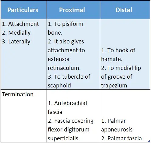
2. Flexor Retinaculum Relations:
Relations of the flexor retinaculum
3. Flexor Retinaculum Importance:
- It gives origin to the thenar and hypothenar muscles.
- The tendon of the palmaris longus is fused in the midline.
- It keeps all the flexor tendons in position.
- It converts the bony gutter into a tunnel.
- It prevents bowstringing of muscles during, flexion thereby leading to effective contraction.
Anterior Forearm Superficial Muscles
Extensor Retinaculum
1. Extensor Retinaculum Definition:
The deep fascia on the back of the wrist is thickened to form the extensor retinaculum. It holds the extensor tendons in place. It is an oblique band, directed downwards and medially. It is about 2 cm broad vertically.
2. Extensor Retinaculum Attachments:
1. Laterally: Lower part of the anterior border of the radius.
2. Medially
- Styloid process of the ulna,
- Triquetral, and
- Pisiform.
Anterior Forearm Superficial Muscles
3. Extensor Retinaculum Compartments:
The retinaculum sends septa and makes various compartments. They are attached to the longitudinal ridges on the posterior surface of the lower end of the radius.
In this way, 6 osseofascial compartments are formed on the back of the wrist. The structures passing through each compartment, from the lateral to the medial side, are
1. 1st Compartment
- Abductor pollicis longus
- Extensor pollicis brevis
2. 2nd compartment
- Extensor carpi radialis longus
- Extensor carpi radialis brevis,
3. 3rd compartment: Extensor pollicis longus,
4. 4th compartment
- Extensor digitorum,
- Extensor indices,
- Posterior interosseous nerve,
- Anterior interosseous artery,
5. 5th compartment: Extensor digit minimi, and
6. 6th compartment: Extensor carpi ulnaris.
Anterior Forearm Superficial Muscles
Question-4: Name The Muscles Supplied By Median Nerve In Hand
Answer:
(LOAF)
1st and 2nd Lumbricals,
Opponens pollicis,
Abductor pollicis brevis, and
Flexor pollicis brevis-superficial head.
Question-5: Describe The Median Nerve Under The Following Heads
1. Median Nerve Root value,
2. Median Nerve Branches, and
3. Median Nerve Applied anatomy.
Answer:
1. Median Nerve Root value:
The median nerve has lateral and medial roots. The Lateral root is the continuation of the lateral cord of the brachial plexus and has C5, C6, and C7 fibers. The medial root is derived from the medial cord and has C8 and T1 fibers.
1. Course and relations: The lateral and medial roots clasp 2nd part of the axillary artery and lie on the respective sides.
2. In the axilla
- The medial root crosses the 3rd part of the axillary artery from the front. It joins the lateral root, and the nerve is formed on the lateral side at the lower border of the axilla.
- It enters the arm. It then passes vertically down lateral to the brachial artery.
- In the middle of the arm, it crosses the brachial artery from the front.
- It remains on the medial side of the brachial artery in the lower part of the arm.
- It courses between the biceps brachii and brachialis.
- In the cubital fossa, it crosses the ulnar artery from medial to lateral.
Note:
The symbol ” ” at the end of the nerve signifies the cutaneous branch
3.In the cubital fossa
1. It is the most medially important structure of the cubital fossa.
2. It passes between two heads of pronator teres.
3. The relations in the lower part of the cubital fossa from superficial to deep are
- Superficial head of pronator teres
- Median nerve
- Deep head of the pronator teres
- Ulnar artery.
4. In the forearm
It runs distally in the midline of the forearm.
It is closely attached to the deep surface of the flexor digitorum superficialis muscle.
It passes between the superficial and deep muscles of the forearm.
It is accompanied by the median artery (branch of an anterior interosseous artery).
5. In the wrist
1. Above the wrist, the nerve comes close to the surface between the tendons of
- Flexor carpi radialis, and
- Flexor digitorum superficialis.
2. It lies deep and partly lateral to the tendon of the palmaris longus.
3. It passes deep to the flexor retinaculum, surrounded by 10 tendons. These are
- Four superficial flexor tendons of digits,
- Four deep flexor tendons of digits,
- A long deep tendon of the thumb, and
- A tendon of the radial side of the wrist.
4. After giving recurrent branch, it divides into
1. Lateral, and
2. Medial branch.
3. The lateral branch subdivides into three palmar digital branches, which supply the
- The skin of both sides of the thumb, and
- Radial side of the index finger.
The branch to the index finger provides a muscular branch to 1st lumbrical.
4. The medial branch subdivides into two common palmar digital nerves. The lateral common digital nerve gives a branch to 2nd lumbrical.
5. Median nerve gives the anterior interosseous nerve which accompanies the anterior interosseous artery and supplies
- Flexor pollicis longus,
- Pronator quadratus, and
- Lateral 1/2 of flexor digitorum profundus.
2. Median Nerve Branches:
They are grouped as branches in the
1. Arm,
2. Cubital fossa,
3. Forearm, and
4. Hand.
5. In the arm, it gives
Sympathetic filaments to the brachial artery, and a twig to the Elbow joint.
6. In cubital fossa, the median nerve gives a branch to pronator teres. After emerging from the pronator teres, it supplies
- Flexor carpi radialis,
- Palmaris longus, and
- Flexor digitorum superficialis
7. In the forearm, gives off
1. Anterior interosseous nerve
2. Palmar cutaneous branch
3. Anterior interosseous nerve: It descends on the interosseous membrane up to the wrist. The nerve ends in the anterior part of the capsule of the wrist and carpal joints.
It is a sensory branch to these joints. Before the nerve ends, it supplies
- Radial 1/2 of flexor digitorum profundus,
- Flexor pollicis longus, and
- Pronator quadratus.
4. Palmar cutaneous branch: It arises about 5 cm proximal to the wrist, and pierces the deep fascia just superficial to the flexor retinaculum. It supplies the skin of the thenar muscles.
7. Hand: It divides into lateral and medial.
1. Lateral, and
2. Medial branches
Note:
1. The symbol ” ” at the end of the nerve signifies the articular branch
2. The symbol ” ” at the end of the nerve signifies the cutaneous branch
3. Lateral branch is muscular. It is short and stout. It recurves around the distal border of the flexor retinaculum to supply the three thenar muscles, namely
- Abductor pollicis,
- Superficial head of flexor pollicis brevis, and
- Opponents policies.
4. Medial branches are cutaneous. They are three in number. They are palmar digital branches.
1. The 1st two supply the skin of
- Sides of the thumb,
- Its web, and
- The distal part of the dorsal surface.
Various positions of hand, muscles involved, and tests carried out for muscle testing
2. The 3rd supplies the skin of
- Radial side of the index finger, and
- 1st lumbrical muscle.
3. Median Nerve Applied anatomy:
- Injury above the elbow joint: The cause of injury is mainly by supracondylar fracture of the humerus.
- Injury at the middle of the forearm: Flexor digitorum superficialis is paralyzed leading to a condition called “Pointing index finger”.
- At wrist: The main cause is compression of the median nerve in the carpal tunnel.
Clinical manifestations of compression of the median nerve in the carpal tunnel
- It is a nerve in humans and other animals in the upper limb.
- It supplies the opponens pollicis muscle which brings the opposition of the thumb.
- The median nerve is called the peripheral eye because it provides sensory innervations to lateral 3 1/2 digits which are used to see the thinness and texture of cloth. It is better appreciated by hand through palpation than with vision.
- It is also called the eye of the hand because the loss of sensations over the thumb and index finger renders the hand virtually useless in buttoning a coat or a shirt.
- To be concise and precise about the median nerve, we can say.
It is called the ‘median nerve’ because it passes down the middle of the front of the arm. It is one of the 5 main nerves of the upper limb originating from the brachial plexus.
It supplies most of the flexor muscles of the forearm and only three muscles, and two lumbricals in the hand.
It is cutaneous to the flexor surfaces, and nails of 3 1/2 digits and a corresponding area of the palm.
It is worth noting that right from its formation, the median nerve enjoys dancing from medial to lateral and vice versa. It literally forgets to give branches in the arm except for the sympathetic branch to the artery and the articular branch to the elbow joint.
I emphasize this piece of information because it is very important to answer the MCQs based on the relations of artery and nerve in the arm and cubital fossa.
In the forearm, it slips between superficial and deep groups of flexor muscles of the forearm and sleeps on the mattress formed by flexor digitorum profundus and blanketed by flexor digitorum superficialis-the muscles of the forearm.
It is interesting to note that here it is accompanied by its fellow brother, i.e. median artery, a branch of the anterior interosseous artery.
Please remember,
It is the nutrient artery to the median nerve.
The anterior interosseous nerve is the nerve of the deep flexor muscles of the forearm.
Apart from the above muscles, it supplies the interosseous membrane and periosteum of the radius and ulna.
Friends, it is important to note here some of the additional features of the common digital nerve.
The lateral common digital nerve supplies 2nd lumbrical.
The medial branch of the common digital nerve gives a communicating branch to the common digital branch of the ulnar nerve.
The pulp pads of the index finger and middle finger are exclusively supplied by the median nerve. Such areas are autonomous sensory areas.
Lastly, the median nerve does give articular branches to the wrist joint-metacarpal-phalangeal and interphalangeal branches.
Question-6: Name The Superficial Flexors Of The Forearm And Their Nerve Supply.
Answer:
Superficial flexors of the forearm
- Pronator teres,
- Flexor carpi radialis,
- Palmaris longus,
- Flexor digitorum superficialis, and
- Flexor carpi ulnaris.
- Nerve supply: All superficial flexors are supplied by the median nerve except flexor carpi ulnaris which is supplied by the ulnar nerve.
Branches Of Ulnar Nerve In Forearm
- Articular,
- Muscular, and
- Cutaneous.
1. Articular branch to the elbow joint. It is given as the nerve lies on the medial collateral ligament.
2. Muscular branches to
- Medial half of flexor digitorum profundus, and
- Flexor carpi ulnaris.
3. Cutaneous branches are palmar cutaneous and dorsal cutaneous.
1. Palmar cutaneous branch.
- It pierces the deep fascia above the flexor retinaculum.
- It supplies the skin over the hypothenar muscles.
2. Dorsal cutaneous branch:
1. It winds around the lower end of the ulna.
2. It is distributed to the skin of the dorsal surface of 1 1/2 fingers except for the skin over the
- The distal phalanx of the little finger, and the
- Middle and distal phalanges of the ring finger.
- It is important to note that the pulp of the little finger is an autonomous area for the ulnar nerve.
An autonomous sensory area is that part of a dermatome that has no overlap from adjacent nerves.
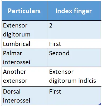
Question-7: Name The Muscles Inserted In The Extensor Expansion Of The Index Finger
Answer:
Muscles inserted in the extensor expansion of the index finger
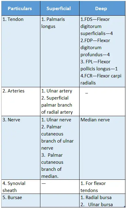
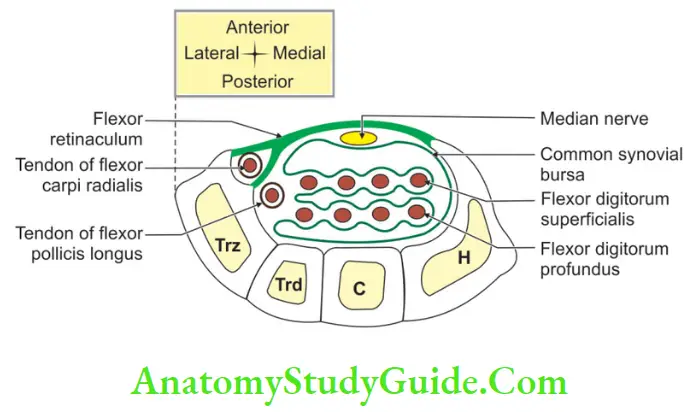
Enumerate The Muscles That Pass Through The Carpal Tunnel
Muscles passing from superficial to deep are
- Four tendons of flexor digitorum superficialis,
- Flexor carpi radialis,
- Four tendons of flexor digitorum profundus, and
- Flexor pollicis longus.
Carpal Tunnel
Carpal Tunnel Introduction:
It is a fibro-osseous tunnel formed by concave palmar surfaces of carpal bones. It is situated in the lower part of the anterior surface of the forearm.
1. Carpal Tunnel Location:
It is located near the wrist joint.
2. Carpal Tunnel Formation:
Pillars
Bones forming the pillars of carpal tunnel
Anterior: Flexor retinaculum.
Posterior: Palmar surfaces of carpal bones.
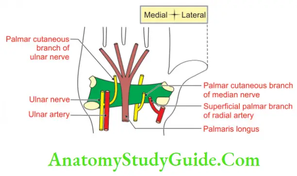
3. Relations:
1. Anteriorly
- Skin,
- Palmaris longus tendon,
- Palmar cutaneous branch of ulnar nerve,
- Palmar cutaneous branch of the median nerve,
- Superficial palmar branch of the radial artery, and
- Ulnar nerve,
- Ulnar vessels.
2. Posteriorly palmar surfaces of carpal bone
4. Carpal Tunnel Contents (From superficial to deep):
- Flexor digitorum superficialis,
- Flexor carpi radialis,
- Flexor digitorum profundus,
- Flexor pollicis longus,
- Median nerve, and
- Radial and ulnar bursa.
5. Carpal Tunnel Applied anatomy:
Carpal Tunnel Syndrome: Compression of the median nerve in the carpal tunnel gives rise to loss of sensations and weakness of the muscles of the thenar eminence, which constitute carpal tunnel syndrome.
Carpal Tunnel Syndrome Etiology: Following are the causes of carpal tunnel syndrome.
Carpal Tunnel Syndrome Median Trap
Myxedema
Edema premenstrual
Diabetes mellitus
Acromegaly
Neoplasia
Trauma
Rheumatoid arthritis
Amyloidosis
Pregnancy
Carpal Tunnel Syndrome Gender variation: Carpal tunnel syndrome is common in females.
Carpal Tunnel Syndrome Age group: Occurs between 40 and 70 years.
1. Carpal Tunnel Syndrome Clinical features: It presents as
·
Intermittent attacks of pain, which are more in the night. It is referred to as the proximal part of the forearm. It may be relieved by dorsiflexion.
Wasting of thenar muscles, namely
- Flexor pollicis brevis,
- Opponens pollicis, and
- Abductor pollicis brevis.
2. Carpal Tunnel Syndrome Treatment
- Pain is relieved by splinting of the wrist in slight dorsiflexion.
- The division of flexor retinaculum is required in severe cases.
Cutaneous Supply Of Palm Of Hand
Palmar surface
- Lateral 3½ fingers-median nerve.
- Medial 1½ fingers-ulnar nerve.
Cutaneous Supply Of Dorsum Of Hand
Dorsal surface
- Lateral 2 ½ fingers-median nerve.
- Medial 2 ½ fingers-radial nerve.
Palmar Aponeurosis
It is a flattened tendon of palmaris longus present in the hand.
1. Palmar Aponeurosis Features:
- Shape: lar
- Apex: Blends with the flexor retinaculum.
- Base: It is directed distally and divides into 4 slips opposite the head of the metacarpals of the medial four fingers. Each slip divides into two parts which provide the passage for digital vessels, nerves, and tendons of lubricants. The palm is divided into compartments by the slips arising from the lateral and medial margins of the palmar aponeurosis.
2. Palmar Aponeurosis Morphology:
Phylogenetically it represents degenerated tendon of palmaris longus. It is homologous to plantar aponeurosis.
3. Palmar Aponeurosis Functions:
- It improves the grip of the hand by fixing the skin.
- It protects the vessels and nerves of the palmar surface of the hand.
4. Palmar Aponeurosis Applied anatomy:
Dupuytren’s contracture: It is an inflammation of the ulnar side of the palmar aponeurosis. There is thickening and contracture of the palmar aponeurosis.
Dupuytren’s Contracture
Dupuytren’s Contracture Introduction:
It is an inflammation of the ulnar side of the palmar aponeurosis. There is thickening and contracture of the palmar aponeurosis.
1. Dupuytren’s Contracture Cause:
It is due to fibrosis and shortening of the palmar aponeurosis
2. Dupuytren’s Contracture Features:
- Usually, the left hand is involved.
- It is more severe towards the ulnar side.
- There is progressive flexion of the 4th and 5th digits.
- There is an involvement of the proximal and middle phalanx because of the insertion of the palmar aponeurosis.
- A high correlation exists between Dupuytren’s contracture and coronary artery disease.
It is possibly due to the result of vasospasm of the arteries innervated by 1st thoracic spinal nerve.
Dorsal Digital Expansion
Dorsal Digital Expansion Introduction:
It is a small aponeurosis formed by each extensor tendon that covers the dorsum of the proximal phalanx and the sides of its base.
1. Dorsal Digital Expansion Shape:
It is lar, base directs proximally and apex directs distally.
2. Formation:
It is mainly formed by the extensor digitorum tendon and it is contributed by interossei and lumbrical muscles. In addition to the above tendon
- In the little finger, it is formed by the extensor digit minimi, and
- In the index finger, by extensor indices.
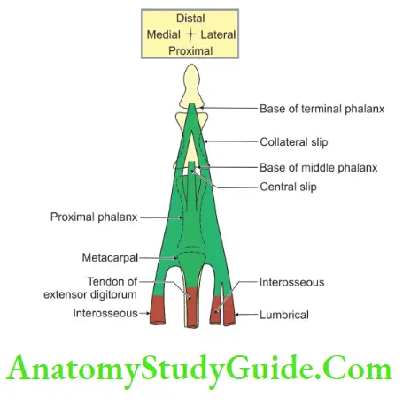
3. Dorsal Digital Expansion Features:
1. Base of dorsal digital expansion
- It covers the dorsal and collateral aspects of the metacarpophalangeal joint.
- It gives attachment to the extensor tendon at the center of the base of digital expansion.
- It forms a hood over the head of the metacarpal.
- It is movable distally and proximally with flexion and extension at the metacarpophalangeal joint.
- Each basal angle is attached to the deep transverse metacarpal ligament.
2. Margins
The lateral margin is thickened by the insertion of the tendon of lumbrical and interossei muscles. The medial margin is thickened by the attachments of interossei only.
3. Wing tendons are of two types
- Proximal wings: Formed by dorsal and palmar interossei.
- Distal wings: Formed by lubricants.
4. Apex of expansion: At the distal end of the proximal phalanx, the apex of the expansion is divided into three bands. They are:
- One central, and
- Two collaterals.
The central band is inserted into the base of the middle phalanx. The two collateral bands unite and finally are inserted into the dorsal aspect of the base of the distal phalanx.
4. Dorsal Digital Expansion Actions:
- It keeps the extensor tendons in the midline.
- It is the only extensor of metacarpophalangeal joints (besides the extensor indices and the extensor digit minimi) for the medial 4 fingers.
- The collateral bands extend the proximal and distal interphalangeal joint through the dorsal digital expansion.
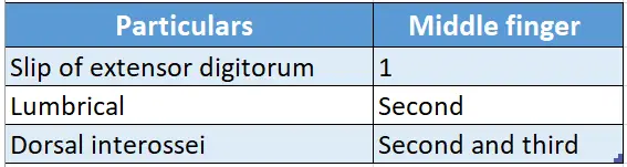
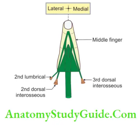
Enumerate The Muscles Inserted In The Extensor Expansion Of The Middle Finger
Muscles inserted in the extensor expansion of the middle finger
Question-8: Describe Interossei Under The Following Heads
1. Interossei Insertion,
2. Interossei Axis,
3. Interossei Strength,
4. Interossei Relation,
5. Interossei Action,
6. Interossei Nerve supply, and
7. Interossei Testing.
Answer:
Interossei Introduction:
These are the small muscles of the hand present in between the metacarpals. They are divided into palmar and dorsal interossei. They are discussed as given in Table.
Palmar and dorsal interossei
1. Interossei Insertion:
Both are inserted on
- Dorsal digital expansion, and
- The base of the proximal phalanx of the respective finger.
2. Interossei Axis:
Passes through middle finger.
3. Interossei Strength:
Palmar interossei are less strong than dorsal interossei.
4. Interossei Relations:
- They are related posteriorly to deep transverse metacarpal ligaments.
- The radial artery passes between the gap produced by two heads of 1st dorsal interosseous.
- Proximal perforating arteries pass between the gap produced by two heads of the 2nd, 3rd, and 4th dorsal interossei.
5. Interossei Actions:
(PAD, DAB. P=palmar, AD=adductor; D=Dorsal, AB=abductor, B-bipennate)
- Palmar interossei are adductors, dorsal interossei are abductors.
- They are powerful flexors of the metacarpophalangeal joint (MP) because their tendons are placed ventrally and distally with respect to the metacarpophalangeal joint.
- They produce an extension of the interphalangeal joint. Both these movements are useful for precision work.
6. Interossei Nerve supply:
They are supplied by a deep branch of the ulnar nerve.
7. Interossei Testing:
Interossei can be tested in the following ways.
- Dorsal interossei: By asking to spread the fingers against resistance.
- Palmar interossei: By asking to hold a piece of paper between the testing finger and the normal finger.
Nerve Supply Of Lubricants
- 1st and 2nd lubricants are supplied by the median nerve.
- 3rd and 4th is supplied by the ulnar nerve.
Actions Of Lubricants
- Flexion of metacarpophalangeal joint of 2nd to 5th digits.
- Extension of interphalangeal joint of 2nd to 5th digits.
Lumbricals
1. Lumbricals Features:
- These are worm-like muscles that arise from tendons of flexor digitorum profundus.
- They are 4 in number and are present on the palmar aspect.
- They are counted from lateral to medial.
- 1st and 2nd lumbricals are unipennate.
- 3rd and 4th lumbricals are bipennate.
- They act as link muscles between the deep flexor and extensor tendons of the hand.
2. Lumbricals Proximal attachments:
- 1st and 2nd lumbricals arise from the radial side of profundus tendon, and
- 3rd and 4th lumbricals arise between neighboring tendons of flexor digitorum profundus.
3. Lumbricals Distal attachments:
They are inserted on the dorsal surface of the base of the middle and distal phalanx, through the lateral border of dorsal digital expansion.
4. Lumbricals Action:
They are flexors of metacarpophalangeal joints and extensors of interphalangeal joints.
5. Lumbricals Nerve supply:
Medial two lumbricals are supplied by the deep branch of the ulnar nerve and lateral two lumbricals are supplied by the median nerve.
6. Lumbricals Relations:
They are dorsal to digital vessels and nerves.
7. Lumbricals Functions:
They produce up-and-down strokes of fingers for skilled work. They produce flexion at the metacarpophalangeal joint and extension of interphalangeal joints.
8. Lumbricals Test:
The muscles are best tested by asking the patient to hold a piece of paper between the sides of two adjacent fingers. If the muscles are acting, the paper will be firmly held and some resistance will be offered to its withdrawal.
Branches Of Superficial Palmar Arch
There are 4 digital branches that supply medial 3 1/2 fingers. Out of these 4 digital branches, lateral 3 digital branches are connected with the deep palmar arch by the palmar metacarpal arteries.
Question-9: Describe The Superficial Palmar Arch Under The Following Heads
1. Superficial Palmar Arch,
2. Superficial Palmar Arch Location,
3. Superficial Palmar Arch Formation,
4. Superficial Palmar Arch Relations,
5. Superficial Palmar Arch Branches, and
6. Superficial Palmar Arch Applied anatomy.
Answer:
Superficial Palmar Arch Introduction:
It is the arterial arch, formed superficial to all the structures in the palm. It is compared with a circle of Willis presents at the base of the brain and plantar arterial arch in the foot.
1. Superficial Palmar Arch:
It may be complete.
2. Superficial Palmar Arch Location:
It is situated in the distal part of the palm and corresponds to the distal palmar crease.
3. Superficial Palmar Arch Formation:
1. It is formed on the medial side of the hand as a continuation of the ulnar artery.
2. On the lateral side, it is completed by one of the following arteries.
- Superficial palmar branch of the radial artery,
- Arteria radialis indices: Artery of index finger (branch of radial artery),
- Arteria princeps pollicis: Artery of thumb (branch of radial artery), and
- Median artery (branch of the anterior interosseous artery which is a branch of the common interosseous artery—ulnar artery).
4. Superficial Palmar Arch Relations:
1. Anterior
- Palmaris brevis, and
- Palmar aponeurosis.
2. Posterior:
- Flexor digitorum superficialis,
- Flexor digitorum profundus,
- Lumbricals,
- Flexor digiti minimi, and
- Digital branches of median nerve.
5. Superficial Palmar Arch Branches:
There are 4 digital branches that supply medial 3 1/2 fingers. Out of these 4 digital branches, lateral 3 digital branches are connected with the deep palmar arch by the palmar metacarpal arteries.
6. Superficial Palmar Arch Applied anatomy:
It is one of the important anastomotic channels for the efficient blood supply of the palm. It is important in case of blockage of the radial or ulnar artery.
Branches Of Deep Palmar Arch
- From convexity-3 palmar metacarpal arteries
- From concavity—recurrent branch
- Dorsally-3 perforating digital arteries.
Question-10: Describe The Deep Palmar Arch Under The Following Heads
1. Deep Palmar Arch Formation,
2. Deep Palmar Arch Location,
3. Deep Palmar Arch Arch,
4. Deep Palmar Arch Relations,
5. Deep Palmar Arch Branches, and
6. Deep Palmar Arch Applied anatomy.
Answer:
Deep Palmar Arch Introduction:
It is an arterial arch present deep in the flexor tendons of digits.
1. Deep Palmar Arch Formation:
It is mainly formed by the radial artery and completed on the medial side by a deep palmar branch of the ulnar artery.
2. Deep Palmar Arch Location:
It is present at the proximal palmar crease.
3. Deep Palmar Arch Arch:
It may be complete.
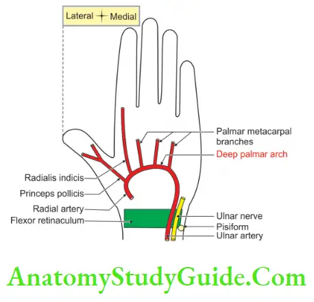
4. Deep Palmar Arch Relations:
1. Anterior
- Oblique head of adductor pollicis,
- Flexor tendons of fingers, and
- Lumbricals.
2. Posterior: Shaft of metacarpal and interossei.
5. Deep Palmar Arch Branches:
1. From the convexity of the arch, 3 palmar metacarpal arteries run distally in the 2nd, 3rd, and 4th spaces. They supply medial 4 metacarpals; terminate in the finger cleft by joining with a common digital branch of the superficial palmar arch.
2. From the dorsal side, 3 perforating arteries pass through medial 3 interosseous spaces to anastomose with dorsal metacarpal arteries.
3. From the convexity of the arch, a recurrent branch arises to supply carpal bones and ends in a palmar carpal arch.
6. Deep Palmar Arch Applied anatomy:
It is one of the important anastomotic channels for the efficient blood supply of the palm. It is important in case of blockage of the radial or ulnar artery.
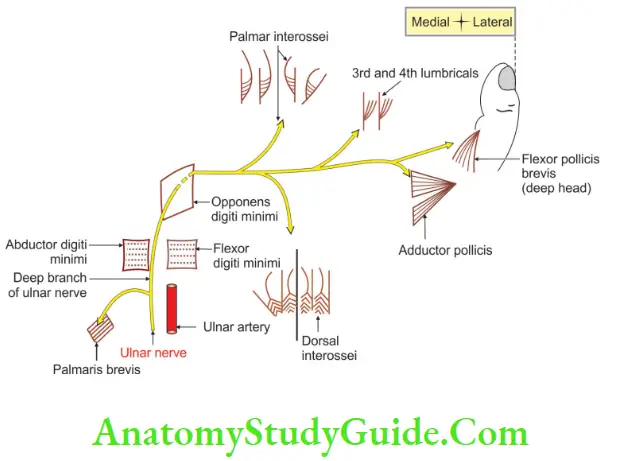
Question-11: Name The Muscles Supplied By Ulnar Nerve In the Hand
Answer:
1. Palmaris brevis,
2. Abductor digit minimi,
3. Flexor digit minimi,
4. Opponens digit minimi,
5. Adductor pollicis,
6. Flexor pollicis brevis (deep head),
7. Interossei,
- 4 palmar, and
- 4 dorsal.
8. Lumbricals: 2 medial.
Question-12: Describe The Ulnar Nerve Under The Following Heads
1. Ulnar Nerve Root value,
2. Ulnar Nerve Course and relations,
3. Ulnar Nerve Course in hand,
4. Ulnar Nerve Branches, and
5. Ulnar Nerve Applied anatomy.
Answer:
Ulnar Nerve Introduction:
It is the most commonly affected nerve in leprosy in the upper limb. It is also called the ‘musician’s nerve’ because fine movements of the hand are required to play musical instruments.
These are executed by small muscles of the hand which are innervated by the ulnar nerve. It is the nerve of the flexor muscles present on the medial side of the forearm, most of the intrinsic muscles of the hand, and skin on the medial side of 1 1/2 fingers.
1. Ulnar Nerve Root value:
It is the largest branch and direct continuation of the medial cord of the brachial plexus. The root value is C7, C8, and 1st T nerve. It picks up additional C7 fibers in the axilla, usually from the lateral cord.
2. Ulnar Nerve Course and relations :
The structures in the course and relation of the ulnar nerve begin with the letter (M)
- The medial cord of the brachial plexus lies on the medial side of 2nd part of the axillary artery.
- The ulnar nerve lies medial to 3rd part of the axillary artery.
- It also lies medial to the brachial artery.
- In the middle of the arm, it pierces the medial intramuscular septum along with the superior ulnar collateral artery.
- It enters the posterior compartment and lies on the medial head of the triceps. It passes behind the medial epicondyle of the humerus and is received by flexor carpi ulnaris.
- It lies on the medial ligament of the elbow joint and rests on the medial side of the flexor digitorum profundus under a thin blanket of flexor carpi ulnaris in the upper 2/3rd of the forearm.
3. Ulnar Nerve Course in the hand:
It divides the flexor retinaculum into superficial and deep branches.
- The superficial branch runs distally beneath palmaris brevis.
- The deep branch is accompanied by the deep branch of the ulnar artery and passes in between the abductor digit minimi and flexor digit minimi then passes through the substance of the opponent digit minimi.
- It passes down to the interossei, grooves the distal border of the hook of the hamate, and arches deeply in the palm within the cavity of the deep palmar arch.
4. Ulnar Nerve Branches:
Distribution of ulnar nerve
Note:
1. The symbol ” ” at the end of the nerve signifies the articular branch
2. The symbol ” ” at the end of the nerve signifies the cutaneous branch
5. Ulnar Nerve Applied anatomy:
1. Ulnar nerve is palpated behind the medial epicondyle of the humerus. This is thickened and is cord-like in Henson’s disease.
2. Ulnar nerve is damaged
- At the elbow joint due to
- Damage in the cubital tunnel is formed by the tendinous arch connecting 2 heads of flexor carpi ulnaris. The ulnar nerve is often entrapped in this tunnel and this entrapment of the ulnar nerve in the cubital tunnel is called cubital tunnel syndrome. It results in radial deviation of the hand.
- Pressure on the ulnar nerve as it passes along the ulnar groove. It produces funny sensations along the medial border of the forearm, hypothenar surface, and little finger. Hence ‘the humerus’ is called “funny bone”.
- At the wrist joint by the compression of the volar carpal ligament and palmaris brevis muscle. Guyon’s canal and volar ligament
- Here all intrinsic muscles of the hand are paralyzed resulting in a typical claw hand (ulnar claw).
- There is a hyperextension of the metacarpophalangeal joint of the ring and a little finger and flexion of the interphalangeal joint. This is because of paralysis of the interossei and lumbrical.
- There is also a loss of sensations on the medial 1 1/2 fingers.
- There is a loss of adduction of the thumb, as the adductor pollicis is paralyzed.
Pulp Space
It is the space between the palmar skin and distal phalanges of all digits of the hand.
1. Pulp Space Situation:
Distal to fibrous sheath of the flexor tendon.
2. Pulp Space Formation:
Formed by fibrous septa connecting skin to the periosteum of the distal phalanx.
3. Pulp Space Contents:
- Subcutaneous fat, and
- Blood vessel.
4. Pulp Space Blood supply of terminal phalanx:
The distal 4/5th part is by digital arteries and the proximal part is by the epiphyseal artery.
5. Pulp Space Applied anatomy:
- Infection of pulp space is called whitlow, which is associated with severe throbbing pain due to increased tension in spaces.
- Abscess is drained by lateral incision and breaking all septa. In neglected cases, it may lead to avascular necrosis of the terminal phalanx.
Contents Of Thenar Space
1. Tendons:
- Flexor digitorum superficialis of index finger,
- Flexor digitorum profundus of index finger,
- Flexor pollicis longus, and
- 1st lumbrical.
2. Vessels:
- Arteria princeps pollicis, and
- Arteria radialis indicis.
3. Nerves of the thumb and index finger.
Muscles Of Thenar Space
- Flexor pollicis brevis,
- Abductor pollicis brevis, and
- Opponents policies.
Nerve Supply Of Muscles Of Thenar Space
All muscles of thenar eminence are supplied by the median nerve except the deep head of the flexor pollicis brevis which is supplied by the deep branch of the ulnar nerve.
Palmar Spaces
Palmar spaces
Extensor Expansion Of Little Finger
Muscles inserted in extensor expansion of little finger
Extensor Expansion Of Ring Finger
Muscles inserted in extensor expansion of ring finger
Posterior Interosseous Nerve
Posterior Interosseous Nerve Introduction:
It is a deep terminal branch of the radial nerve and arises in front of the lateral epicondyle.
1. Posterior Interosseous Nerve Peculiarity:
It bears a pseudoganglion.
2. Posterior Interosseous Nerve Course and relation:
The nerve appears in the back of the forearm by piercing the supinator muscle and winds around the lateral side of the radius.
3. Posterior Interosseous Nerve Branches:
1. Before piercing the supinator
Extensor carpi radialis brevis, and
Supinator.
2. After piercing the supinator, the following branches are given
1. Muscular branches
Short branches to
- Extensor digitorum,
- Extensor digiti minimi, and
- Extensor carpi ulnaris.
Long branches are divided into
- Medial set, which supplies
- Extensor pollicis longus, and
- Extensor indices.
Lateral set, which supplies
- Abductor pollicis longus, and
- Extensor pollicis brevis.
2. Articular branches to
- Wrist joint,
- Inferior radioulnar joint, and
- Intercarpal joints.
3. Sensory branches
- Interosseous membrane,
- Radius, and
- Ulnar.
Leave a Reply