Front Of Thigh
Name the muscles forming boundaries of the femoral triangle
Table of Contents
1. Femoral Triangle Lateral boundary is formed by the medial border of the sartorius.
2. Femoral Triangle Medial boundary is formed by the medial border of the adductor longus.
3. Femoral Triangle Base is formed by the inguinal ligament.
4. Femoral Triangle Apex is formed by the meeting point of the medial borders of the adductor longus and sartorius.
Name the muscles forming the floor of the femoral triangle
The muscles forming the floor of the femoral (from medial to lateral) are
Read And Learn More: Anatomy Notes And Important Question And Answers
1. Medial sloping wall is formed by
- Adductor longus medially, and
- Pectineus laterally.
2. Lateral sloping wall is formed by
- Psoas major medially, and
- Iliacus laterally.
Name the structures forming the boundaries of the femoral ring
- Femoral Ring Anteriorly: Inguinal ligament,
- Femoral Ring Posteriorly: Pectineus and fascia covering it,
- Femoral Ring Medially: Concave margin of lacunar ligament, and
- Femoral Ring Laterally: Septum separating it from the femoral vein.
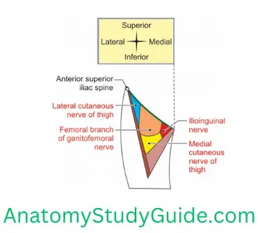
Name the cutaneous nerves seen in the roof of the femoral triangle
1. Femoral branch of the genitofemoral nerve: It supplies the skin over the upper part of the femoral triangle.
2. Branches of the ilioinguinal nerve.
3. Medial cutaneous nerve of thigh it crosses the femoral artery from the lateral to medial.
4. Lateral cutaneous nerve of thigh is seen lateral of femoral triangle.
Name the muscles of the anterior compartment of the thigh
The muscles of the anterior compartment of the thigh are
1. Sartorius,
2. Quadriceps femoris which includes
- Rectus femoris,
- Vastus lateralis,
- Vastus medialis, and
- Vastus intermedium.
3. Articularis genu.
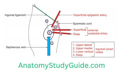
Superficial Inguinal Lymph Nodes
Superficial Inguinal Lymph Nodes Introduction: The lymph vessels draining all the structures superficial to deep fascia of lower limb drain into superficial inguinal lymph nodes. They accompany the great saphenous vein.
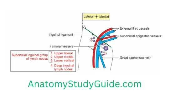
1. Superficial Inguinal Lymph Nodes Arrangement: They are arranged in the form of a “T”.
2. Superficial Inguinal Lymph Nodes Location
- Along the great saphenous vein is the vertical group of inguinal lymph nodes, and
- Parallel to an inguinal ligament is the horizontal group of lymph nodes.
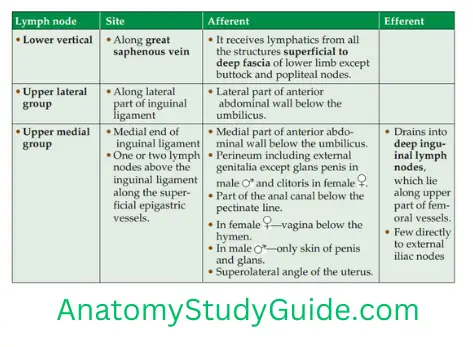
3. Afferent and efferent lymphatics
4. Superficial Inguinal Lymph Nodes Applied anatomy
The upper group of the superficial inguinal lymph nodes are enlarged due to pathology of the following structures. It may be an infection or malignant growth of one of the following structures.
- Superolateral part of the uterus,
- The skin of shaft of penis,
- Part of the vagina below the hymen, and
- Part of the anal canal below the pectinate line.
- Syphilitic lesion of prepuce of penis.
Lateral group lymph nodes of superficial inguinal lymph nodes are enlarged due to pathology of the gluteal region.
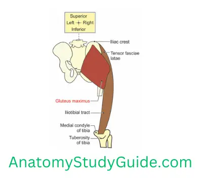
Fascia Lata
(Lata—deep and broad)
Fascia Lata Introduction: It is a tough, fibrous, deep fascia which envelops the thigh like a stocking.
1. Fascia Lata Modifications
- Iliotibial tract, and
- Saphenous opening.
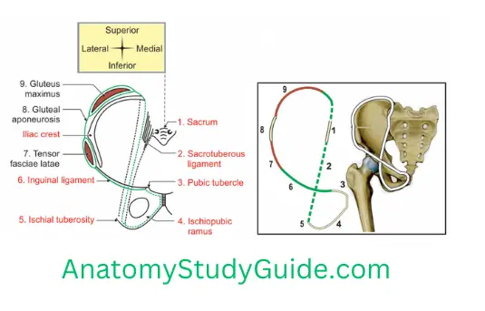
2. Fascia Lata Gives attachments
1. Proximally is attached to
- Pubic tubercle,
- Inguinal ligament,
- Outer lip of the entire iliac crest (it splits to enclose the tensor fascia lata and gluteus maximus muscles),
- Dorsal surface of the sacrum,
- Coccyx,
- Sacrotuberous ligament, and
- Ischial tuberosity.
2. Anteromedially attached to
- Ischiopubic ramus,
- Anterior margin of pubic symphysis,
- Pubic crest,
- Pubic tubercle, and
- Pectin pubis.
3. Distally attached to the
- Patella,
- Inferior margin of the tibial condyles, and
- Head of the fibula.
3. Fascia Lata Encloses
- Gluteus maximus
- Tensor fascia lata.
4. Fascia Lata Functions
- Protects the deeper structures,
- Keeps deeper structures in position, and
- It acts as tight stockings and helps in venous return.
Iliotibial Tract
It is a modified thick fascia of the thigh (fascia lata). It is a 2” wide band.
1. Iliotibial Tract Situation: It is present on the lateral aspect of thigh.
Extends from iliac crest to lateral condyle of tibia.
1. Superiorly it has two laminae.
- Superficial lamina is attached to the tubercle of iliac crest, and
- Deep lamina is attached to the capsule of hip joint.
2. Inferiorly it is attached to smooth area on the anterior surface of lateral condyle of the tibia.
2. Iliotibial Tract Features: It gives insertion to
- Greater part of gluteus maximus,
- Tensor fascia lata.
3. Iliotibial Tract Nerve supply: Superior gluteal nerve.
4. Iliotibial Tract Functions
- It stabilizes the knee joint both in extension and partial flexion. Hence, it is used constantly during walking and running.
- It is the main support of knee joint against gravity.
- It is used as suturing material.
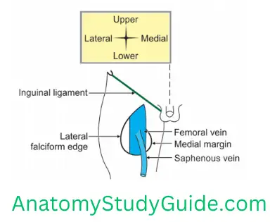
Saphenous Opening
(Saphenous—easily seen)
Saphenous Opening Introduction: It is an oval opening present in the fascia lata.
1. Saphenous Opening Location: 4 cm below and 4 cm lateral to pubic tubercle. The vertical height is 4 cm. It has two margins
- Upper, lateral and lower, and
- Medial margin.
1. The lateral margin is superficial and well-defined.
- It is half-moon shaped, and
- It is present in front of the femoral sheath. It is also called falciform margin.
2. Saphenous Opening Medial margin is deep and ill-defined due to cribriform fascia.
2. Opening: It is closed by an areolar membrane called cribriform fascia. The fascia is pierced by a number of structures and gives a sieve-like appearance, hence it is called cribriform fascia.
3. The structures passing through the saphenous opening are the following
1. Vein: Great saphenous vein. It receives the following veins before it opens in saphenous openings.
- Superficial external pudendal vein,
- Superficial epigastric vein, and
- Superficial circumflex iliac vein
2. Arteries
- Superficial external pudendal artery,
- Superficial epigastric artery, and
- Deep external pudendal artery—deep branch of femoral artery
3. Lymph vessels: Lymphatics from superficial lymph nodes converge to the saphenous opening. They pass through the cribriform fascia and enter deep inguinal lymph nodes.
4. Comparative anatomy: Saphenous opening is present in fascia lata only in man.
Name the branches of the femoral artery in the femoral triangle
1. Superficial branches
- Superficial epigastric,
- Superficial circumflex iliac, and
- Superficial external pudendal.
2. Deep branches
1. Deep external pudendal artery. It supplies deeper structures in the perineal region
2. Profunda femoris and its branches
- Medial circumflex femoral artery and its branches
- Lateral circumflex femoral artery and its branches
3. Muscular branches to muscles of anterior and medial compartment of thigh.
4. The upper two perforating branches of profunda femoris artery arise in the femoral tiangle.
Describe femoral triangle (triangle of Scarpa) under following heads:
1. Femoral Triangle Site,
2. Femoral Triangle Boundaries,
3. Femoral Triangle Roof,
4. Femoral Triangle Floor,
5. Femoral Triangle Contents,
6. Femoral Triangle Relations, and
7. Femoral Triangle Applied anatomy
Introduction: It is subfascial lar depression present in upper part of front of thigh.
1. Femoral Triangle Site
- It is present in front of the upper 1/3rd of the thigh below the inguinal ligament.
- In living person, it appears as a lar depression inferior to the inguinal ligament.
- It is best appreciated when the thigh is flexed, abducted, and laterally rotated.
2. Femoral Triangle Boundaries
- Laterally: Medial border of sartorius.
- Medially: Medial border of adductor longus. It can be felt as a distinct ridge when the thigh is adducted against resistance.
- Base: Inguinal ligament.
- Apex: It is formed by the meeting point of the medial borders of adductor longus and sartorius. It is directed downward.
3. Femoral Triangle Roof
1. Skin
2. Superficial fascia containing
1. Arteries: Superficial branches of the femoral artery
- Superficial circumflex iliac,
- Superficial external pudendal, and
- Superficial epigastric artery.
2. Veins: Tributaries of the femoral vein
- Superficial circumflex iliac,
- Superficial external pudendal, and
- Superficial epigastric vein.
3. Nerves
- The femoral branch of the genitofemoral nerve runs downwards. It is anterior to the femoral artery in the anterior wall of femoral sheath. This nerve supplies an area of skin over the femoral.
- The lateral cutaneous nerve of the thigh runs near the lateral angle of the femoral.
3. Deep fascia and structures within it. These include
- Saphenous opening,
- Cribriform fascia,
- Terminal part of the saphenous vein, and
- Superficial inguinal lymph nodes.
4. Floor: It is muscular and hollow. It is best seen when thigh is flexed. The muscles are grouped as muscles forming medial and lateral sloping wall. They are described from medial to lateral.
1. Medial sloping wall is formed by
- Adductor longus medially. The medial border forms the boundary of the femoral, hence the whole muscle is included in the floor.
- Pectineus laterally. It is called key muscle of femoral.
1. Lateral sloping wall is formed by
- Psoas major medially, and
- Iliacus laterally. It is covered by fascia iliacus.
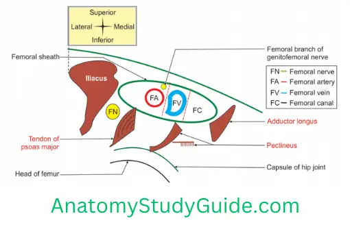
5. Femoral Triangle Contents
1. Femoral sheath and its contents (from medial to lateral)
1. Femoral Canal and its Contents, namely
- Fibrofatty tissue,
- Cloquet lymph nodes (deep inguinal lymph node, or Rosenmüller lymphnode), and
- Cloquet lymphatics (afferent and efferent lymphatics of these lymph nodes).
2. Femoral vein and its tributaries
1. Great saphenous vein and its tributaries
- Superficial epigastric,
- Superficial circumflex iliac, and
- Superficial external pudendal.
2. Muscular veins
3. Profunda femoris vein and its tributaries
- Medial circumflex femoral vein and its tributaries
- Lateral circumflex femoral vein and its tributaries
3. Femoral artery is central and dominant structure within the femoral and the branches of femoral artery within the femoral are grouped as
1. Superficial branches
- Superficial epigastric,
- Superficial circumflex iliac, and
- Superficial external pudendal.
2. Deep branches are
1. Profunda femoris and its branches,
- Medial circumflex femoral artery and its branches
- Lateral circumflex femoral and its branches
2. Muscular branches, and
3. Deep external pudendal supplies muscles of thigh.
4. The upper two perforating branches of profunda femoris artery arise in the femoral triangle.
2. Nerves
1. Femoral nerves and its divisions
1. Anterior division and its branches
- Muscular branch to Sartorius
- Medial CUTaneous nerve of thigh, and
- Intermediate CUTaneous nerve of thigh.
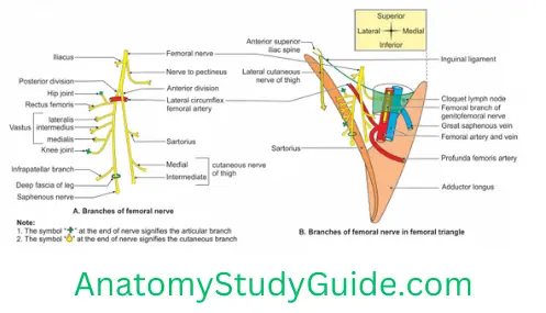
2. Posterior division and its branches
1. Muscular branches to QUADRIceps
- Rectus femoris,
- Vastus medialis,
- Vastus intermedius, and
- Vastus lateralis.
2. Cutaneous branch—SAPHenous nerve
3. Articular branch to
- Hip joint, and
- Knee joint
2. The nerve to the pectineus arises from the femoral nerve just above the inguinal ligament. It passes behind femoral sheath to reach the anterior surface of pectineus.
3. The femoral branch of the genitofemoral nerve occupies the lateral compartment of the femoral sheath. It runs along with the femoral artery. It supplies most of the skin over the femoral .
4. The lateral cutaneous nerve of the thigh crosses the lateral angle of the .It runs on the lateral side of the thigh. It ends by dividing into anterior and posterior branches. These supply the anterolateral aspect of the front of thigh and lateral aspect of the gluteal region respectively.
6. Relations of vessels
1. At the base (from medial to lateral)
- Femoral Vein,
- Femoral Artery, and
- Femoral Nerve.
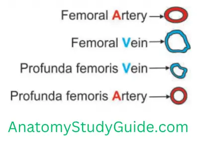
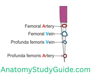
2. At apex (from anterior to posterior) AVVA
- Femoral Artery,
- Femoral Vein,
- Profunda femoris Vein, and
- Profunda femoris Artery
7. Femoral Triangle Applied anatomy
Femoral Aartery
Pulsations of femoral artery are felt in femoral . They are felt at the midinguinal point against
- Head of femur, and
- Tendon of psoas major.
Stab wound injury at the apex of femoral may be fatal as it cuts all the vessels of lower limb.
Femoral vein
- Blood can be drawn from the femoral vein by feeling the pulsations of femoral artery and going just medially to it.
- Femoral vein catheterization is used when rapid access to a large vein is needed.
Saphenous vein
Varicosities of saphenous veins are treated by Trendelenburg’s operation. The tributaries of saphenous vein are tied in the femoral.
Deep inguinal lymph nodes in femoral are enlarged in the
- Infection of the skin of
- Abdomen below umbilicus.
- Gluteal region
- Leg, sole and external genitalia.
- Malignancy of fundus of uterus.
Stab wounds at the apex of femoral may be fatal as it cuts all vessels of lower limb.
Pus may appear in the femoral behind the femoral sheath. It may travel to
- The back of thigh along the perforating branches of the profunda femoris artery.
- The adductor canal and even to the popliteal fossa along the femoral vessels.
Femoral hernia: The femoral ring is a weak area in the anterior abdominal wall. It is of the size of the little finger. The femoral ring is the usual site of a femoral hernia. It is a protrusion of abdominal viscera (often a loop of small intestine) through the femoral ring into the femoral canal. A femoral hernia appears as a mass, often tender, in the femoral, inferolateral to the pubic tubercle.
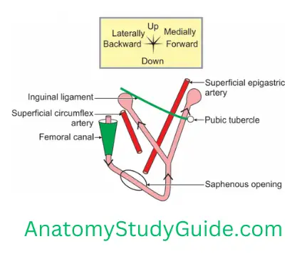
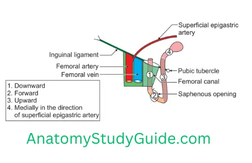
Direction of course of femoral hernia: As the herniasac enlarges, it passes through the saphenous opening, and then turns upwards. Here it may pass along the course of
- Superficial circumflex iliac vessels, or
- Superficial epigastric vessels.
Note: Depending upon the pathway, it may be
- Above the inguinal ligament, if it chooses the pathway along superficial epigastric vessels.
- Below the inguinal ligament, if it chooses the pathway along superficial circumflex iliac vessels.
Name the fascia forming the femoral sheath
1. Anterior wall: Transversalis fascia.
2. Posterior wall: Fascia iliaca.
3. Lateral and medial wall: Fusion of fascia transversalis and fascia iliaca.
Enumerate the contents of the femoral sheath
Contents of the femoral sheath (from medial to lateral) are described as the contents of each compartment.
1. Medial compartment is the femoral canal. Femoral Canal and its Contents are
- Fibrofatty tissue,
- Cloquet lymph node (deep inguinal lymph node, or Rosenmüller lymph node),and
- Cloquet lymphatics (afferent and efferent lymphatics of these lymph nodes).
2. Intermediate compartment is venous compartment. It contains femoral vein and its tributary—great saphenous vein.
3. Lateral compartment is arterial compartment. It contains
- Femoral artery, and
- Femoral branch of genitofemoral nerve in anterior wall.
Leave a Reply