Occlusion In The Permanent Dentition Notes
Occlusion
It is important for a practitioner to have complete knowledge about the masticatory system. It is also vital to know about the possible errors and its implication and ways to avoid them to achieve the desired occlusion.
Table of Contents
Occlusion is defined as the relationship between the mandible and maxilla when the jaw is closed and the teeth are in maximal intercuspal position.
Every individual has a different occlusal pattern and there is a change in occlusal pattern with age. For the ease of understanding, it can be studied under the following headings:
- Occlusion in the deciduous dentition
- Occlusion in the mixed dentition
- Occlusion in the permanent dentition
| Body Fluids | Muscle Physiology | Digestive System |
| Endocrinology | Face Anatomy | Neck Anatomy |
| Lower Limb | Upper Limb | Nervous System |
Read And Learn More: Oral Anatomy Notes
Occlusion In The Deciduous Dentition
By 3 years of age, 20 primary teeth are present in the oral cavity. The relationship of thesecond primary molar indicates the occlusion that would develop. It can also be determined if there might be malocclusion in the near future aiding in better treatment planning if necessary.
1. Mesiodistal relationship in primary second molars:
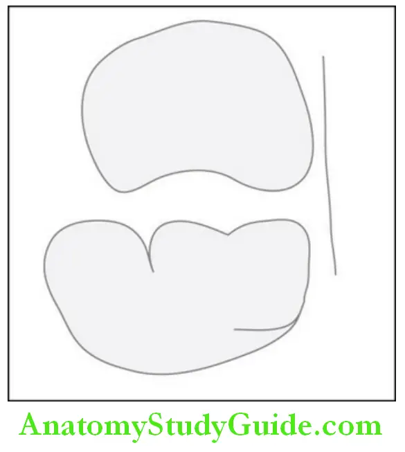
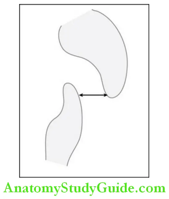
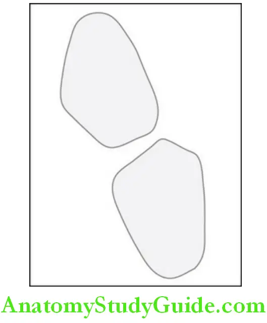
1. Flush terminal/vertical plane type:
The distal surface of the maxillary and mandibular teeth is in a straight line. Both the maxillary and mandibular second primary teeth have the same vertical plane in their distal aspect. This relation is very favorable for the development of class I molar relationship in permanent dentition.
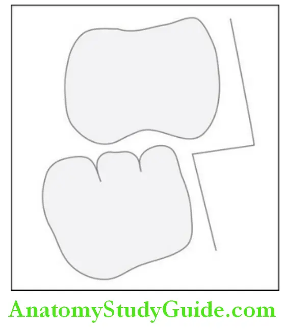
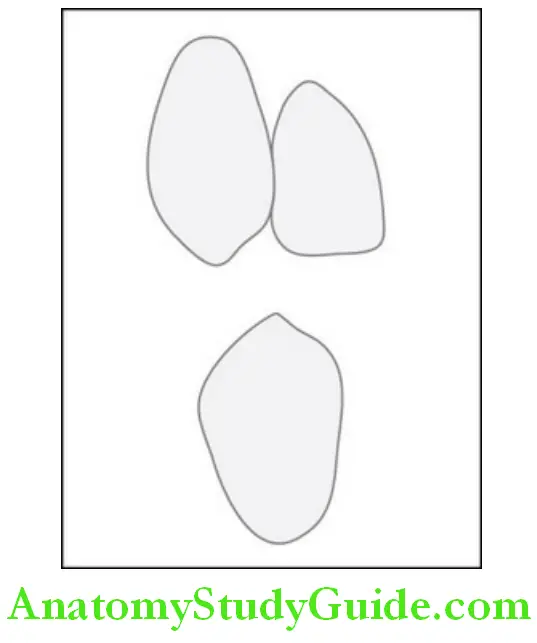
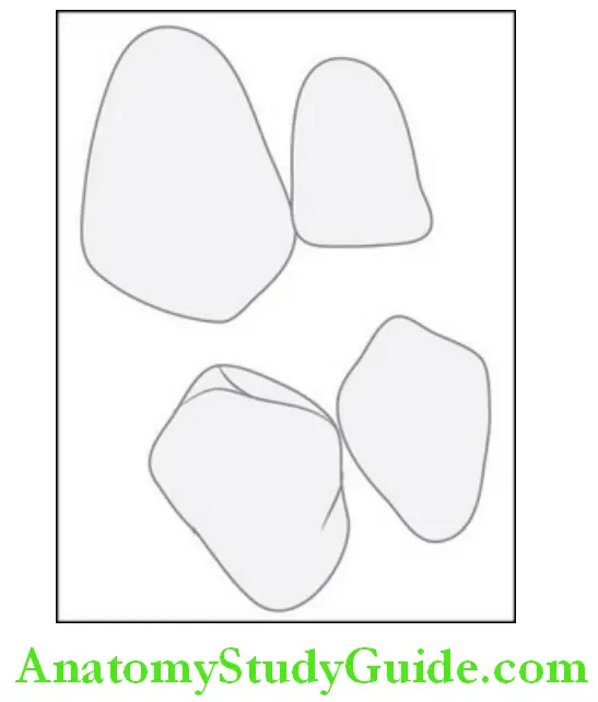
2. Mesial step type:
The distal surface of the mandibular primary second molar Is more mesial to that of the maxillary primary second Molar. This occlusal relationship may guide the permanent Molars to the angle’s class I molar relation or sometimes into a class III molar relationship.
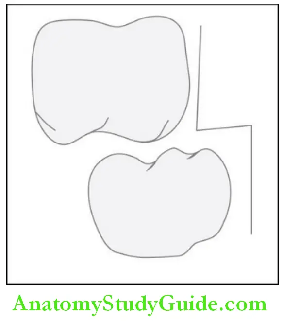
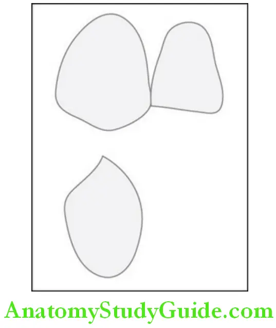
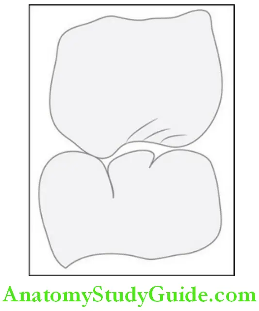
3. Distal step type:
The distal surface of the mandibular second molar is more distal to the maxillary counterpart. This relationship opposes the developing occlusion to obtain class I relation in permanent molars and may lead to class II molar relationship in the permanent dentition.
2. Anterior teeth relationship:
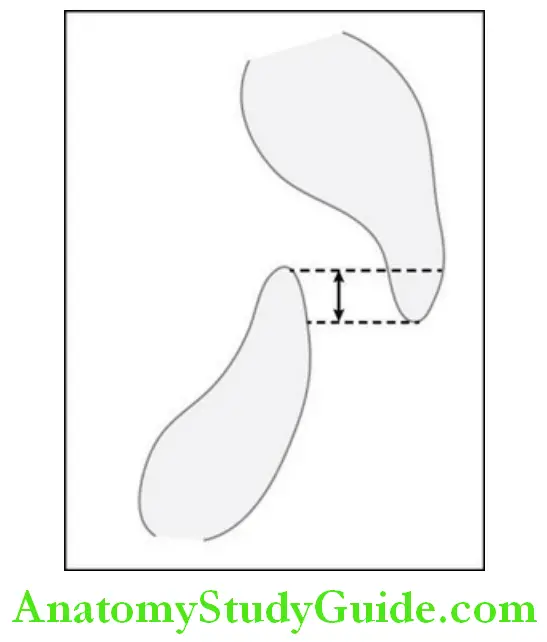
1. overbite:
The distance from the incisal edge of the maxillary incisors to the incisal edges of the mandibular incisors, i.e. The distance of vertical overlap of the anterior teeth is called overbite. It is about 2 mm.
2. overjet:
The horizontal distance between the lingual aspect of the maxillary incisor and the labial aspect of the mandibular incisor due to overlap is called overjet. The overjet is about 1–2 mm and it varies from 2 to 6 mm with age.
3. Canine relation:
The canine relationship is one of the most stable relationships in the deciduous dentition.
1. If the primary mandibular canine approximates the embrasure of the maxillary lateral incisor and canine, it is a class I relationship.
2. If the primary mandibular canine approximates distal to the embrasure, it is a class II relationship.
Occlusion In The Mixed Dentition
- The stage when primary and permanent teeth function together in the oral cavity is called mixed dentition.
- The first permanent tooth to erupt in the oral cavity is the first molar at the age of 6 years.
- The age of 6–13 years is termed as the mixed dentition period and this is the period when there is a transition of primary dentition to permanent dentition.
- The transition occurs in three phases:
1. First transitional phase/ugly duckling phase
- Emergence of the first permanent molars
- Shift/transition of incisors
- Occlusal relationship obtained in molars
2. Intertransitional phase
- Shedding of deciduous incisors and eruption of permanent incisors
- Ugly duckling to mature phase
3. Second transitional phase
- The eruption of cuspids and bicuspids
- Occlusal relation accomplished
-
- The permanent teeth are larger in dimension in comparison to the primary teeth.
- The growth of the maxilla and mandible with age leads to spacing in the primary dentition.
- The interdental space between the maxillary primary teeth is about 4 mm and 3 mm in the mandibular teeth.
- The shedding of the primary teeth and the space achieved with the growth of the jaws aid to accommodate the permanent teeth.
- The permanent canine and premolars are smaller in width than the primary canine and primary molars and this difference of space is called leeway space.
- Leeway space aids to accommodate permanent canines and moves the Mandibular molar mesially as the bicuspids are smaller in width.
- The interdental spaces and leeway space play an important role in establishing occlusion in permanent dentition.
Occlusion In The Permanent Dentition
When all the permanent teeth have erupted in the oral cavity, then it is called a permanent dentition.
The angulation of the teeth depends on the angulation of the alveolar bone.
The angulation changes with the growth pattern of the individual.
The angulation of the alveolar bone is more in permanent dentition compared to the deciduous and mixed dentition due to the growth of the maxilla and mandible.
1. Occlusal relationship in the anterior teeth:
1. Overjet: Overjet is the horizontal overlap of the anterior teeth.
Class I: 2–4 mm
Class II: 4–6 mm
Class III: 0–0.5 mm
2. Overbite: The vertical overlap of anterior teeth is called overbite; it is about 0.5–2 mm.
3. Canine relation:
This is classified as:
- Class I: The mesial slope of the maxillary canine is in approximation with the distal slope of the mandibular canine.
- Class II: The distal slope of the maxillary canine is in approximation with the mesial slope of the mandibular canine.
- Class III: The mesial slope of the maxillary canine is in approximation with a mesial slope of the mandibular first premolar.
2. Occlusal relationship in posteriors:
- The first permanent tooth to erupt into the oral cavity is the first molar and is used to classify the occlusal relation. The classification was proposed by angle.
- Angle’s class i: The mesiobuccal cusp of the maxillary first molar occludes into the mesiobuccal groove of the mandibular first molar.
- Angle’s class ii: The distobuccal cusp of the maxillary first molar occludes into the mesiobuccal groove of the mandibular first molar.
- Angle’s class iii: The mesiobuccal cusp of the maxillary first molar occludes with the interdental space between the mandibular first and second molar.
- Cusp to fossa contact: This contact provides excellent function, stability, and distribution of forces during mastication and occlusion. The functional cusps aid in cutting and grinding the bolus.
- The functional cusps are those which occlude with the fossa of the opposing teeth. Palatal cusps of the maxillary posteriors and buccal cusps of the mandibular posterior are the functional cusps.
- The slopes of the cusps guide the occlusion and support in stabilization of occlusion.
Theories Of Occlusion
1. Bonwill theory: This theory is called the equilateral triangle theory. The triangle is formed by joining the condyles as the base of the triangle to the midline of the mandibular central incisors towards the incisal edge. The equilateral triangle measures 4 inches in length.
2. Conical theory: Hall proposed the conical theory stating that the lower teeth move over to form a 45° angle between the occlusal plane and long axis of the cone.
3. Spherical theory: The spherical theory states that each cusp and incisal edge conforms to a segment of the surface of a sphere of 8 inches in diameter with the center of the glabella during the closure of the mouth.
Occlusal Curves
There are a few natural curves in the dentition to aid in maximum occlusion. These curves improve masticatory efficiency.
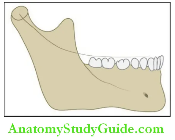
1. The curve of speed: The curve of Spee is defined as an imaginary curve that begins from the tip of the canine and runs through the buccal cusps of premolars and molars and along the ramus of the mandible to the condyle of the mandible.
- This is a concave curve and the maxillary counterpart will be convex to have centric occlusion.
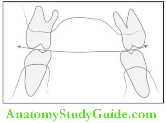
2. The curve of Wilson: The curve of Wilson is an imaginary curve that starts from the buccal cusp of mandibular posteriors to the buccal cusp of the contralateral side.
- The glabella is the center of the arc of the sphere. The curve is concave in the mandible and convex in the maxilla when viewed from the frontal aspect.
To achieve centric relation, non-deflected closure, distribution of stress/load, occlusal and incisal plane, and functional and aesthetic harmony, it is important for us to value the concepts of occlusion. The lack of knowledge of occlusion and application could be a detriment to the success of the restoration and rehabilitation.
Occlusion Conclusion
- Occlusion is defined as the relationship between the mandible and maxilla when the jaw is closed and the teeth are in the maximal intercuspal position.
- It can be studied under the following headings i.e. occlusion in the deciduous, mixed, and permanent dentition.
- In the deciduous dentition, the mesiodistal relationship in primary second molars may be of the flush terminal/vertical plane type, mesial step type, and distal step type.
- Anterior teeth relationship includes overbite and overjet.
- Canine relation may be class I or class II.
- Occlusion in the mixed dentition: The stage when primary and permanent teeth function together in the oral cavity is called mixed dentition which is between 6 and 13 years.
- This is the period when there is a transition from primary dentition to permanent dentition.
- The transition occurs in three phases, the first transitional phase/ugly duckling phase, the inter-transitional phase, and the second transitional phase
- Occlusion in the permanent dentition: When all the permanent teeth (28 teeth) have erupted in the oral cavity, then it is called permanent dentition.
- Occlusal relationship in posteriors: The first permanent tooth to erupt into the oral cavity is the first molar and is used to classify the relation.
- The classification was proposed by angle which includes class I, class II, and class III and cusp to fossa contact.
- Theories of occlusion include the bonwill theory, the conical theory, and the spherical theory.
- Occlusal curves: There are a few natural curves in the dentition to aid in maximum occlusion.
- These curves improve masticatory efficiency. The curve of Spee and the curve of Wilson are a few
Leave a Reply