Cementum
- Cementum is a mineralized dental hard connective tissue that covers the anatomic roots of the human teeth. It has some physical, chemical and structural properties similar to compact bone and is considered a specialized connective tissue.
- The fibres of the periodontal ligament (PDL) are inserted into the cementum which is one among the four tissues that form the supporting apparatus of the tooth (PDL, alveolar bone and gingiva being the rest)
Read And Learn More: Oral Histology Notes
Table of Contents
Salient features of cementum
- Cementum begins at the cementoenamel junction (CEJ) in the cervical portion of the tooth and continues till the apex of the tooth.
- The cementum is firmly attached to the entire surface of radicular dentin.
- It varies with the teeth, location, structure, rate of formation, same tooth in different regions and mineralization.
- It surrounds the apical foramen and may extend into the inner wall of dentin.
Physical Properties of Cementum
- Cementum Colour
- Cementum is pale yellow and has a dull surface.
- It is darker than enamel and slightly lighter in colour than dentin.
However, it is tough to distinguish between cementum and dentin
under clinical conditions only based on the colour.
- Cementum Permeability
- Cementum is permeable to certain substances and varies with age.
- It is more permeable than enamel and dentin.
- The permeability decreases with advancement in age.
- Cellular cementum is more permeable than acellular cementum.
- Cementum Hardness
- It is the least calcified among the dental hard tissues.
- It is softer than dentin and enamel.
- Cementum Thickness
- Cementum varies in thickness from the cervical region of the tooth to the apex.
– Thinnest in the cervical region – (20 to 50 µ).
– Thickest at the apex and inter-radicular area of multirooted teeth – 150 to 200 µ, it may exceed 600 µ
- Cementum varies in thickness from the cervical region of the tooth to the apex.
- Cementum Resorption
- Cementum is more resistant to resorption than bone. This could be due to the avascularity of cementum. This property of cementum is helpful in orthodontic treatment. The application of force thus moves the tooth rather than resorb the root surface.
- It does not have the capacity to remodel.
- Cementum Lack of innervation
- As cementum is not innervated, it is not sensitive to pain and thus the patient does not experience pain during scaling. (Sensitivity might be present if the layer of cementum sealing the dentinal tubules is lost.)
Chemical Composition Of Cementum
The chemical composition is similar to bone with certain differences mentioned in Table
Differences Between Cementum and Bone
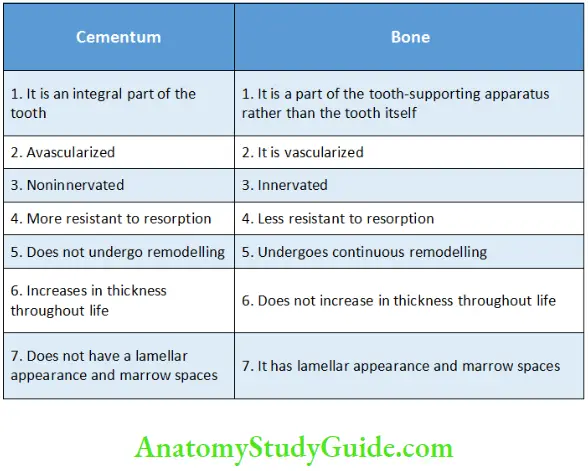
Cementum has more fluoride content than other mineralized tissues in the body. The chemical constituents of cementum by weight and volume are described in Table
Chemical Constituents of Cementum by Volume and Weight

Inorganic And Organic Components Of Cementum
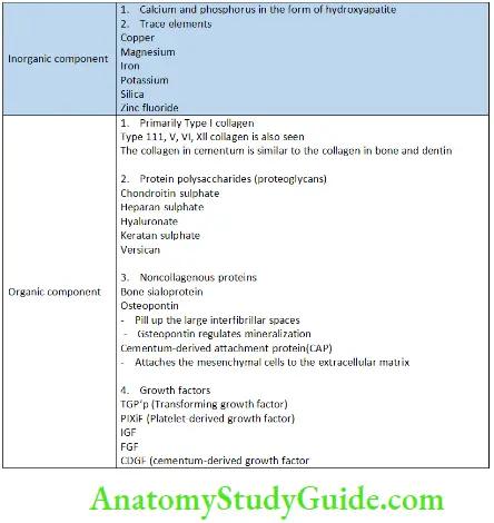
Inorganic and Organic Constituents of Cementum
Cementogenesis
The process of formation of cementum is called cementogenesis. It is
derived from the dental follicle. It begins at the cervical margin and extends downwards with the growth of the root.
The dental sac comprises three layers during the early bell stage.
- Inner layer – cellular and vascular – forms the cementum.
- Intermediate layer – It forms the PDL.
- Outermost layer – It forms the alveolar bone.
Cementogenesis can be discussed in the following steps:
- Formation of the hyaline layer
- Differentiation of cementoblasts and formation of precementum
- Formation of intrinsic and extrinsic fibres
- Mineralization
1. Formation of the hyaline layer:
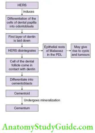
- The formation of cementum begins after the formation of enamel is complete. The future root outline is being laid down by the HERS (Hertwig’s epithelial root sheath).
- The root sheath induces the differentiation of the cells of the dental papilla into odontoblasts which lay down predentin, the first formed dentin comprising of organic substance and collagen fibrils. As the odontoblastic processes are not seen in this thin layer of tissue it appears structure less and is called the hyaline layer.
2. Differentiation of cementoblasts and formation of precementum
- The HERS breaksdown and cells of the inner layer of the dental follicle come in contact with the newly formed dentin. These cells differentiate into the cementoblasts and form cementum.
- There is loss of the basal lamina on the cemental side due to the degeneration of the HERS. It is followed by the appearance of cementoblasts and collagen fibrils between the epithelial cells and the root sheath.
- Some cells of the root sheath
– Move towards the developing PDL and remain as the cells rests of Malassez.
– Some are incorporated into the cementum.
3. Formation of intrinsic and extrinsic fibres
- The cells of the dental sac differentiate into cementoblasts. These cells lay down the organic matrix of collagen comprising collagen and protein polysaccharides.
- The collagen fibrils at the superficial surface produced by the
cementoblasts form a fibrous fringe perpendicular to the periodontal
space. The cementoblasts then retract and intermingle with the
fibroblasts of the PDL. - The Type I collagen fibres secreted by the cementoblasts are called the intrinsic fibres. They are parallel to the dentin and loosely arranged.
- The collagen bundles of the developing PDL that get inserted obliquely into the cementum are called the extrinsic fibres.
4. Mineralization
- The mineralization of the first formed dentin begins within the matrix vesicles a few microns deep to the outermost surface of the hyaline layer and spreads inwards towards the pulp and outwards towards the PDL. (Matrix vesicles are membrane-bounded organelle which contain a variety of enzymes and are rich in phosphates and initiate mineralization.) There is delayed mineralization in the outermost part of the hyaline layer.
- The delayed mineralization in the hyaline layer spreads outwards until it is fully mineralized and spreads into the fibrous fringe secreted by the cementoblast. The first few microns of acellular cementum are now firmly attached to the root dentin.
Acellular extrinsic fibre cementum and acellular intrinsic fibre cementum
- In later stages, the principal fibres of the PDL become continuous with the fibrous fringe of the cementum. This type of cementum is called acellular extrinsic fibre cementum. It occurs in permanent teeth after two-third development of the root.
- The cementum lining the tooth before this is called acellular intrinsic fibre cementum.
Incremental lines of Salter
- Incremental lines are formed due to alternate periods of rest and periods of activity in cementogenesis and are associated with decrease in activity or rest. These are called Incremental lines ofSalter.
Intermediate cementum
- The epithelial rests may be trapped in the cementum near the cementodentinal junction (CDJ). This is called intermediate cementum.
- It is seen in the apical half of molars.
Formation Of Acellular Afibrillar Cementum
- Acellular afibrillar cementum is deposited at the cervical margin as a thin layer.
- Due to the early loss of the reduced enamel epithelium covering the newly formed cervical enamel, the connective tissue cells of the adjacent dental follicle make contact with enamel and differentiate into cementoblasts. These secrete a matrix devoid of collagen fibres but containing noncollagenous proteins, which later undergoes calcification
Formation Of Cellular Fibrillar Cementum
- After the formation of acellular cementum, the cellular cementum is formed
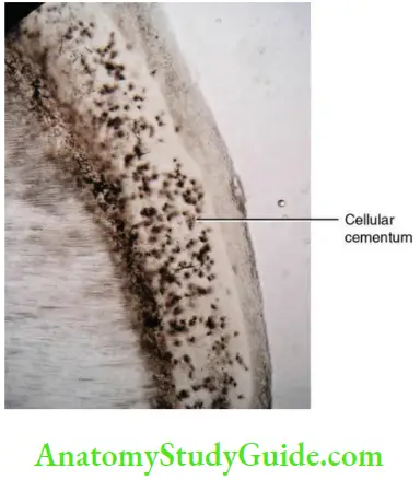
- The cementoblasts secrete the collagen fibrils and organic matrix that form the intrinsic fibres. The fibres are parallel to the root. Some cementoblasts are entrapped in lacunae in their own matrix. These entrapped cells are called cementocytes and this type of cementum is referred to as cellular cementum.
- Due to the increase in distance from the surface and decrease in the diffusion of the nutrients, the cementocytes in the deeper layer are not viable. The incremental lines are not closely placed due to the rapid rate of formation.
- Mineralization:
- The mineralization of the cementum matrix is initiated by the
hydroxyapatite crystals in the adjacent dentin. - Mineralization progresses slowly in a linear manner but is less than in acellular cementum.
- The mineralization of the cementum matrix is initiated by the
- The cellular intrinsic fibre cementum provides attachment to the extrinsic fibres and this is called as cellular mixed fibre cementum.
- The cellular intrinsic fibre cementum alternates with acellular extrinsic fibre cementum to form the cellular mixed stratified cementum.
Cementoid
Cementum grows in a rhythmic manner. As the old layer of cementum calcifies a new thin layer of cementoid is formed. A mineralization front is always present on the cemental surface. Cementoblasts line the cementoid tissue. The connective tissue fibres of the PDL pass between the cementoblasts and are embedded into the cementum are called Sharpey’s fibres. They attach the tooth to the bone. Cementoid is absent in acellular extrinsic fibre cementum.
Types Of Cementum On The Basis Of The Presence Or Absence Of Cells: Acellular And Cellular Cementum
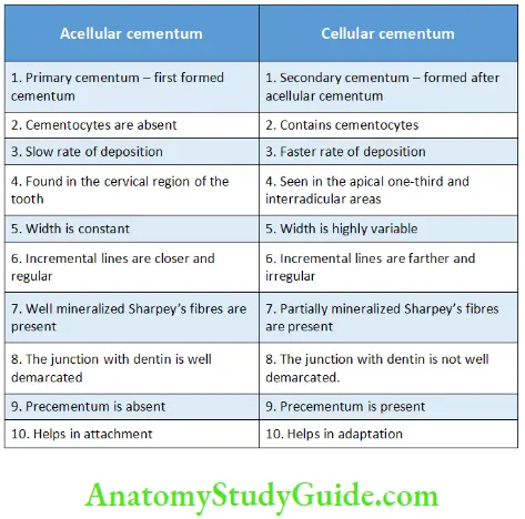
As the name suggests, cellular cementum contains entrapped cementocytes and the acellular cementum does not. The differences are mentioned in the table
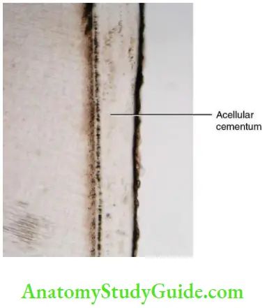
Differences between Acellular and Cellular Cementum
Types Of Cementum On The Basis Of The Origin And Nature Of Organic Matrix
The organic matrix of cementum is derived from two sources.
- Extrinsic fibre cementum
- When the fibres are derived from the Sharpey’s fibres of the PDL.
- The fibres run in the direction as the principal fibres
- Intrinsic fibre cementum
- When the fibres are derived from the cementoblasts.
- The fibres are parallel to the root and perpendicular to the extrinsic fibres
Type Of Cementum On The Basis Of The Presence And Absence Of Cells And The Origin And Nature Of The Organic Matrix
- Acellular extrinsic fibre cementum
- Cellular intrinsic fibre cementum
- Cellular mixed fibre cementum
- Cellular mixed stratified cementum.
Cementum can be classified based on various criteria as mentioned in Flowchart.
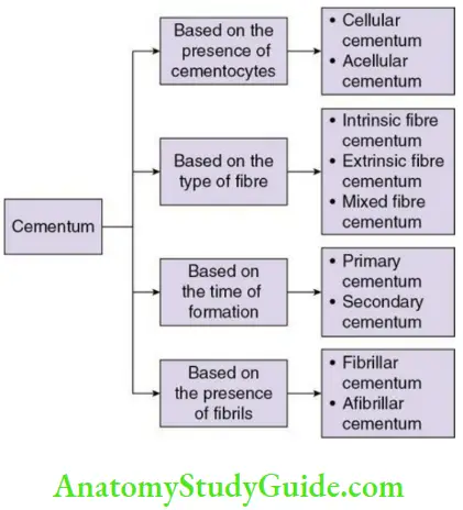
Classification of cementum:
Acellular extrinsic fibre cementum:
- Location: cervical margin to the apical one-third
- Teeth: single-rooted tooth
- Origin of fibres: Collagen fibres are derived from the Sharpey’s fibres and thus extrinsic
- Orientation: The extrinsic fibres are perpendicular to the cementum and known as Sharpey’s fibres.
- Rate of formation: Slow
- Root surface: Smooth
- Incremental lines: Parallel to the surface and closer
- Cementoid: Absent
- Function: Anchorage
Cellular cementum:
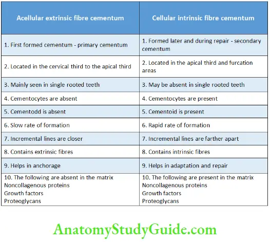
Cellular cementum is also called secondary cementum and formed after the formation of AEFC. Differences between acellular extrinsic fibre cementum and cellular intrinsic fibre cementum are given in Table
1. Cellular intrinsic fibre cementum
- It contains cells (cellular) and intrinsic fibres.
- It is seen in the apical third and the furcation areas of the tooth.
- It has no role in attachment.
- It plays a role in repair.
2. Cellular mixed fibre cementum
- The collagen fibres are derived from the cementoblasts (intrinsic) and
the PDL (extrinsic). - The intrinsic and extrinsic fibres run in between each other at different orientations and sometimes perpendicular to each other.
- The number of intrinsic fibres are less than the extrinsic fibres. The
intrinsic fibres are smaller (1–2 microns) in diameter than the extrinsic (5–7 microns); the bundles of extrinsic fibres are ovoid. - The fibres are less well mineralized if the rate of formation is faster.
3. Cellular mixed stratified cementum
- When the acellular extrinsic fibre cementum and the cellular intrinsic
fibre cementum are present as alternate layers the cementum is called cellular mixed stratified cementum. - Location: apex of the root and furcation areas.
- Thickness: 100–1000 microns.
Differences Between Acellular Extrinsic Fibre Cementum And Cellular Intrinsic Fibre Cementum
Cementodentinal junction
The surface of dentin upon which cementum is deposited is smooth in permanent teeth. In deciduous teeth, the CDJ is scalloped. The attachment of cementum to dentin in both deciduous and permanent teeth is firm. The CDJ appears more distinct under the light microscope than under the electron microscope.
In decalcified sections, the collagen fibres of cementum are in distinct bundles compared to the haphazard arrangement of collagen fibres of dentin. The CDJ contains huge amount of collagen and ground substance such as chondroitin sulphate and dermatan sulphate, which absorb water and make it stiff. This helps in redistributing the occlusal load to the alveolar bone.
A zone that does not exhibit features of either dentin or cementum may be present in between dentin and cementum. This zone is called the intermediate cementum layer. This zone is structure less zone and appears hyaline, thus called the hyaline layer. This is more common in the apical two-thirds of the premolars and molars.
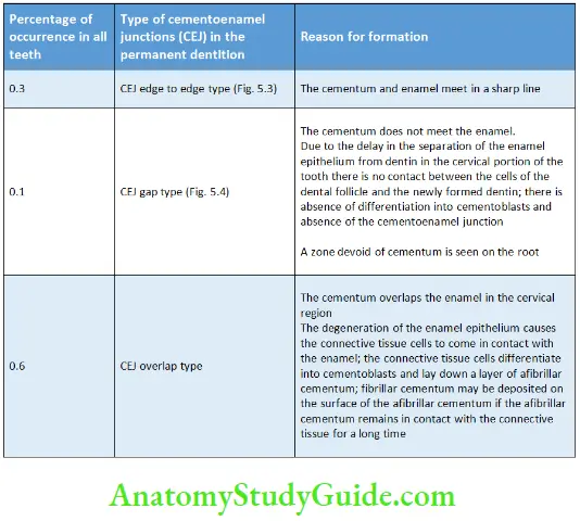
Cementoenamel Junction
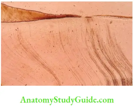
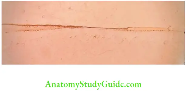
There are three types of junctions between the cementum and the enamel in the cervical portion of the tooth. The types of Cementoenamel junctions are described in Table
Recent advance:
A new pattern of CEJ has been seen where the enamel overlaps the cementum.
Types of CEJ in the deciduous dentition
- CEJ edge to edge – most common
- CEJ overlap – second most common
- CEJ gap and enamel overlapping the cementum – rare
Types of Cementoenamel Junction
Functions Of Cementum
1. Cementum Anchorage and attachment
- Primary function of cementum.
- The fibres of the PDL are attached into the cementum and thus the tooth is attached to the alveolar bone.
Clinical consideration: Hypophosphatasia – genetic disorder characterized by the absence of cementum leads to premature exfoliation of anterior deciduous teeth.
2. Cementum Adaptation to function
- Cementum helps in functional adaptation of the teeth.
- Cementum does not resorb under normal conditions. The loss of tooth structure from the occlusal surface due to mastication is compensated by the deposition of cementum on the root. This deposition of cementum helps in maintaining occlusion.
3.Cementum Repair
- Cementum is newly formed to help repair the root surface in cases of fracture and resorption. The newly formed cementum is rapidly laid down. It is similar to cellular cementum with smaller apatite crystals and a wide cementoid zone.
Functions Of Cementum
- Anchorage and attachment
- Adaptation to function
- Repair
Hypercementosis
Hypercementosis is the abnormal thickening of cementum due to excess deposition on the root. The types of hypercementosis are mentioned in
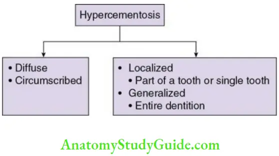
Few of the causes of hypercementosis are mentioned
- Localized hypertrophy
- Seen in teeth that are under stress.
- A spur or small extension of cementum is formed that increases the surface area for the attachment of fibres. This leads to better and firm anchorage of the tooth in the alveolar bone.
- Localized hypercementosis
- It involves a single tooth.
- The enamel drops formed on dentin are covered by hyperplastic cementum which is irregular and may contain calcified epithelial rests.
- Similar calcified bodies are seen in localized areas of hyperplastic
cementum. These knob-like projections are called excementoses and are
seen around degenerated epithelial rests.
- Excess formation of cementum is seen around the tooth in areas of periapical inflammation.
- Teeth that are not in function also exhibit localized thickening of cementum with decrease in the number of Sharpey’s fibres
- Cementum is thick in embedded and newly erupted teeth, at the apex of all teeth, in the furcation of multirooted teeth.
- Hyperplasia of cementum increases with age
- Generalized hypercementosis is seen in Paget disease
- Localized hypercementosis is seen in benign cementoblastoma, acromegaly, calcinosis
Causes Of Hypercementosis
Causes of hypercementosis:
- Periapical inflammation (inflammation at the apex of a tooth)
- Teeth under stress
- Supraeruption of a tooth
- Injury to the cementum
- Teeth that are not in function
- Systemic factors such as Paget disease, hyperpituitarism, benign cementoblastoma, acromegaly, calcinosis
Hypercementosis Clinical significance:
- The hypercementosed tooth is firmly attached to the bone. Extraction of such teeth might cause fracture of the jaw. Thus, radiographic examination needs to be done. In most of the cases, extraction of the hypercementosed teeth is done by the surgical method which requires removal of bone. Fragments of the tooth that remain after the extraction can be left behind in the jaw. These would get covered by cementum and cause no disturbance
Clinical Considerations of Cementum
- Cementum in orthodontic treatment:
- Cementum is more resistant to resorption than bone.
- The bone is resorbed on the pressure side and formed on the tension side during orthodontic treatment. The resorption of cementum is minimum or absent even though the pressure applied on the bone and the cementum is equal on the pressure side. This is due to the avascular nature of cementum.
- Anatomic repair:
- Trauma or excess occlusal forces can cause resorption of cementum and sometimes dentin. This damage might be repaired by the formation of acellular or cellular cementum and the former outline of the root may be established. This is referred to as anatomic repair.
- Functional repair
- In cases of deep resorption, a thin layer of cementum may be laid down and the outline of the root is not formed again. The defect may be filled by bone and the PDL space is maintained and the function is restored. This is called functional repair.
- Severe trauma
- It can cause loss of fragments of cementum from the dentin.
- Root fractures (transverse) – heal by new cementum formation.
- Periodontal pockets and plaque
- The physical, chemical and structural properties of the surface of cementum are altered due to the by-products of the microorganisms present in plaque. The surface of the cementum is hypermineralized due to the incorporation of calcium, phosphorus and fluoride from the oral cavity.
- Healing after periodontal therapy depends on the surface of the cementum. Surface alteration of exposed cementum might delay healing. The altered cementum must be removed during periodontal surgical procedures to fasten the regeneration of periodontium.
Products that promote formation of new cementum and periodontal regeneration such as enamel matrix-related proteins may be applied onto the cleaned and planed root surface. The enamel matrix proteins help in differentiation of the cementoblasts that produce cementum. The newly formed cementum is of the cellular type.
- Cementum and systemic conditions:
- Generalized hypercementosis: It is seen in Paget disease.
- Complete absence of cementum is seen in Hypophosphatasia
- Thickening of cementum is seen in Type 2 diabetes
- Ankylosis:
- Ankylosis is the fusion of cementum and bone.
- Cemental caries
- Cemental caries occur in older individuals where gingival recession leads to exposure of cementum.
- Cementum and age estimation
- The incremental lines in cementum help in the estimation of age of an individual.
Cementum Synopsis
- Cementum is a mineralized dental hard connective tissue that covers the anatomic roots of the human teeth. The fibres of the PDL are inserted into the cementum which is one among the four tissues that form the supporting apparatus of the tooth (PDL, alveolar bone and gingivae being the rest.
- Cementum begins at the CEJ in the cervical portion of the tooth and continues till the apex of the tooth.
- Cementum contains 45%–50% inorganic and 50%–55% organic constituents.
- The formation of cementum is called cementogenesis. It is derived from the dental follicle. It begins at the cervical margin and extends downwards with the growth of the root
- Cementogenesis can be discussed in the following steps
-
- Formation of the hyaline layer
- Differentiation of cementoblasts and formation of precementum
- Formation of Intrinsic and extrinsic fibres
- Mineralization
- On the basis of the presence and absence of cells and the origin and nature of the organic matrix
-
- Acellular extrinsic fibre cementum
- Cellular intrinsic fibre cementum
- Cellular mixed fibre cementum
- Cellular mixed stratified cementum
- Cellular cementum is also called secondary cementum and formed after the formation of AEFC.
- In cellular mixed fibre cementum, the collagen fibres are derived from the cementoblasts (intrinsic) and the PDL (extrinsic).
- When the acellular extrinsic fibre cementum and the cellular intrinsic fibre cementum are present as alternate layers, the cementum is called cellular mixed stratified cementum.
- There are three types of CEJs: CEJ edge to edge type, CEJ gap type and CEJ overlap type. A new pattern of CEJ has been seen where the enamel overlaps the cementum.
- The functions of cementum are anchorage and attachment, adaptation to function and repair.
- The causes of hypercementosis are periapical inflammation, teeth under stress, supraeruption of a tooth, injury to the cementum, teeth that are not in function, systemic factors such as Paget disease, hyperpituitarism, benign cementoblastoma, acromegaly, calcinosis.
- Cementum is more resistant to resorption than bone.
- Trauma or excess occlusal forces can cause resorption of cementum and sometimes dentin.
- Severe trauma can cause loss of fragments of cementum from the dentin.
- Generalized hypercementosis is seen in Paget disease.
- Ankylosis is the fusion of cementum and bone.
- The incremental lines in cementum help in the estimation of age of an individual.
Leave a Reply