Oral Histology Shedding Of Teeth Notes
Shedding Of Teeth
The human dentition usually has two different generations. The first being the primary dentition and the other being the permanent dentition. The first set of teeth that erupt in the oral cavity are called the primary or the deciduous dentition, whereas the second set of teeth that erupt following the primary dentition are called the secondary dentition or the permanent dentition.
Table of Contents
Read And Learn More: Oral Histology Notes
Significance Of Shedding
- The deciduous teeth cannot withstand jaw growth from childhood to adult, thus another set of larger teeth is needed for larger jaws. In order to accommodate the larger-sized permanent dentition, the oral cavity follows a physiologic process resulting in the elimination of the deciduous dentition. This process of elimination of deciduous teeth is called shedding or exfoliation.
- Secondly, the growth of masticatory muscles leads to increased forces of mastication which the deciduous teeth cannot withstand. It also leads to trauma to their periodontal ligament (pdl) necessitating another set of larger teeth.
Pattern Of Shedding
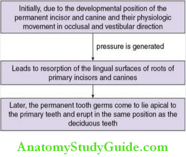
The basic concept behind the shedding of deciduous teeth is the progressive resorption of their roots and other tooth-supporting apparatus, especially the periodontal ligament.
The resorption of the dental hard tissues is carried out by multinucleated cells similar to osteoclasts. The pattern of shedding of deciduous teeth depends on the pressure generated by the erupting permanent teeth.
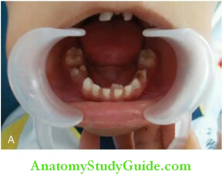
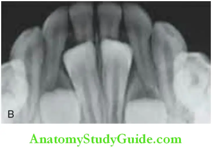
Schematic Representation Of Pattern Of Shedding For Deciduous Maxillary Incisors And Canine:
Pattern Of Shedding Of Deciduous Maxillary Incisors And Canines:
Note: The tooth germs of the permanent mandibular incisors do not occupy an apical position, instead erupt lingual to the still functional primary tooth.
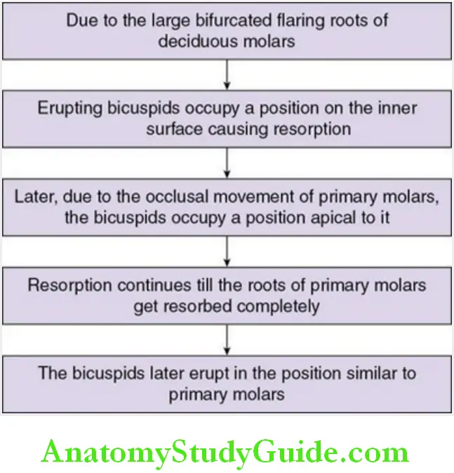
For Deciduous Molars:
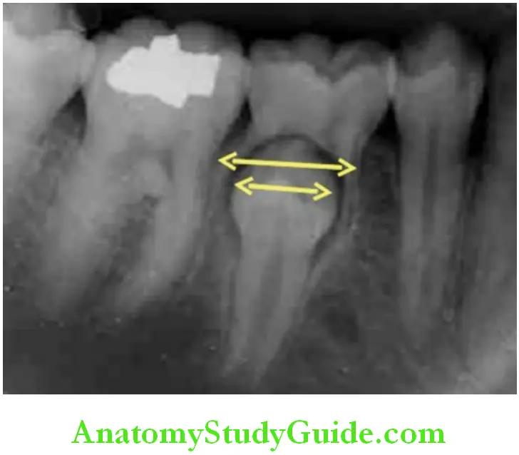
Pattern Of Shedding Of Deciduous Molars:
Histology Of Shedding
The shedding of deciduous teeth occurs mainly due to the pressure generated by the underlying permanent tooth germs which causes resorption of the roots. The resorption takes place due to the recruitment of multinucleated cells similar to an osteoclast called the odontoclast.
Odontoclasts:
Origin: Derived from monocytes
Histology:
- Large, multinucleated cells.
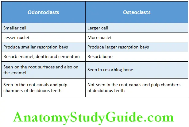
- They are smaller in size than the osteoclasts and contain lesser nuclei. The differences between osteoclasts and odontoclasts are given in.
- The cytoplasm appears vacuolated and forms a ruffled border with the surface of the dental hard tissue where resorption takes place. The ruffled border is the extensive folding of the cell membrane into a series of 2–3 micron deep invaginations. The invaginations contain mineral crystallites.
- A clear zone with the cytoplasm devoid of organelle is seen peripheral to the ruffled border. This zone is rich in actin and myosin filaments and is the attachment apparatus of the odontoclast.
- The cytoplasm adjacent to the ruffled border contains numerous mitochondria and vacuoles.
- The vacuoles are rich in acid phosphatase.
- Multinucleated giant cells are formed due to the fusion of the odontoclasts.
Differences Between Odontoclasts And Osteoclasts:
Location: They are present in the resorption bays of the Dental Hard Tissue.
Distribution:
- They are found on the root surface in relation to the erupting permanent tooth.
- Also seen in the root canals and pulp chambers of the resorbing deciduous teeth
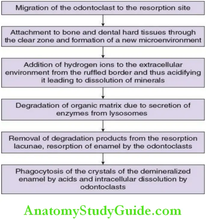
Odontoclasts may even resorb the enamel in some cases in addition to the dental hard tissues. The dentinal tubules provide a passage for the extension of the odontoclast processes. The odontoclastic activity during shedding is described in the flowchart. The key points of odontoblasts are given in the box.
Odontoclast:
Odontoclast is a multinucleated cell that resorbs dentin and cementum
Origin: Fusion of monocytes
Site: Site of root resorption in a depression termed Howship’s lacunae
Functions:
- Shedding of deciduous tooth
- Resorption of remaining root
Resorption And Shedding
Pressure from the erupting succedaneous tooth plays an important role in initiating shedding as the odontoclasts appear at the site of pressure. Resorption involves cellular activities by the odontoclasts which occur as follows:
Apart from the pressure from the successional tooth, the other factor involved in initiating root resorption is the forces of mastication. The growth of the facial muscles exerts forces on the pdl of the deciduous teeth greater than it can withstand thus leading to trauma to the pdl. At this point, the resorption stops, and repair occurs. The rate of eruption is determined by resorption and repair.
The resorption of the periodontal ligament occurs due to apoptotic cell death.
Clinical Considerations
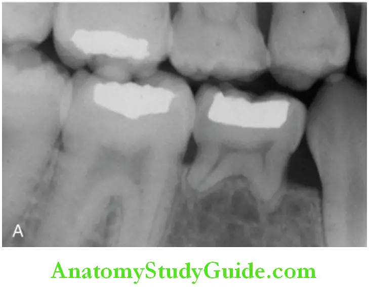
Submerged Deciduous Teeth:
- Ankylosis is the bony union between the alveolar bone and roots of the teeth.
- Mild trauma may result in damage to the periodontal ligament leading to ankylosis of a deciduous tooth rather than its loss. The ankylosed tooth fails to erupt.
- Due to the continued eruption of the neighboring teeth and increased height of the alveolar process, the ankylosed tooth may be either ‘shortened’ or submerged in the alveolar process (below the plane of occlusion by 1 mm or more).
- Submerged deciduous teeth may prevent the eruption of their permanent successors or force them away from their position and therefore should be removed at the earliest.
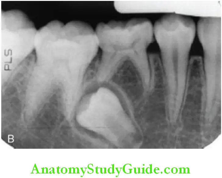
Retained Deciduous Teeth:
- Deciduous teeth may be retained beyond their scheduled shedding period.
- Retention may be due to local or systemic causes.
- Congenital absence and impaction of the permanent successors are the common causes for this retention.
- The most commonly retained teeth in the oral cavity are the upper lateral incisors, the second deciduous molar, and the lower central incisors in decreasing order of occurrence.
- Fate of retained deciduous teeth:
- Retained deciduous teeth may remain for many years in a good functioning condition. However, resorption of the roots and continued active and passive eruption cause their loosening and final loss in most cases.
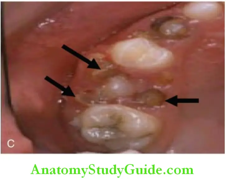
Remnants Of Deciduous Teeth:
- Sometimes, segments of the roots of deciduous teeth that are not in the path of the erupting successors may escape resorption.
- These fragments are most frequently found in the region of the premolars, especially the lower second premolar region. This happens as the mesiodistal diameter of the second premolar is lesser than the maximum distance between the roots of the deciduous molar
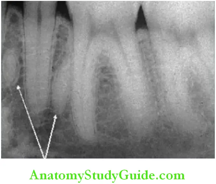
Fate Of Root Remnants:
- Root Remnants May Remain Embedded In The Bone Completely Surrounded By And Ankylosed.
- Frequently They Are Cased In Heavy Layers Of Cellular Cementum.
- When They Are Close To The Surface Of The Jaw, They May Ultimately Be Exfoliated.
- Progressive Resorption Of The Root Remnants And Replacement By Bone May Cause Their Disappearance.
Shedding Of Teeth Synopsis
- Shedding is a physiological process of elimination of deciduous teeth and is carried out by multinucleated giant cells called odontoclasts.
- Mainly occurs due to the pressure exerted by the underlying permanent tooth germ.
- Resorption of anterior teeth usually occurs on the lingual surface, whereas the posteriors are usually resorbed in the area between the roots.
- Resorption is not continuous, during repair the cementoblasts deposit cementum-like tissue, whereas the enamel and dentin continue to get resorbed.
- The pdf undergoes apoptotic cell death.
- Odontoclasts are similar to osteoclasts except few cytological differences like an increased number of vacuoles and mitochondria near the ruffled border.
- Both originate from the bloodstream monocyte. Rankl (receptor activator of nuclear factor kappa β ligand) and opg (osteoprotegerin) expression is required for activation and formation of odontoclasts.
- A few parts of teeth that do not lie in the eruption pathway may escape resorption and remain retained as a part of the entire deciduous tooth.
- The union between the tooth and the bone may not allow the deciduous teeth to erupt and thus it may remain submerged or ankylosed without undergoing shedding.
Leave a Reply