Origin Of Thrombi At Different Sites
Thrombi may arise from the heart, arteries, veins or in microcirculation by different mechanisms.
Cardiac Thrombi:
Thrombi may form in any of the chambers of the heart and on the valve cusps. They are more common in the atrial appendages, especially of the right atrium, and on mitral and aortic valves such as vegetations seen in infective endocarditis and non-bacterial thrombotic endocarditis.
Read And Learn More Homeostasis
Cardiac thrombi are mural (non-occlusive) as are the mural thrombi encountered in large vessels such as the aorta in atherosclerosis and in aneurysmal dilatations.
Rarely, large round thrombus may form and obstruct the mitral valve and is called ball-valve thrombus. Agonal thrombi are formed shortly before death and may occur in either or both the ventricles. They are composed mainly of fibrin.
Arterial Thrombi:
The examples of major forms of thrombi formed in the arteries are as under:
- Aorta: Aneurysms, Arteritis.
- Coronary arteries: Atherosclerosis.
- Mesenteric artery: Atherosclerosis, arteritis.
- Arteries of limbs: Atherosclerosis, Diabetes mellitus, Buerger’s disease, Raynaud’s disease.
- Renal artery: Atherosclerosis, Arteritis.
- Cerebral artery: Atherosclerosis, Vasculitis.
Venous Thrombi:
A few common examples of these are as under:
- Veins of lower limbs: deep veins of legs, varicose veins.
- Popliteal, femoral and iliac veins: Postoperative stage, postpartum.
- Pulmonary veins: CHF, Pulmonary hypertension.
- Hepatic and portal vein: Portal hypertension.
- Superior vena cava: Infections in head and neck.
- Inferior vena cava: Extension of thrombus from hepatic vein.
- Mesenteric veins: Volvulus, intestinal obstruction.
- Renal vein: Renal amyloidosis.
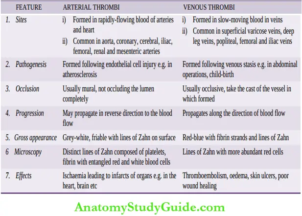
Distinguishing features between thrombi formed in rapidly-flowing arterial circulation and slow-moving venous blood are given in Table.
Capillary Thrombi:
Minute thrombi composed mainly of packed red cells are formed in the capillaries in acute inflammatory lesions, vasculitis and in disseminated intravascular coagulation (DIC).
Morphologic Features:
The general morphologic features of thrombi formed in various locations are as under:
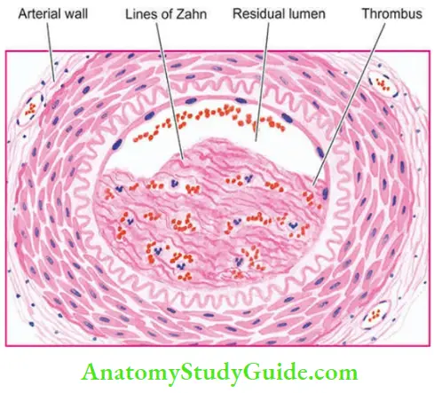
- Grossly: Thrombi may be of various shapes, sizes and composition depending upon the site of origin. Arterial thrombi tend to be white and mural while the venous thrombi are red and occlusive. Mixed or laminated thrombi are also common and consist of alternate white and red layers called lines of Zahn. Red thrombi are soft, red and gelatinous whereas white thrombi are firm and pale.
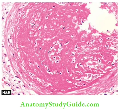
- Microscopically: The composition of a thrombus is determined by the rate of flow of blood i.e. whether it is formed in the rapid arterial and cardiac circulation, or in the slow-moving flow in veins. The lines of Zahn are formed by alternate layers of light-staining aggregated platelets admixed with fibrin meshwork and a dark-staining layer of red cells.
Red (venous) thrombi have more abundant red cells, leucocytes and platelets entrapped in fibrin meshwork. Thus, red thrombi closely resemble blood clots.
Distinguishing features of arterial and venous thrombi:
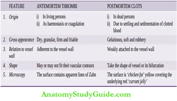
Red thrombi (antemortem) have to be distinguished from postmortem clots.
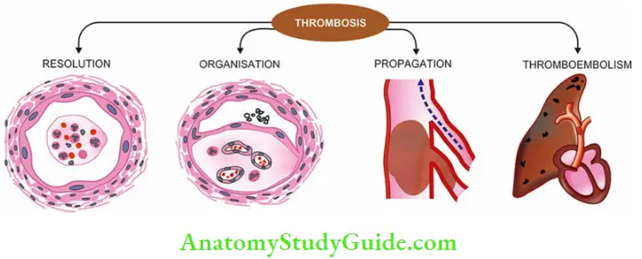
Fate of Thrombus:
The outcome of thrombi can be as under :
- Resolution:
- Thrombus activates the fibrinolytic system with consequent release of plasmin which may dissolve the thrombus completely resulting in resolution. Usually, lysis is complete in small venous thrombi while large thrombi may not be dissolved.
- Fibrinolytic activity can be accentuated by administration of thrombolytic substances (for example Urokinase, streptokinase), especially in the early stage when fibrin is in monomeric form for example Thrombolytic therapy in early-stage acute myocardial infarction.
- Organisation: If the thrombus is not removed, it starts getting organised. Phagocytic cells (neutrophils and macrophages) appear and begin to phagocytose fibrin and cell debris. The proteolytic enzymes liberated by leucocytes and endothelial cells start digesting the coagulum.
- Capillaries grow into the thrombus from the site of its attachment and fibroblasts start invading the thrombus.
- Thus, fibrovascular granulation tissue is formed which subsequently becomes dense and less vascular and is covered over by endothelial cells.
- The thrombus in this way is excluded from the vascular lumen and becomes part of vessel wall.
- The new vascular channels in it may be able to re-establish the blood flow, called recanalisation.
- The fibrosed thrombus may undergo hyalinisation and calcification for example, Phleboliths in the pelvic veins.
- Propagation: The thrombus may enlarge in size due to more and more deposition from the constituents of flowing blood. In this way, it may ultimately cause obstruction of some important vessel.
- Thromboembolism: The thrombi in early stage and infected thrombi are quite friable and may get detached from the vessel wall. These are released in part or completely in the bloodstream as emboli which produce ill effects at the site of their lodgement
Clinical Effects:
Besides differences in mechanism of thrombosis at different sites, clinical effects depend upon not only the site but also on rapidity of formation and nature of thrombi.
- Cardiac thrombi: Large thrombi in the heart may cause sudden death by mechanical obstruction of blood flow or through thromboembolism to vital organs.
- Arterial thrombi: These cause ischaemic necrosis of the deprived part (infarct) which may lead to gangrene. Sudden death may occur following thrombosis of the coronary artery.
- Venous thrombi (Phlebothrombosis): These may cause the following effects:
- Thromboembolism
- Oedema of area drained
- Poor wound healing
- Skin ulcer
- Painful thrombosed veins (thrombophlebitis)
- Painful white leg (phlegmasia alba dolens) due to ileofemoral venous thrombosis in postpartum cases
- Thrombophlebitis migrans in cancer.
Distinguishing features of antemortem thrombi and postmortem clots:
- Capillary thrombi: Microthrombi in microcirculation may give rise to disseminated intravascular coagulation (DIC).
Thrombosis:
Thrombosis is the process of the formation of solid mass in circulation from the constituents of flowing blood; the mass itself is called a thrombus.
Thrombogenesis involves interplay of 5 events:
- Endothelial injury
- Platelets and their release reaction
- coagulation system
- Alterations in the flow of blood, and
- Role of certain predisposing conditions and factors causing hypercoagulable states (or thrombophilia).
Thrombi may originate in the chambers of the heart, lumina of arteries, veins and microcirculation. The effects of thrombi depend upon their anatomic location, rapidity of formation and nature of thrombi. In general, thrombi produce life-threatening harmful effects by ischaemia and by thromboembolism.
- Grossly: Thrombi are of various shapes, size, consistency and colour.
- Microscopically: All types of thrombi show lines of Zahn formed by alternate layers of light-staining aggregated platelets and dark-staining red cells.
The possible fates of thrombi are resolution, organisation, propagation and thromboembolism.
Leave a Reply