Visual Process Introduction
- The visual process is the series of actions that take place during visual perception. During the visual process, the image of an object seen by the eyes is focused on the retina resulting in the production of visual perception of that object.
- When the image of the object in the environment is focused on the retina, the energy in the visual spectrum is converted into electrical potentials (impulses) by rods and cones of the retina through some chemical reactions.
Read And Learn More: Medical Physiology Notes
Table of Contents
- The impulses from rods and cones reach the cerebral cortex through the optic nerve. And, the sensation of vision is produced in the cerebral cortex. Thus, the process of visual sensation is explained on the basis of image formation, and neural, chemical, and electrical phenomena.
Image Forming Mechanism
- While looking at an object, the light rays from the object are refracted and brought to a focus upon the retina. The image falls on the retina in an inverted position and is reversed side to side. In spite of this, the object is seen in an upright position. It is because of the role played by the cerebral cortex.
- The light rays are refracted by the lens and cornea. The refractory power is measured in diopter (D) A diopter is the reciprocal of focal length expressed in meters.
- The focal length of the cornea is 24 mm and the refractory power is 42D. The focal length of the lens is 44 mm and the refractory power is 23D.
Neural Basis Of Visual Process
- The retina contains visual receptors which are also called light-sensitive receptors, photoreceptors or electromagnetic receptors. The visual receptors are rods and cones. There are about 6 million cones and 12 million rods in the human eye.
- The distribution of the photo-receptors varies in different areas of the retina. Fovea has only cones and no rods. While proceeding from the fovea towards the periphery of the retina, the rods increase and the cones decrease in number. At the periphery of the retina, only rods are present and cones are absent.
1. Structure Of Rod Cell: Rod cells are cylindrical structures with a length of about 40-60 μ and a diameter of about 2 p. Each rod is composed of four structures:
- Outer segment
- Inner segment
- Cell body
- Synaptic terminal
1. Outer Segment:
- The outer segment of the rod cell is long and slender. So it gives a rod-like appearance. It is in close contact with the pigmented epithelial cells. The outer segment of the rod cell is formed by the modified cilia and it contains a pile of freely floating flat membranous disks. There are about 1000 disks in each rod. The disks in rod cells are closed structures and contain the photosensitive pigment, rhodopsin.
- The rhodopsin is synthesized in inner segments and inserted into newly formed membranous disks at the inner portion of the outer segment. The new disks push the older disks toward the outer tip.
- The older disks are engulfed (by phagocytosis) from the tip of the outer segment by the cells of the pigment epithelial layer. Thus, the outer segment of the rod cell is constantly renewed by the formation of new disks. The rate of formation of new disks is 3 or 4 per hour.
2. Inner Segment: The inner segment is connected to the outer segment by means of modified cilium. The inner segment contains many types of organelles with a large number of mitochondria.
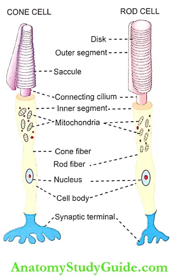
3. Ceil Body: A slender fiber called rod fiber arises from the inner segment of the rod cell and passes to the outer nuclear layer through an external limiting membrane. In the outer nuclear layer, the enlarged portion of this fiber forms the ceil Docfy or rod granule that contains the nucleus.
4. Synaptic Terminal: A thick fiber arising from the cell body passes to the outer plexiform layer and ends in a small and enlarged synaptic terminal or body. The synaptic terminal of the rods synapses with dendrites of bipolar cells and horizontal cells. The synaptic vesicles present in the synaptic terminal contain the neurotransmitter, glutamate.
2. Structure Of Cone Cell:
The cone cell is the visual receptor with a length of 35-40 p and a diameter of about 5 p. Generally, the cone cell is flask-shaped. The shape and length of the cone vary in different parts of the retina. The cones in the fovea are long, narrow, and almost similar to rods. Near the periphery of the retina, the cones are short and broad.
Like rods, cones are also formed by four parts:
- Outer segment
- Inner segment
- Cell body
- Synaptic terminal
1. Outer Segment:
- The outer segment is small and conical. It does not contain separate membranous disks as in rods. In cone, the infoldings of the cell membrane form the saccules, which are the counterparts of rod disks.
- The photopigment of the cone is synthesized in the inner segment and incorporated into the folding of the surface membrane forming the saccule. The renewal of the outer segment of the cone is a slow process and it differs from that in rods. It occurs at many sites of the outer segment of the cone.
2. Inner Segment: As in the case of rods, in cones also, the inner segment is connected to the outer segment by a modified cilium. Though various types of organelles are present in this segment, the number of mitochondria is more.
3. Cell Body: The cone fiber arising from the inner segment is thick and it enters the inner nuclear layer through the external limiting membrane. In the inner nuclear layer, the cone fiber forms the cell body or cone granule that possesses the nucleus.
4. Synaptic Terminal: The fiber from the cell body of the cone leaves the inner nuclear layer and enters the outer plexiform layer. Here, it ends in the form of an enlarged synaptic terminal or body. The synaptic vesicle present in the synaptic terminal of the cone cell also possesses the neurotransmitter, glutamate.
2. Functions Of Rods And Cones:
- Functions of Rods:
- Rods are extremely sensitive to light and have a low threshold. So, the rods are responsible for dim light vision or night vision or scotopic vision. But, rods do not take part in resolving the details and boundaries of objects (visual acuity) or the color of the objects (color vision).
- The vision by the rod is black, white or in a combination of black and white namely, gray. Therefore, the colored objects appear faded or grayish in twilight.
- Functions of Cones:
- Cones have a high threshold for light stimulus. So, the cones are sensitive only to bright light. Therefore, the cone cells are called receptors of bright light vision or photopic vision or daylight vision. The cones are also responsible for the acuity of vision and color vision.
- Achromatic Interval:
- When an object is placed in front of a person in a dark room, he cannot see the object. When there is slight illumination, the person can see the object but without color. It is because, at this level, only rods are stimulated.
- When the illumination is increased, the threshold for cones is reached. Now, the person can see the object in finer detail and in color. The interval between the threshold for rods and cones, i.e. the interval from when the object is first seen, and the time when the object is seen with color is called achromatic interval.
Chemical Basis Of Visual Process
Photosensitive pigments present in rods and cones are concerned with the chemical basis of the visual process. The chemical reactions involved in these pigments lead to the development of electrical activity in the retina and the generation of impulses (action potentials) which are transmitted through the optic nerve. The photochemical changes in the visual receptor cells are called Wald’s visual cycle.
1. Rhodopsin: Rhodopsin or visual purple is the photosensitive pigment of rod cells. It is present in the membranous disks tooted In the outer segment of rod cells.
- Chemistry of Rhodopsin:
- Rhodopsin is a conjugated protein with a molecular weight of 40,000. It is made up of a protein called opsin and a chromophore. The opsin present in rhodopsin is known as scotopsin. Chromophore is a chemical substance that develops color in the cell. The chromo¬phore present in the rod cells is called retinal. The retinal is the aldehyde of vitamin A or retinol.
- Retinal is derived from food sources and it is not synthesized in the body. It is derived from carotenoid substances like (3 carotene present in carrots.
- Retinal is present in the form of 11-cis retinal known as retinyl. Retininel is present in human eyes. It is different from creatinine 2 which is present in the eyes of some animals. The significance of the 11-c/s form of retinal is that only in this form, it combines with scotopsin to synthesize rhodopsin.
- Photochemical Changes In Rhodopsin-Wald’S Visual Cycle:
- When the retina is isolated and examined in the dark, the rods appear in red because of rhodopsin. During exposure to light, rhodopsin is bleached and the color becomes yellow.
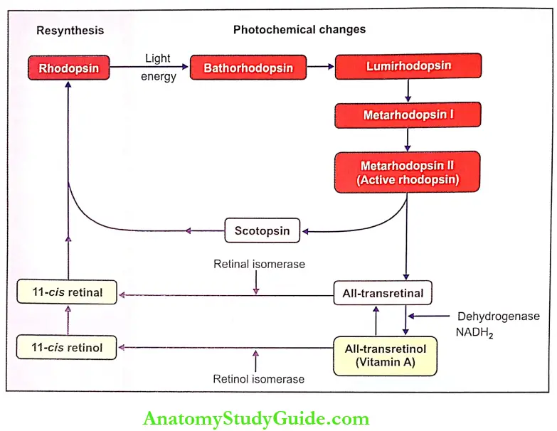
- When rhodopsin absorbs the light that falls on the retina, it is split into creatinine and the protein called opsin through us?’, various intermediate photochemical reactions. Following changes occur due to the absorption of light energy by rhodopsin:
- First, rhodopsin is decomposed into bathorhodopsin which is very unstable
- Bathorhodopsin is converted into lumirhodopsin
- Lumirhodopsin decays into metarhodopsin 1
- Metarhodopsin 1 is changed to metarhodopsin 2
- Metarhodopsin 2 is split into scotopsin and all- trans retinal
- The all-trans-retinal is converted into all-trans-retinol (vitamin A) by the enzyme dehydrogenase in the presence of reduced nicotinamide adenine dinucleotide (NADH2).
Metarhodopsin is usually called activated rhodopsin since it is responsible for the development of receptor potential in rod cells.
- Resynthesis of Rhodopsin:
- First, the all-trans-retinal derived from metarhodopsin II is converted into 11-c/s retinal by the enzyme retinal isomerase. 11-cis retinal immediately combines with scotopsin to form rhodopsin.
- The all-trans-retinol (vitamin A) also plays an important role in the resynthesis of rhodopsin. The all¬transretinol is converted into 11 -cis retinol by the activity of enzyme retinol isomerase. It is converted into 11-c/s retinal, which combines with scotopsin to form rhodopsin. The all-trans retinol is also reconverted into all-transactional.
- Rhodopsin can be synthesized directly from all-cis retinol (vitamin A) in the presence of nicotinamide adenine dinucleotide (NAD). However, the synthesis of rhodopsin from 11-c/s retinal (creatinine) is faster than from 11-cis retinol (vitamin A).
2. Phototransduction:
- Visual or phototransduction is the process by which light energy is converted into receptor potential in visual receptors.
The resting membrane potential in other sensory receptor cells is usually between -70 and -90 mV. However, in the visual receptors in the dark, the negativity is reduced and the resting membrane potential is about- 40 mV. - It is because of the influx of sodium ions. Normally in the dark, the sodium ions are pumped out of the inner segments of rod cells to ECF. However, these sodium ions leak back into the rod cells through the membrane of the outer segment and reduce the electro¬negativity inside the rod cell.
- Thus, the sodium influx maintains a decreased negative potential up to – 40 mV. This potential is constant and it is also called the dark current.
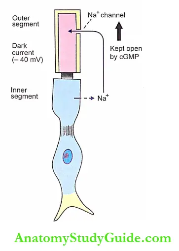
- The influx of sodium ions into the outer segment of the rod cell occurs mainly because of cyclic guanosine monophosphate (cGMP) present in the cytoplasm of the cell. The cGMP always keeps the sodium – channels open. The closure of sodium channels occurs due to the reduction in cGMP.
- The concentration of sodium ions inside the rod cell is regulated by the sodium-potassium pump. When light falls on the retina, the rhodopsin is excited leading to the development of receptor potential in the rod cells. Following is the phototransduction cascade of receptor potential:
- When a photon (the minimum quantum of light energy) is absorbed by rhodopsin, the 11-c/s retinal is decomposed into metarhodopsin through a few reactions mentioned earlier. Metarhodopsin 2 is considered as the active form of rhodopsin. It plays an important role in the development of receptor potential.
- Metarhodopsin 2 activates a G-protein called trans- during that is present in rod disks.
- The activated transducin activates the enzyme called cyclic guanosine monophosphate phospho¬diesterase (cGMP phosphodiesterase), which is also present in the rod disks.
- The activated cGMP phosphodiesterase hydrolyzes cGMP to 5’-GMP.
- Now, the concentration of cGMP is reduced in the rod cell.
- The reduction in the concentration of cGMP immediately causes the closure of sodium channels in the membrane of visual receptors.
- The sudden closure of sodium channels prevents the entry of sodium ions leading to hyperpolarization. The potential reaches – 70 to – 80 mV. It is because of the sodium-potassium pump.
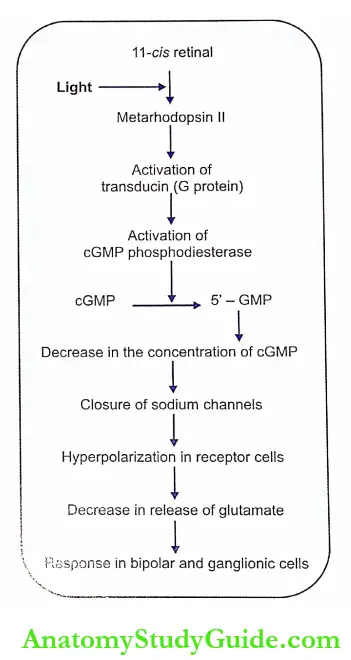
Thus, the process of receptor potential in visual receptors is unique in nature. When other sensory receptors are excited, the electrical response is in the form of depolarization (receptor potential). But, in visual receptors, the response is in the form of hyperpolarization.
- Significance of Hyperpolarization: Hyperpolarization in visual receptor cells reduces the release of synaptic transmitter glutamate. It leads to the development of response in bipolar cells and ganglionic cells so that, the action potentials are transmitted to the cerebral cortex via an optic pathway.
3. Photosensitive Pigments In Cones:
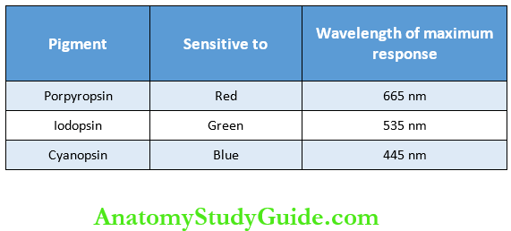
- The photosensitive pigment in the cone cells is of three types, namely porpyropsin, iodopsin and cyanopsin. Only one of these pigments is present in each cone. The photopigment in cone cells also is a conjugated protein made up of a protein and chromophore.
- The protein in cone pigment is called photopsin, which is different from scotopsin, the protein part of rhodopsin. However, the chromophore of cone pigment is the retinal that is present in rhodopsin.
- Each type of cone pigment is sensitive to a particular light and the maximum response is shown at a particular light and wavelength. The details are given in the.
- The various processes involved in phototransduction in cone cells are similar to those in rod cells.
4. Dark Adaptation:
Dark Adaption Definition: Dark adaption is the process by which the person is able to see objects in dim light. If a person enters a dimly lighted room (darkroom) from a bright-lighted area, he is blind for some time, i.e. he cannot see any object. After some time his eyes get adapted and he starts seeing the objects slowly. The maximum duration for dark adapt¬ation is about 20 minutes.
Causes for Dark Adaptation:
The dark adaptation is due to the following changes in the eyeball:
1. Increased sensitivity of rods as a result of the resynthesis of rhodopsin:
- The time required for dark adaptation is partly determined by the time to build up rhodopsin. In bright light, much of the pigment is being bleached (broken down).
- But in dim light, it requires some time for the regeneration of a certain amount of rhodopsin, which is necessary for optimal rod function. The dark adaptation occurs in cones also.
2. Dilatation of pupil:
- The dilatation of the pupil during dark adaptation allows more and more light to enter the eye. Radiologists, aircraft pilots, and others, who need maximal visual sensitivity in dim light, wear red glass before entering dimly lighted areas, because, the red of the light spectrum stimulates the rods slightly while the cones are allowed to function well.
- Thus, the person wearing red goggles can see well in bright light areas and also can see objects clearly, as soon as he enters the dim light area.
Dark Adaptation Curve: It is the curve that demonstrates the relationship between the threshold of light stimulus (illumination) and the time spent in the dark.
- Dark Adaptation Curve Procedure: The experiment to obtain the dark adaptation curve is done in a completely darkroom. First, the subject is exposed to a bright light in order to bleach (breakdown) most of the photopigment in the retina. The subject looks directly at a bright flashing light with a wavelength of 420 nm against a dark background for about 5-7 minutes.
- Then the bright light is switched off and the subject is in the dark. Now a small dim light (stimulus) is produced. Immediately, the absolute threshold (minimum strength of stimulus – the minimum intensity of light stimulus) for detecting this dim light is determined by adjusting the intensity of light (illumination).
- The time interval between the switching off bright light and the detection of dim light is noted. After a short time, the absolute threshold is measured again and elapsed time is noted. This procedure is repeated for about 30 minutes.
- When the experiment is completed, the results are plotted and the dark adaptation curve is obtained.
- The curve: The dark adaptation curve is biphasic. The first part of the curve represents the threshold of photographic vision which indicates the cone adaptation. The second part of the curve represents the threshold of scotopic vision which indicates the rod adaptation.
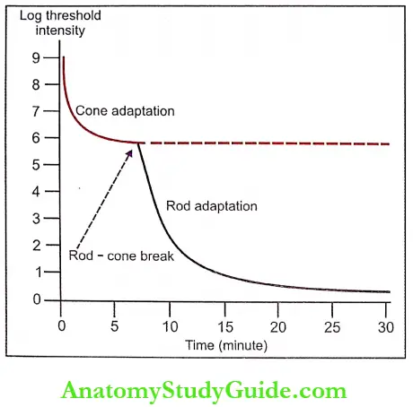
- Cone adaptation: This first phase is rapid and it is completed in 8-10 minutes. During this time the threshold decreases by 2-3 log units. That is the sensitivity of the eye in the darkroom increases by 1000 times within 8-10 minutes. By this time, the cones get adapted.
- Rod-cone break: After the first phase, there is a sudden change in the slope of the curve and this point of the curve is called rod-cone break. The rod-cone break represents the point where rod sensitivity begins to exceed cone sensitivity and the remaining part of the curve is determined by the continuing adaptation of rods.
- Rod adaptation: The second phase of the curve is slow. During this phase, there is a gradual decrease in the threshold and it is completed in 20-30 minutes. During this phase, the threshold decreases further by 5-6 log units. That is the sensitivity of the eye in the dark room increases by 1,00,000-10,00,000 times within 20-30 minutes. By this time, the rods get adapted completely.
5. Light Adaptation:
Light Adaptation Definition:
Light adaptation is the process in which eyes get adapted to increased illumination. When a person enters a bright lighted area from a dimly lighted area, he feels discomfort due to the dazzling effect of bright light. After some time, when the eyes become adapted to light, he sees the objects around him without any discomfort. It is the mere disappearance of dark adaptation. The maximum period for light adaptation is about 5 minutes.
Causes of Light Adaptation: There are two causes of light adaptation:
- Reduced sensitivity of rods: During light adaptation, the sensitivity of rods decreases. It is due to the breakdown of rhodopsin.
- Constriction of pupil: Constriction of the pupil reduces the number of light rays entering the eye.
6. Night Blindness:
Night blindness definition: Night blindness is defined as the loss of vision when the light in the environment becomes dim. It is otherwise called nyctalopia or defective dim light (scotopic) vision.
Causes o f Night Blindness: It is due to the deficiency of vitamin A, which is essential for the function of rods. The deficiency of vitamin A occurs because of any of the following causes:
- A diet containing less amount of vitamin A
- Decreased absorption of vitamin A from the intestine.
Vitamin A deficiency causes defective cone function. Prolonged deficiency leads to anatomical changes in rods and cones, and finally, the degeneration of other retinal layers occurs. So, retinal function can be restored, only if treatment is given with vitamin A before the visual receptors start degenerating.
Electrical Basis Of Visual Process Electroretinogram
Electroretinogram Definition:
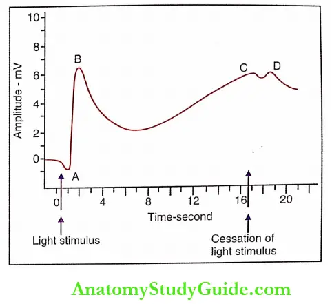
- Electroretinogram (ERG) is the record of the electrical activity in the retina. When light rays stimulate the retina, a characteristic sequence of potential changes occurs which can be recorded in the form of an electroretinogram.
- This diagnostic procedure is useful in determining retinal disorders such as cone dystrophy (degeneration of cones) and retinitis pigmentosa (hyperactivity of the pigmented retinal epithelial cells, leading to damage of photo¬receptors and blindness).
1. Method Of Recording Erg: ERG is recorded by using a galvanometer or a suitable recording device. The recording electrode is placed on the cornea of the eye in its usual forward up-looking position. The indifferent electrode is placed over any moist surface of the body, like inside the mouth.
2. Waves Of Erg: ERG has 4 waves namely ‘A:, ‘B’, ‘C’, and ‘D’. ‘A’ is the only negative wave and the other three are positive waves. ‘A’, ‘B’, and ‘C’ waves occur when a light stimulus falls on the retina. ‘D’ wave occurs when the light stimulus is stopped. ‘A’ and ‘B’ waves arise from rods and cones. The ‘C’ wave arises from the pigment epithelial layer and the ‘D’ wave arises from the inner nuclear layer.
Acuity Of Visiom
1. Acuity Of Vision Definition: Acuity of vision is the ability of the eye to determine the precise shape and details of the object, it is also called visual acuity. The acuity of vision is also defined as the ability to recognize the separateness of two objects placed together. The cones of the retina are responsible for the acuity of vision. Visual acuity is highly exhibited in the fovea centralis, which contains only cones. It is greatly reduced during refractory errors.
2. Test For Visual Acuity: Acuity of vision is tested for distant vision as well as near vision. If there is any difficulty in seeing the distant object or the near object, the defect is known as an error of refraction. The refractive errors are described separately in.
- Distant Vision: Snellen’s chart is used to test the acuity of vision for distant vision in the diagnosis of refractive errors of the eye.
- Near Vision: Jaeger’s chart is used to test the visual acuity for near vision.
Leave a Reply