Pupillary Reflexes Introduction
Pupillary reflexes are the visceral reflexes, which alter the size of the pupil. Pupillary reflexes are classified into three types:
Table of Contents
- Light reflex
- Ciliospinal reflex
- Accommodation reflex.
Light Reflex
It is the reflex in which the pupil constricts when light is flashed into the eyes. It is also called the pupillary light reflex. Light reflex is of two types:
- Direct light reflex
- Indirect light reflex.
Read And Learn More: Medical Physiology Notes
1. Direct Light Reflex: It is the reflex in which there is constriction of the pupil in an eye when light is thrown into that eye. It is also called the direct pupillary light reflex.
2. Indirect Light Reflex: Indirect light reflex is the reflex that involves constriction of the pupil in both eyes when light is thrown into one eye. If the light is flashed into one eye, the constriction of the pupil occurs in the opposite eye also even though no light rays fall on that eye. It is otherwise called consensual light reflex.
3. Pathway For Light Reflex:
- The pathway for light reflex is slightly deviated from the visual pathway. The fibers of the light reflex pathway and the fibers of the visual pathway are the same up to the optic tract. Beyond that, these two sets of fibers are separated.
- When light falls on the eye, the visual receptors are stimulated. The afferent (sensory) impulses from the receptors pass through the optic nerve, optic chiasma, and optic tract. At the midbrain level, few fibers get separated from the optic tract and synapse on the neurons of the pretectal nucleus, which lies close to the superior colliculus.
- The pretectal nucleus of the midbrain forms the center for light reflexes. The efferent (motor) impulses from this nucleus are carried by short fibers to the Edinger-Westphal nucleus (parasympathetic nucleus) of the oculomotor nerve (third cranial nerve). From Edinger- Westphal nucleus, the preganglionic fibers pass through the oculomotor nerve and reach the ciliary ganglion.
- The postganglionic fibers arising from the ciliary ganglion pass through the short ciliary nerves and reach the eyeball. These fibers cause contraction of the constrictor pupillae muscle of the iris.
- The reason for the consensual light reflex is that, some of the fibers from the pretectal nucleus of one side cross to the opposite side and end on the opposite Edinger- Westphal nucleus.
4. Ciliqspinal Reflex:
- Ciliospinal reflex is the dilatation of the pupil in the eyes caused by painful stimulation of skin over the neck. It is due to the contraction of the dilator pupillae muscle. Sensory impulses pass through a cutaneous afferent nerve.
- The center is in the first thoracic spinal segment. The efferent impulses pass through sympathetic fibers and reach dilator pupillae.

Accommodation Reflex
1. Accommodation Reflex Definition: Accommodation is the adjustment of the eye to see either near or distant objects clearly. It is the process, by which light rays from near objects or distant objects are brought to focus on the sensitive part of the retina. It is achieved by various adjustments made in the eyeball.
2. Mechanism Of Accommodation: Light rays from distant objects are approximately parallel and are less refracted while getting focused on the retina. But, the light rays from near objects are divergent. So, to be focused on the retina, these light rays should be refracted (converged) to a greater extent. There are three possible ways by which, accom¬modation occurs:
- The retina must be moved towards or away from the lens. It is done by shortening or elongation of the eyeball. So, the divergent, parallel, or convergent rays are focused accurately. This mechanism is present only in some mollusks and not in human beings
- The lens must be moved towards or away from the retina. It is done in photography. This mechanism exists in some fishes
- The convexity of the lens must be altered, so that the refractory power of the lens is altered according to the need. This mechanism is present in the human eye. The mechanism was first suggested by Young and later supported by Helmholtz.

- Young-Helmholtz Theory:
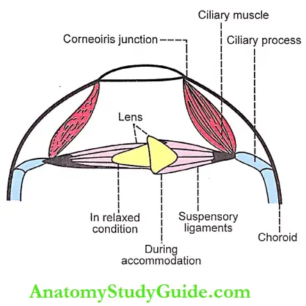
- This theory describes how the curvature of the lens increases and thereby, the refractive power of the lens is enhanced. When the eyes are fixed on a distant object (distant vision) lens is flat due to the traction of suspensory ligaments which extend from the capsule of the lens and are attached to the ciliary processes.
- The ciliary processes are attached to the choroid through the ciliary muscle. When the vision is shifted from a distant object to a near object (near vision), the ciliary muscle contracts and draws the choroid forward. The ciliary processes are brought closer to the lens, i.e. these processes form a small circle. The suspensory ligaments are slackened.
- Now, the tension on the lens is released. The lens, due to its elastic property, bulges forward. The anterior curvature (convexity) of the lens increases greatly. Very little change occurs in posterior curvature. This can be demonstrated by using Purkinje-Sanson images.
- In the resting eye, the intraocular pressure sets up tension in the choroids and pulls the ciliary processes backward and outward. The suspensory ligaments are tensed up and the lens becomes flat.
- Purkinje-Sanson Images:
- The Purkinje-Sanson images are used to demonstrate the change in the convexity of the lens during accom¬modation for near vision. A subject is made to sit in a darkroom. A lighted candle is held in front. One eye is opened and the other eye is closed. Three images of the flame are seen in the opened eye.
- The first image is upright and bright. It shines from the surface of the cornea, which acts as a mirror. The second image is upright but dim. It is reflected from the anterior convex surface of the lens. The third image is inverted and small. It is formed by the posterior surface of the lens, which acts as a concave mirror.
- When the person looks at a distant object, the second image reflected from the anterior surface of the lens is near the third image from the posterior surface. During accommodation for near vision, no change occurs either in the first image or the third image. But, the second image becomes smaller and moves towards the first image.
- Thus, the increased convexity of the anterior surface of the lens during accommodation for near vision is evident by the change in the size and position of the second image.

- Other Adjustments in Eyeball during Accommodation: In addition to the increase in anterior curvature of the lens, two more adjustments are made in the eyeball during accommodation for near vision.
-
- Convergence of both eyeballs: It is necessary to bring the retinal images onto the corresponding points
- Constriction of pupil: It is necessary to:
- Increase the visual acuity by reducing lateral chromatic and spherical aberrations
- Reduce the quantity of light entering the eye
- Increase the depth of focus through the more central part of the lens as its convexity is increased.
3. Accommodation Reflex:
Accommodation is a reflex action. When a person looks at a near object after seeing a far object, three adjustments are made in the eyeballs:
- Convergence of the eyeballs due to contraction of the medial recti.
- Constriction of the pupil due to the contraction of constrictor pupillae of the iris
- Increase in the anterior curvature of the lens due to contraction of the ciliary muscle.

- Thus, the accommodation reflex involves both skeletal muscle (medial recti) and smooth muscle (ciliary muscle and sphincter pupillae).
- During accommodation, all the adjustments are carried out simultaneously. Although accommodation is a reflex action, it can be controlled by willpower to a certain extent.
4. Pathway For Accommodation Reflex:
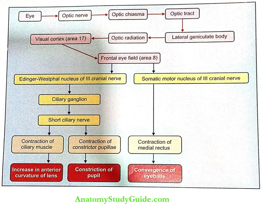
- Afferent Pathway: Visual impulses from the retina pass through the optic nerve, optic chiasma, optic tract, lateral geniculate body, and optic radiation to the visual cortex (area 17) of the occipital lobe. From here, the association fibers carry the impulses to the frontal lobe.
- Center: The center for accommodation lies in the frontal eye field (area 8) that is situated in the frontal lobe of the cerebral cortex.
- Efferent Pathway:
- Efferent fibers to ciliary muscle and sphincter pupillae:
- From area 8, the corticonuclear fibers pass via the internal capsule to the Edinger-Westphal nucleus of the 3 cranial nerves. From here, the preganglionic fibers pass through the third cranial nerve to the ciliary ganglion.
- The postganglionic fibers from the ciliary ganglion pass via the short ciliary nerves and supply the ciliary muscle and the constrictor pupillae.
- Efferent fibers to medial rectus: Some of the fibers from the frontal eye field terminate in the somatic motor nucleus of the oculomotor nerve. The fibers from the motor nucleus supply the medial rectus.
- Efferent fibers to ciliary muscle and sphincter pupillae:
5. Range And Amplitude Of Accommodation:
- The farthest point from the eye at which the object can be seen is called the far point or punctum remotum. In the normal eye, it is infinite, i.e. at a distance beyond 6 meters or 20 feet. It is limited only by the size of the object, the clearness of the atmosphere, and the curvature of the earth.
- The nearest point from the eye at which the object is seen clearly is called the near point or punctum proximum. It is about 7-40 cm, depending on the age. The distance between the far point and the near point is called the range of accommodation.
- Since the focal length of the eye is different in near vision and far vision, the refractive power of the eye is also altered. The refractive power during far vision is called static refraction (R) and that during near vision is called dynamic refraction (P).
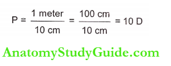
- The difference between these two refractive powers (P – R) is called the amplitude of accom¬modation, which is expressed in diopter. The refractive power is reciprocal of focal length and the unit for focal length is 1 meter or 100 cm. The refractory power is expressed as diopter (D). For example, in a normal eye, if the near point is 10 cm, the dynamic refraction is

- In the emmetropic (normal) eye since the far point is at infinite distance, the static refraction is taken as zero.
- Now the amplitude of accommodation = P – R = 10-0 = 10 D
The amplitude of Accommodation at Different Ages: The amplitude of accommodation varies with age. The amplitude of accommodation at different age groups is:
10 years = 11.0 D
20 years = 9.5 D
30 years = 7.5 D
40 years = 5.5 D
50 years = 2.0 D
60 years = 1.2 D
70 years = 1.0 D
Applied Physiology
1. Argyll Robertson Pupil: It is a clinical condition in which the light reflex is lost but the accommodation reflex is present. It is common in syphilis. It also occurs because of lesions in the Edinger-Westphal nucleus, diabetes, and alcoholic neuropathy.
2. Horner’S Syndrome: Horner’s syndrome is a disease characterized by ptosis (drooping of eyelids), constriction of the pupil, exophthalmos, and absence of sweating on one side of the face. It occurs due to injury to the cervical sympathetic nerve that is a common dunno, accident involving the neck region.
3. Presbyopia: In old age, the amplitude of accommodation is decreased point is away from the eye. This condition is called presbyopia.
Leave a Reply