Sole Of Foot
Name the muscles of 1st layer of the sole
Table of Contents
They are tendons of
- Abductor hallucis,
- Abductor digit minimi, and
- Flexor digitorum brevis.
Name the muscles of 2nd layer of sole
They are tendons of
- Flexor hallucis longus,
- Flexor digitorum longus,
- Flexor accessorius, and
- Lumbricals.
Read And Learn More: Anatomy Notes And Important Questions And Answers
Name the muscles of 3rd layer of the sole
They are tendons of
- Adductor hallucis,
- Flexor hallucis brevis, and
- Flexor digiti minimi brevis.
Mention the structures in the 4th layer of the sole of the foot.
The structures in the 4th layer of the sole of the foot
- Tibialis posterior,
- Palmar and dorsal interossei, and
- Peroneus longus.
Plantar Aponeurosis Anatomy
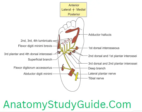
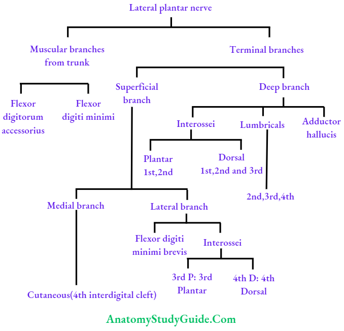
Muscles supplied by lateral plantar nerve
These are branches from the main trunk and from terminal branches.
1. Branches from the main trunk
- Flexor digitorum accessories, and
- Abductor digiti minimi.
2. Terminal branches
1. Superficial branch
1. Lateral branch
- Flexor digiti minimi brevis,
- 3rd plantar interossei, and
- 4th dorsal interossei.
2. Medial branch is cutaneous supplies 4th intern left.
2. Deep branches supply the following 9 muscles
- Three lubricants
- 2nd,
- 3rd, and
- 4th lumbrical.
2. Five interossei
1. Three dorsal interossei:
- 1st,
- 2nd, and
- 3rd
2. Two plantar interossei
- 1st, and
- 2nd plantar interossei.
3. Adductor hallucis.
Muscles supplied by the medial plantar nerve
The medial plantar nerve supplies four muscles of the sole. They can be grouped as muscles from
1. First layer
- Abductor hallucis, and
- Flexor digitorum brevis.
2. 2nd layer: 1st lumbrical.
3. 3rd layer: Flexor hallucis brevis.
Nerve supply of lubricants of sole
- 1st lumbrical is supplied by the medial plantar nerve, and
- 2nd, 3rd, and 4th lubricants are supplied by the deep branch of the lateral plantar nerve.
Plantar Aponeurosis Anatomy
Actions of dorsal interossei of foot
1. Axis of movement: The longitudinal axis of the foot lies along the 2nd metatarsal bone and the phalanges of the 2nd toe.
2. The actions of the interossei of the foot are indicated by the formula
- Dorsal interossei are abductors. They abduct away from the axis line.
- The first and 2nd interossei cause medial and lateral abduction of the second toe. Third and 4th are abductors of 3rd and 4th toes.
Actions of plantar interossei of foot
1. Axis of movement: The longitudinal axis of the foot lies along the 2nd metatarsal bone and the phalanges of the 2nd toe.
2. The actions of the interossei of the foot are indicated by the formula
- The plantar interossei are adducting muscles. They adduct towards the 2nd toe.
- The 1st toe has its adductors. (Oblique and transverse heads of adductor hallucis). The lateral three toes require adductors.
- 1st, 2nd, and 3rd plantar interossei arise only from the metatarsal bone of its digit. They are inserted by tendons into the medial sides of the 3rd, 4th and 5th digits.
Plantar Aponeurosis Anatomy
Plantar Aponeurosis Introduction: It is a thickened central part of the deep fascia.
- Plantar Aponeurosis Formation: It is formed by longitudinal displayed compact bundles of collagen fibers.
- Plantar Aponeurosis Features: It consists largely of white fibrous connective tissue. They are glistening in nature.
- Plantar Aponeurosis Parts: Deep fascia in the sole has the following parts
Foot Anatomy Phalanx
1. Lateral part (calcaneometatarsal ligament): It covers the abductor digiti minimi. It extends from the lateral tubercle of the calcaneum to the base of the proximal phalanx of the 5th toe.
2. Medial part: It covers abductor hallucis. It extends from the lateral tubercle of the calcaneum to the base of the proximal phalanx of the great toe. Both lateral and medial parts are thin.
3. Central part: It is the thickest part and most prominent part and in reality, it is the plantar aponeurosis. These medial and lateral parts are, sometimes, called plantar fascia.
4. Shape: It is large in shape having the base and apex.
1. The proximal part is the apex and is attached to the medial tubercle of the calcaneum.
2. The distal part is the base and divides opposite the head of metatarsal bones into 5 slips or processes which are connected by transverse fibers. There are 5 gaps in the processes. The digital vessels, nerves, and lumbricals pass through it.
3. Each slip or process is divided into
Foot Anatomy Phalanx
Plantar Aponeurosis Anatomy
1. Superficial part: It is attached to the transverse sulcus of the skin. It is present at the toes of the foot.
2. Deep part: It splits into two parts and transmits the flexor tendons. They are attached to
- Transverse metatarsal ligament,
- Fibrous sheaths of flexor tendons, and
- Lateral and medial borders of proximal and middle phalanx.
4. Plantar Aponeurosis Relations
1. Superficial
- Thick heavily keratinized skin, and
- Cutaneous branches/tributaries of medial plantar vessels and nerves.
2. Deep
- Abductor hallucis,
- Flexor digitorum brevis, and
- Abductor digiti minimi.
5. Plantar Aponeurosis Structures piercing: Cutaneous branches of the medial and lateral plantar nerves penetrate the aponeurosis to supply the overlying skin and fascia.
6. Plantar Aponeurosis Morphology: It is a continuation of the plantaris muscle and homologous to palmar aponeurosis.
Foot Anatomy Phalanx
7. Plantar Aponeurosis Functions
- Plantar Aponeurosis maintains the stability of medial and lateral longitudinal arches.
- Plantar Aponeurosis acts as a tie beam which prevents the separation of the anterior and the posterior pillars underweight on the top.
- Plantar Aponeurosis provides attachment to superficial plantar muscles.
- Plantar Aponeurosis protects plantar vessels and nerves.
- The skin ligaments also anchor the skin to the underlying deep fascia (plantar aponeurosis), improving the “grip” of the sole.
- Plantar Aponeurosis gives proximal attachments to the muscles of the 1st layer of the sole.
- Plantar Aponeurosis bears greater stress than the longitudinal arches of the foot.
- Plantar Aponeurosis acts like a superficial ligament.
8. Plantar Aponeurosis Anatomy Applied anatomy
1. Plantar fasciitis
- Definition: Inflammation of the aponeurosis is called plantar fasciitis.
Plantar Aponeurosis Anatomy
2. Infection of the sole
- The collection of pus under plantar aponeurosis is well localized.
- Normally, it cannot come out through aponeurosis, because of its tough nature.
- The condition is very painful and causes more damage to the structures underneath.
3. High arched foot (pes cavus) occurs when the longitudinal arch becomes unduly elevated. It is due to
- Shortening of plantar aponeurosis, or
- Contracture of the intrinsic muscles of the foot.
Comparison Between The Plantar And Paimar Apeoneurosis
Layers of sole
Layers Of Sole Introduction: The sole of the foot contains long tendons and a number of small muscles which are arranged in four layers. Unlike the hand, the big toe is prevented from coming into contact with the other toes. However, the toes do have the capability to be used for grasping. This can be seen in people born without arms, who can learn to write, draw, and manipulate objects with their feet.
Plantar Aponeurosis Anatomy
1. Muscles of each layer
1. 1st layer or most inferior layer or superficial layer.
1. Muscles
- Abductor hallucis,
- Abductor digiti minimi, and
- Flexor digitorum brevis.
2. Proximal attachments: All these muscles arise from the tuberosity of the calcaneus.
3. Distal attachments
- The abductor hallucis inserts on the proximal phalanx of the big toe.
- The abductor digiti minimi inserts on the proximal phalanx of the little toe.
- The flexor digitorum brevis separates into four tendons that insert onto the middle phalanges of the four lateral toes.
2. 2nd layer
1. Tendons of
- Flexor digitorum longus, and
- Flexor hallucis longus.
2. Muscles
1. Flexor accessories
- Proximal attachment: It has two heads from the calcaneus.
- Distal attachments: Into the tendon of the flexor digitorum longus muscle.
- Action: Align the pull of the tendons of the flexor digitorum longus by straightening the diagonal vector of the long tendon.
2. Lumbricals: 1st lumbrical is only unipennate, 2nd, 3rd and 4th lumbricals are bipennate.
1. Proximal attachments: Arise from the tendons of the flexor digitorum longus muscle.
Plantar Aponeurosis Anatomy
2. Distal attachments
- The medial side of the proximal phalanx of the lateral four toes, and
- Dorsal extensor hoods of the toes.
3. 3rd layer: The 3rd layer of plantar muscles consists of two flexors and an adductor (with two heads), in contrast to the 1st layer, which contains two flexors and an abductor
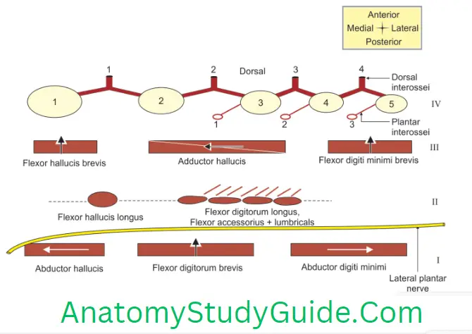
- Flexor hallucis brevis,
- Flexor digiti minimi brevis, and
- Adductor hallucis.
4. 4th layer or uppermost layer
Tibialis posterior.
Interossei: 3 plantar and 4 dorsal.
Tendon of peroneus longus.
Plantar Aponeurosis Anatomy
2. Nerve supply
1. The medial plantar nerve supplies 4 intrinsic muscles. (Refer to OLA-46)
2. The lateral plantar nerve supplies 14 intrinsic muscles. (Refer to SN-20:1 and 2)
3. Extrinsic muscles are supplied by the nerve of the respective compartment of the leg.
1. 2nd layer
- Flexor digitorum longus
- Flexor hallucis longus—tibial nerve
2. 4th layer
- Tibialis posterior—branch of nerve to popliteus (tibial nerve)
- Peroneus longus—superficial peroneal nerve
3. Plantar Aponeurosis Functions
1. They are chiefly concerned with supporting the arches of the foot.
2. Although their names would suggest control of individual toes, this function is rarely used in most people.
4. Plantar Aponeurosis Applied anatomy
- Injury to the tibial component of the sciatic nerve causes paralysis of the muscles of the sole. It leads to an inability to stand on the toes (due to loss of plantar flexion of the foot) and loss of Achilles tendon reflex.
- There will be sensory loss on the sole. Trophic ulcers tend to develop in the sole due to loss of sensation.
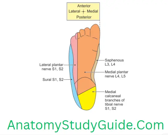
Cutaneous Nerve Supply Of Sole Of Foot
1. Medial calcaneal branches of the tibial nerve supply the posterior and medial portions of the foot.
2. Branches from the
- Medial plantar nerve to the larger anteromedial portion including the medial 3½ digits,
- Lateral plantar nerve to the smaller anterolateral portion. It includes the lateral 1½ digits.
- Saphenous and sural nerves supply small areas on the medial and lateral sides of the foot.
Leave a Reply