Spermatogenesis
Spermatogenesis Introduction: It is the complex series of changes by which spermatogonium is
transformed into spermatozoa.
Table of Contents
1. Spermatogenesis Site:
- It takes place in seminiferous tubules.
- It is controlled by testosterone.
- It begins at puberty and continues up to old age.
2. Spermatogenesis Duration:
64 days are required to complete one cycle of spermatogenesis. These complex series of changes do not take place in all parts of seminiferous tubules simultaneously.
3. Spermatogenesis Stages: The following are the stages.
Read And Learn More: General Histology Question And Answers
Spermatocytosis:
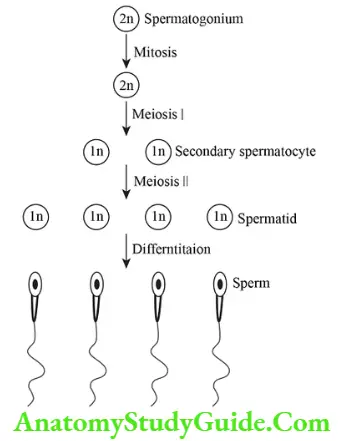
It involves series of mitotic divisions from which spermatogonium A is converted to primary spermatocyte. Primary spermatocyte is comparatively larger cell due to increased nuclear and cytoplasmic material.
Meiosis:
Primary spermatocyte (diploid) soon enters into 1st meiotic division, transforms into secondary spermatocyte (haploid). It is changed into spermatid by 2nd meiotic division. Spermatids have haploid number of chromosomes.
Spermiogenesis:
It involves series of changes by which the spermatid (non-motile) is converted into an elongated (motile) sperm.
4. End result of spermatogenesis: In one cycle, 4 motile sperms are formed from one primary spermatocyte which possess haploid number of chromosomes.
5. Amount of ejaculation is 2-3 ml:
1. Each ml contains nearly 100 million sperms.
2. The life of the sperm is 24 – 48 hours.
3. Rate of motility is 1.5 to 3 mm/min.
4. Contents of semen are
1. Sperm

2. Chemicals and enzymes:
- Citric acid
- Lactic acid
- Pyruvic acid
- Fructose
- Proteolytic enzymes
- Prostaglandins
- Inositol, and
- Sorbitol.
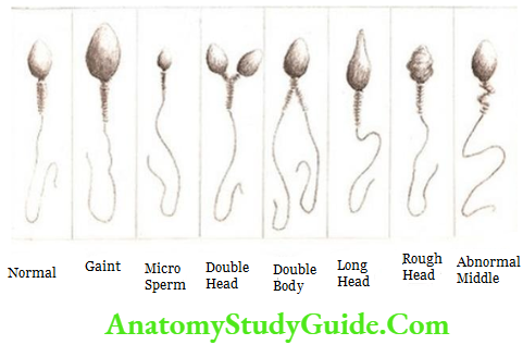
6. Anomalies: They can be grouped in
Morphological:
- Double heads
- Poorly motile
Numerical:
- Oligospermia: Less than 10 million sperms/ml.
- Azoospermia: Zero sperm count.

Spermiogenesis
1. Spermatogenesis: It involves series of changes by which a spermatid (non-motile) is converted into an elongated (motile) sperm. The cell organelle transforms into the following structure.
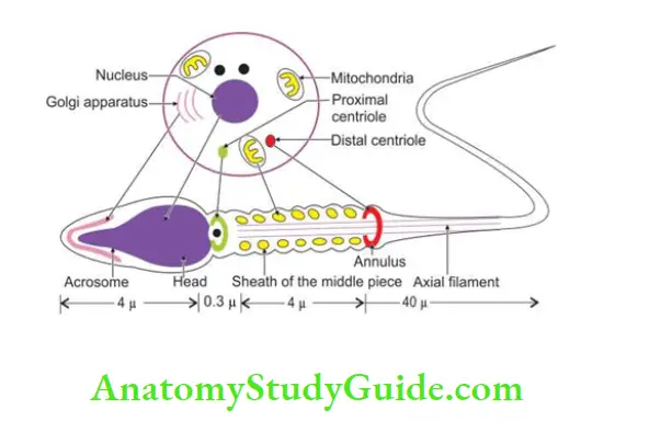
2. Golgi complex: Forms acrosomal cap on upper two-thirds of circumference of the head of the sperm. It helps in penetration of the cells present in corona radiata and zona pellucid a (secondary oocyte). It contains enzymes, namely
- Acrosin
- Acid phosphatase
- Proteases, and
- Hyaluronate.
3. Nucleus: The nucleus is converted into head of the sperm. The specific number of chromosome is maintained, i.e. haploid number of chromosome.
4. Mitochondria: Mitochondria are arranged in helical form around the axoneme of the middle piece. It provides energy for motility.
5. There are two centrioles present at neck and tail of the sperm.
- The proximal centriole is present at the neck.
- The distal centriole is present in tail of sperm. It helps in motility.
6. Residual body: The excess of cytoplasm is shed off which is engulfed by cells of Sertoli.
Introduction: It is a male ♂ gamete, smaller than the female ♀ gamete, i.e. ovum, and is about 45-50 pm in length. It is a motile cell.
Parts: It has the following parts.
Sperm
1. Head:
Shape:
- Ovoid

- Pyriform

Size: 4 μ m.
2. Tail: Principal piece
- Neck: It is constricted part distal to the head.
- Middle piece: It contains a motor element—axoneme (central doublet with peripheral doublet) which is surrounded by the course fibers in middle piece and principal piece. The mitochondria are absent in principal piece.
- The proximal piece, and
- Terminal piece.

Capitation of sperm
Introduction: It is the final stage of maturation of sperm.
1. Duration: 7 hours.
2. Site: Female ♀ genital tract.
A glycoprotein coat and seminal plasma proteins are removed from the plasma membrane. They are present in the acrosomal region.
3. Result:
- No morphological changes in the capacitated sperm.
- There is increase of the activity.
4. Capacitated sperm:
The capacitated sperm comes in contact with corona radiata because of following facts.
- There is an interaction between the zona pellucida and head of the sperm. It includes release of acrosin which helps in digestion of zona pellucida.
- The action of tubal mucosal enzyme.
Oogenesis
Introduction: It is a process involving complex changes by which oogonia are converted into ova, the female gamete ♀.
1. Site: It takes place in the ovary and fallopian tubes.
2. Onset of oogenesis: It begins when the child in the womb of the mother.
3. End of oogenesis: It ends at menopause.
4. Duration: It takes approximately 12–60 years to complete one cycle of oogenesis. The minimum average duration of an oogenesis cycle is 12 years. The maximum duration of an oogenesis cycle will be 60 years.
5. Ovary: The female ♀ gonad is the ovary. It has an outer part—the cortex, and inner part—medulla. The cortex contains large cells called oogonia. All oogonia are produced before birth.

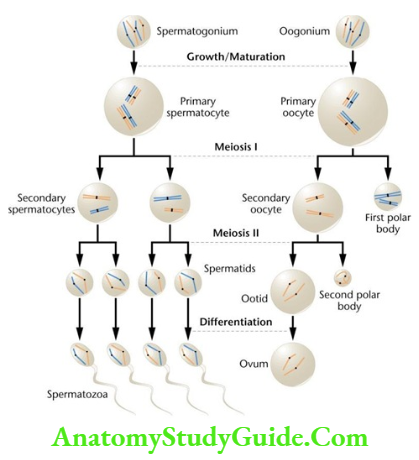
The oogonium enlarges and is called the primary oocyte.
1. Primary oocyte: Divides by 1st meiotic division (reduction) into two secondary oocytes. One of two secondary oocytes contains 1/2 number of chromosomes and almost all of its cytoplasm.
Second of two secondary oocytes contains only 1/2 number of chromosomes and no cytoplasm. It is called 1st polar body. The 1st polar body is, therefore, formed merely to get rid of unwanted chromosomes.
2. Secondary oocyte: Gets arrested into metaphase. It divides into the ovum and 2nd polar body by the 2nd meiotic division.
This occurs only at the time of fertilization.
Number of germ cells at different stages of life

6. Ovulation: The liberation of the secondary oocyte from the ovary is called ovulation.
Leave a Reply