Muscle Mass
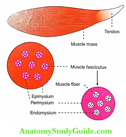
- The muscle mass (or tissue) is made up of a large number of individual muscle cells or myocytes. The muscle cells are commonly called muscle fibers because these cells are long and slender in appearance. The skeletal muscle fibers are multinucleated and arranged parallel to one another with some connective tissue in between.
- The muscle mass is separated from the neighboring tissues by the thick fibrous tissue layer known as fascia. Beneath the fascia, the muscle is covered by a connective tissue sheath called epimysium. In the muscle, the muscle fibers are arranged in various groups called bundles or fasciculi.
- The connective tissue sheath that covers each fasciculus is called perimysium. Each muscle fiber is covered by the connective tissue layer called the endomysium.
Read And Learn More: Medical Physiology Notes
Table of Contents
Muscle Fiber
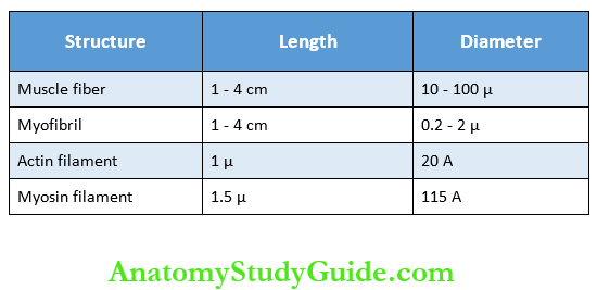
- Each muscle cell or muscle fiber is cylindrical in shape. The average length of the fiber is 3 cm. And, it varies between 1 and 4 cm depending upon the length of the muscle. The diameter of the muscle fiber varies from 10 to 100 μ. The diameter varies in a single muscle.
- The muscle fibers are attached to à tough cord of connective tissue called a tendon. Tendon is in turn attached to the bone. The tendon of some muscles is thin, flat, and stretched but tough. Such a type of tendon is called aponeurosis.
- Each muscle fiber is enclosed by a cell membrane called a plasma membrane that lies beneath the endomysium. It is also called sarcolemma. The cytoplasm of the muscle is known as sarcoplasm. Many structures are embedded within the sarcoplasm:
- Nuclei
- Myofibril
- Golgi apparatus
- Mitochondria
- Sarcoplasmic reticulum
- Ribosomes
- Glycogen droplets
- Occasional lipid droplets.
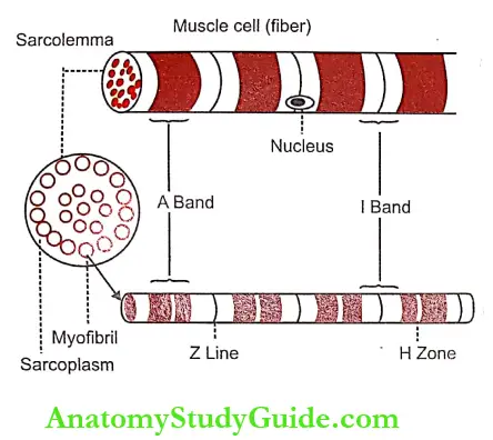
Nuclei: There are one or more nuclei in each muscle fiber. In long muscle fibers, many nuclei are seen. Nuclei are oval or elongated and situated just beneath sarcolemma. Usually, in other cells, the nucleus is in the interior of the cell. All the organelles of muscle fiber have the same functions as those of other cells.
Myofibril
- Myofibrils or myofibrillar are the fine parallel filaments present in the sarcoplasm of the muscle cell. The myofibrils run through the entire length of the muscle fiber.
- In the cross-section of a muscle fiber, the myofibrils appear like small distinct dots within the sarcoplasm. The diameter of the myofibril is 0.2-2 p. And, the length of a myofibril varies between 1 and 4 cm depending upon the length of the muscle fiber.
- In some muscle fibers, some of the myofibrils are arranged in groups called Cohnheim’s areas or fields.
Microscopic Structure Of A Myofibril:
Light microscopic studies show that each myofibril consists of a number of two alternating bands which are also called sections, segments or disks. The two bands are:
- Light band or ‘I’ band
- Dark band or ‘A’ band.
1. Light Band or ‘I’ Band: The light band is isotropic in nature. When the polarized light is passed through the muscle fiber at this area the light rays are refracted at the same angle. So, this band is called ‘I’ (isotropic) band.
2. Dark Band or ‘A’ Band:
- The dark band is anisotropic in nature. When the polarized light is passed through the muscle fiber at this area, the light rays are refracted at different directions (An not; iso it; trops = turning). So, this band is otherwise called ‘A’ (anisotropic) band. The dark band is also called Q disk (Querscheibe cross disk).
- In an intact muscle fiber, ‘I’ band and ‘A’ band of the adjacent myofibrils are placed side-by-side. It gives the appearance of characteristic cross striations in the muscle fiber.
- I band is divided into two portions by means of a narrow and dark line called the ‘Z’ line or ‘Z’ disk (in German zwischenscheibe = between disks). The ‘Z’ line is formed by a protein disk which does not permit the passage of light. The portion of myofibril in between two ‘Z’ lines is called sarcomere.
Sarcomere
- Sarcomere Definition: Sarcomere is the structural and functional unit of the skeletal muscle. It is also called the basic contractile unit of the muscle.
- Sarcomere Extent: Each sarcomere extends between two ‘Z’ lines of to the original position. myofibril. Thus, each myofibril contains many sarcomeres arranged in series throughout its length. When the muscle is in a relaxed state, the average length of each sarcomere is 2-3 microns.
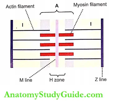
- Sarcomere Components: Each myofibril consists of an alternate dark ‘A’ band and light ‘I’ band (Fig. 29-3). In the middle of ‘A’ band, there is a light area called ‘H’ zone (H= hell = light-in German, H Henson discoverer). In the middle of ‘H’ zone lies the middle part of the myosin filament. This is called ‘M’ line (in German Mittel middle). ‘M’ line is formed by myosin-binding proteins.
Electron Microscopic Study Of Sarcomere:
The electron microscopic studies reveal, that the sarcomere consists of many thread-like structures called myofilaments. Myofilaments are of two types:
- Actin filaments
- Myosin filaments.
1. Actin Filaments: Actin filaments are thin filaments with a diameter of 20 A and a length of 1 μ. These filaments extend from either side of the ‘Z’ lines, run across ‘I’ band, and enter into ‘A’ band up to the ‘H’ zone.
2. Myosin Filaments:
- Myosin filaments are thick filaments with a diameter of 115 Å and a length of 1.5 μ. These filaments are situated in the A band.
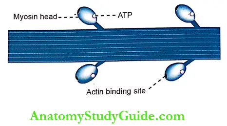
- There are some lateral processes (projections) or cross bridges arising from myosin filaments. These bridges have enlarged structures called myosin heads at their tips. The myosin heads attach themselves to actin filaments. These heads pull the actin filaments during the contraction of the muscle by means of a mechanism called the sliding mechanism or ratchet mechanism.
- During the contraction of the muscle, the actin filaments glide down between the myosin filaments towards the center of the ‘H’ zone and approach the corresponding actin filaments from the next ‘Z’ line.
- The ‘Z’ lines also approach the ends of myosin filaments, so that, the ‘H’ zone and ‘I’ bands are shortened during contraction of the muscle. During the relaxation of the muscle, the actin filaments and ‘Z’ lines come back to the original Position.

Contractile Elements(Proteins) Of Muscle
The myosin filaments are formed by myosin molecules. The actin filaments are formed by three types of proteins called actin, tropomyosin and troponin. These four proteins together constitute the muscle proteins or the contractile elements of the muscle.
1. Myosin Molecule:
- Each myosin filament consists of about 200 myosin molecules. About 17 classes of myosin are identified so far. Myosin II is present in sarcomere.
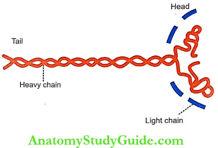
- Myosin II is a globulin with a molecular weight of 4,80,000. Each myosin molecule is made up of 6 polypeptide chains of which two are heavy chains and four are light chains.
- The molecular weight of each heavy chain is 2,00,000 (2 x 2,00,000 = 4,00,000). The molecular weight of each light chain is 20,000 (4 x 20,000 80,000). Thus, the total molecular weight of each myosin molecule is 4,80,000 (4,00,000 + 80,000).
- The two heavy chains twist around each other to form a double helix. At one end, the two chains remain twisted around one another and form the tail portion. At the other end, both the chains turn away in opposite directions and form the globular head portion. To each part of this head, are attached two light chains.
- Thus, there are four light chains in each myosin molecule. Each myosin head has two attachment sites. One site is for actin filament and the other one is for one ATP molecule. In the central part of the myosin filament, i.e. in the ‘H’ zone, the myosin head is absent.
2. Actin Molecule:
- Actin molecules are the major constituents of the thin actin filaments. Each actin molecule is called F actin and it is the polymer of a small protein known as G actin. There are about 300-400 actin molecules in each actin filament.
- The molecular weight of each molecule is 42,000. The actin molecules in the actin filament also are arranged in the form of a double helix. Each F actin molecule has an active site to which the myosin head is attached.
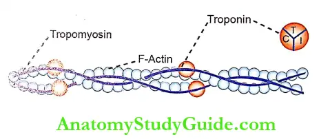
3. Tropomyosin:
- There are about 40-60 tropomyosin molecules situated along the double helix strand of the actin filament. Each tropomyosin molecule has a molecular weight of 70,000.
- In the relaxed conditions of the muscle, the tropomyosin molecules cover all the active sites of F actin molecules.
4. Troponin:
It is formed by three subunits:
- Troponin I – Attached to F actin
- Troponin T- Attached to tropomyosin
- Troponin – Attached to calcium ions.
Other Proteins Of The Muscle:
In addition to the contractile proteins, the sarcomere contains some more proteins such as:
- Actinin- Attaches actin filament to ‘Z’ line
- Desmin – Binds ‘Z’ line with the sarcolemma
- Nebulin- Runs in close association with and parallel to actin filaments
- Titin – A large protein connecting the ‘M’ line and the ‘Z’ line. Each titin molecule forms scaffolding (framework) for the sarcomere and provides elasticity to the muscle. When the muscle is stretched, the titin unfolds itself. However, if the stretching is more, it offers resistance and protects the sarcomere from overstretching
- Dystrophin-A rod-shaped large protein that connects actin filament to dystroglycan. Dystroglycan is a transmembrane protein present in the sarcolemma. Dystrophin and dystroglycan form dystrophin-dystroglycan or dystrophin-glycoprotein complex
Leave a Reply