Introduction To Dental Anatomy And Landmarks
Tissues Of The Tooth
The tissues of a tooth are enamel, dentin, cementum, and pulp. Enamel, dentin, and cementum are the hard tissues and are calcified. Pulp is a soft connective tissue.
Table of Contents
Enamel is the tissue that forms the outermost protective covering of the crown of the tooth. It is highly mineralized and rich in calcium hydroxyapatite. Enamel is laid down by ameloblasts.
Dentin is a hard tissue that is yellow in color and is present in between enamel and pulp. It makes up the bulk of the crown. It is less mineralized than enamel and is laid down by odontoblasts.
Read And Learn More: Oral Anatomy Notes
Pulp is the soft connective tissue present in the pulp cavity enclosed by dentin. It is present in the crown and in the root within the pulp chamber and the root canal. The pulp communicates with the tissue at the apex of the tooth via the apical foramen.
Cementum is the tissue that covers the root of the tooth and is present over dentin. It varies in thickness from the neck of the tooth to the apex of the root. It is a mineralized tissue which is harder than bone and softer than enamel. It is laid down by cementoblasts.
Crown Root And Alveolar Bone
A tooth has a crown and root:
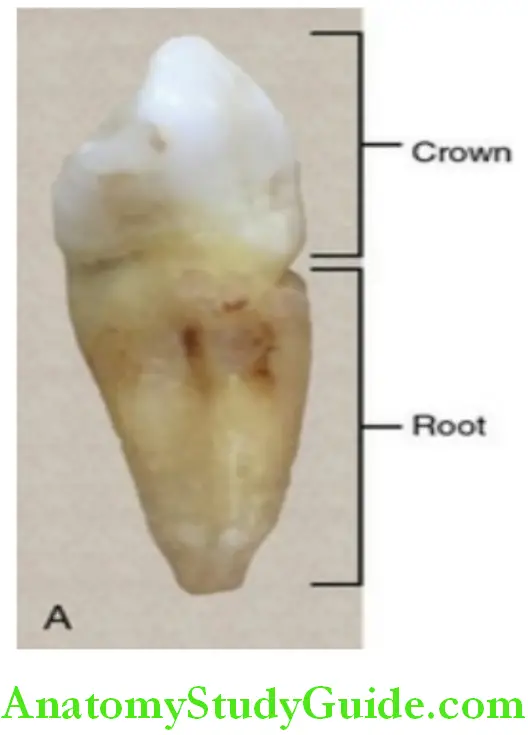
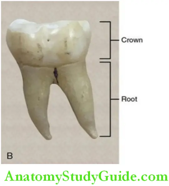
Crown:
The crown is the part of the tooth covered by enamel.
Root:
The root is the part of the tooth covered by cementum.
The junction of the crown and root is the cervical/neck area of the tooth and is called the cementoenamel junction. The junction is marked by the cervical line.
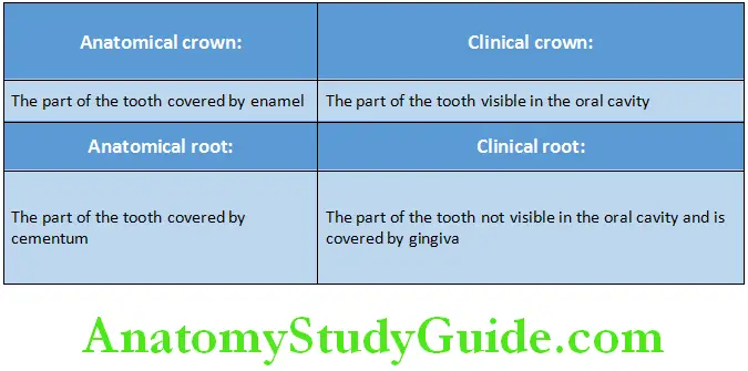
Crown Root And Alveolar Bone Clinical Consideration
- In a newly erupted tooth, the clinical crown is shorter than the anatomical as a portion of the anatomical crown is still covered by gingiva.
- With age, the clinical crown may become longer than the anatomical crown due to the recession of the gingiva and thus increased exposure of the tooth in the oral cavity.
- In a newly erupted tooth, the clinical root is longer than the anatomical root as the unexposed part of the root is considered a part of it.
- With advancing age, the clinical root becomes shorter than the anatomical root as the part of the root that is exposed is more due to the recession of the gingiva.
Alveolar bone:
The part of the jaw that supports the teeth is called the alveolar bone.
Anterior And Posterior Teeth
Anterior teeth:
The teeth present close to the midline in each arch are termed the anterior teeth. They are present in the front of the mouth.
Anterior: Incisors and canines
Deciduous and permanent dentition: 12 anterior teeth
Six anterior teeth in each arch: Central incisor, lateral incisor, and canine – 2 in no
Posterior teeth:
The teeth present away from the midline in each arch are termed the posterior teeth. They are present in the back of the mouth.
Posteriors: Premolars and molars
Deciduous dentition: Eight posterior teeth Two molars in each quadrant, four in each arch, and thus eight in total
Permanent dentition: 20 posterior teeth Two premolars, three molars in each quadrant, 10 in each arch, and thus 20 in total
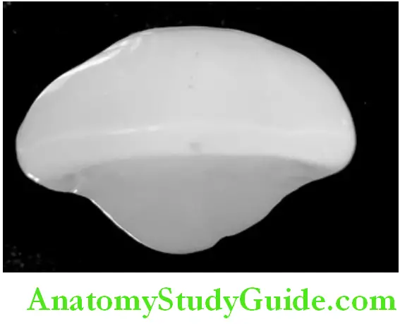
Surfaces Of The Tooth
The surfaces of the teeth are named based on the position, location, and uses of the tooth.
Facial surfaces:
Labial surface: The surface of the tooth facing the lips is the labial surface.
It is applicable to the anterior teeth i.e. The incisors and canines.
Buccal surface: The surface of the tooth facing the cheek is called the buccal surface.
It is applicable to the posteriors, i.e. The premolar and molars.
The labial and buccal surfaces are called collectively called facial surfaces.
Lingual surface: The surface of the tooth facing the tongue is called the lingual surface.
Palatal surface: The surface of the tooth facing the palate is called the palatal surface.
The terms lingual surface and palatal surface are used interchangeably; however, lingual surface is more appropriate for the mandibular teeth and palatal surface for the maxillary teeth.
Proximal surfaces:
The imaginary vertical line that passes in between the maxillary and mandibular central incisors is considered the midline which is the reference for the mesial and distal surfaces.
Mesial surface: The surface of the tooth that is closer to the midline of the arch is called the mesial surface.
Distal surface: The surface of the tooth that is away from the midline of the arch is called the distal surface.
Generally, the mesial surface of each tooth comes in contact with the distal surface of the adjacent tooth.
Exception: Maxillary and mandibular incisors and the maxillary and mandibular third molars. The mesial surfaces of the incisors come in contact with each other, whereas the distal surfaces of the third molars have no teeth present distal to them the mesial and distal surfaces are collectively called the proximal surfaces.
Incisal and occlusal surfaces:
The surfaces of the teeth that come in contact with those in the opposite arch when the jaws are closed are called the incisal or the occlusal surfaces.
The incisal surface is applicable to the interiors, i.e. Incisors and the canines.
The occlusal surface is applicable to the posteriors, i.e. Premolars and molars.
The depressions, grooves and other land marks are mentioned in the boxes below.
Depressions and grooves in teeth:
- Fossa
- Sulcus
- Grooves
- Pit
- Fissure
Other landmarks:
- Apex
- Root trunk
- Furcation
- Cervical line
Landmarks Of A Tooth
The landmarks of a tooth may be broadly grouped under elevations and ridges.
Elevations and ridges:
- Lobe
- Mamelon
- Cusp
- Tubercle
- Cingulum
- Ridge
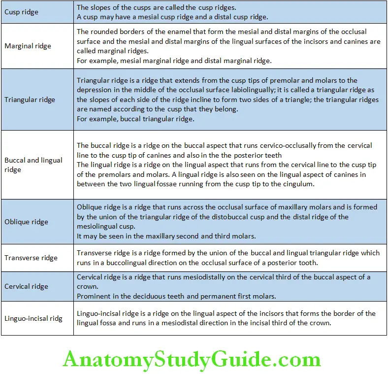
- Cusp ridge
- Marginal ridge
- Triangular ridge
- Buccal and lingual ridge
- Oblique ridge
- Transverse ridge
- Cervical ridge
- Perikymata
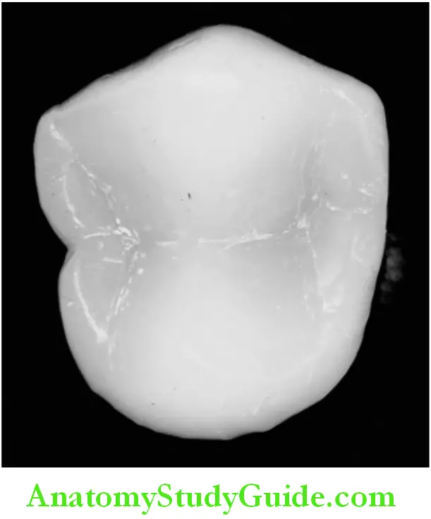
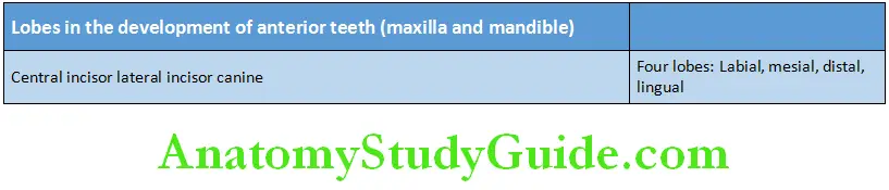
Lobe:
The lobe is one of the primary sections of formation in the development of the crown. The cusps and mamelons are representative of the lobes. Permanent teeth develop from a minimum of four lobes.
Anterior:
The incisors and canines develop from four lobes.
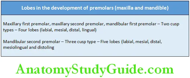
Posteriors:
The premolars develop from four lobes. However, the permanent mandibular second premolar of the three cusp type develops from five lobes.
Mamelons:
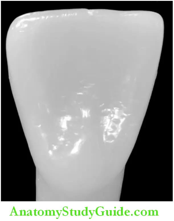
Mamelon is any one of the three rounded protuberances or tubercles found on the incisal edges of the newly erupted incisor teeth.
- They are worn out with mastication once the tooth erupts.

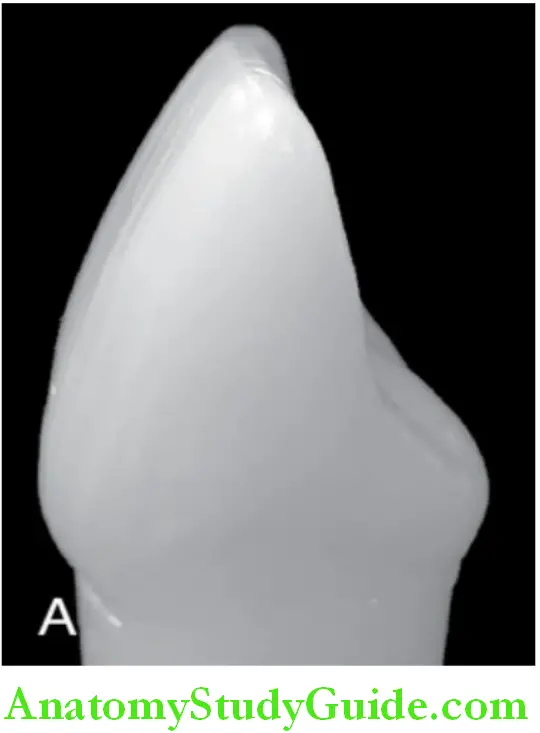
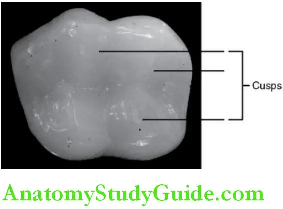
Cusp:
A cusp is a pyramidal elevation or mound on the crown portion of the tooth making up a divisional part of the occlusal surface. Cusps are seen in canines and the posterior teeth. The cusps are named according to the location.
Canines: One cusp, hence called cuspid.
Premolar: Two cusps – bicuspid; sometimes a three-cusp form may be seen in the mandibular second premolar.
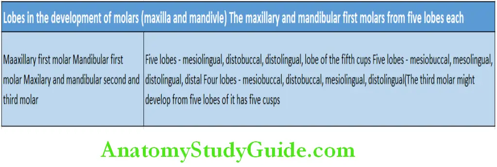
Molars: Four or five cusps may be seen
Each cusp has:
- One mesial slope or mesial cusp ridge
- One distal slope or distal cusp ridge
- One buccal cusp ridge (labial ridge on the canines)
- One lingual cusp ridge
- Triangular ridge of the cusp
Tubercle:
A tubercle is a smaller elevation on some portion of the crown produced by an extra formation of enamel.
- Most often seen on the lingual aspect of the maxillary lateral incisor.
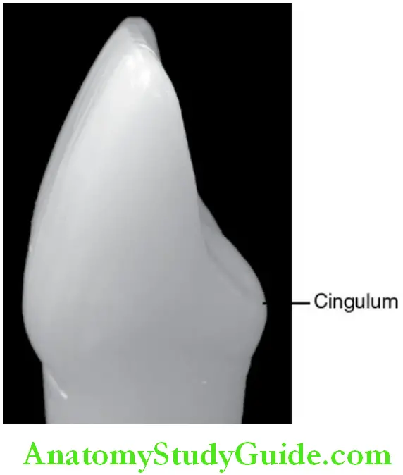
Cingulum:
Cingulum is the bulge on the cervical third of the lingual surface of the anterior teeth.
- It makes up the bulk of the cervical third.
Ridge:
A linear elevation on the surface of a tooth is called a ridge and is named according to the location.
The ridges as named according to the location are as follows:
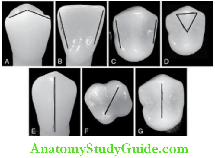
A. Cusp ridges.
B. marginal ridges in an anterior tooth.
C. marginal ridges in a posterior tooth.
D. Triangular ridge in a posterior tooth.
E. Buccal ridge in a posterior tooth.
F. Oblique ridge.
G. Transverse ridge in a posterior tooth.
Perikymata:
Perikymata are multiple and minute horizontal ridges on the surface of the enamel of newly erupted permanent teeth.
- More closely packed in the cervical third
- More pronounced in younger individuals
- Lost with age due to tooth brushing and mastication.
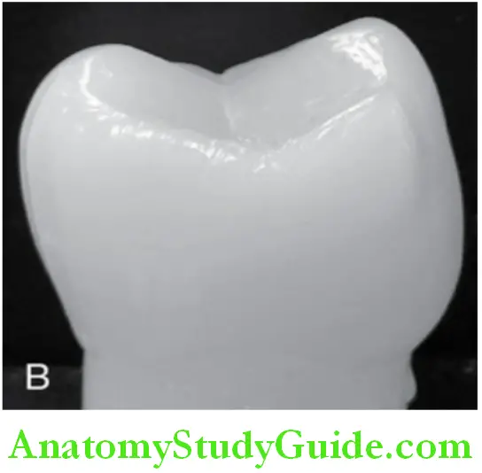
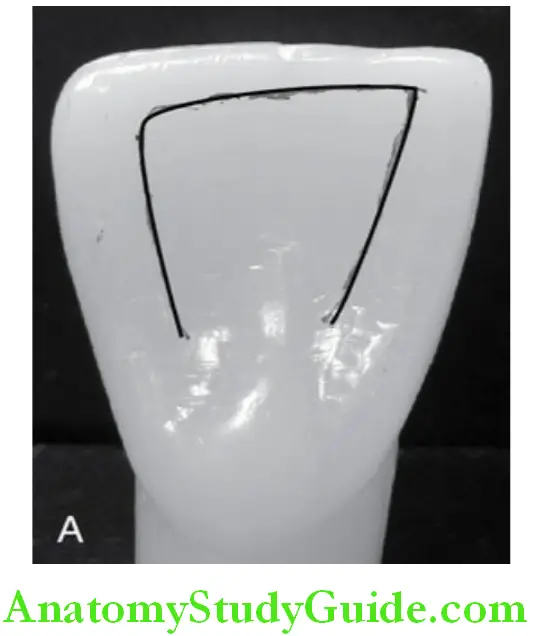
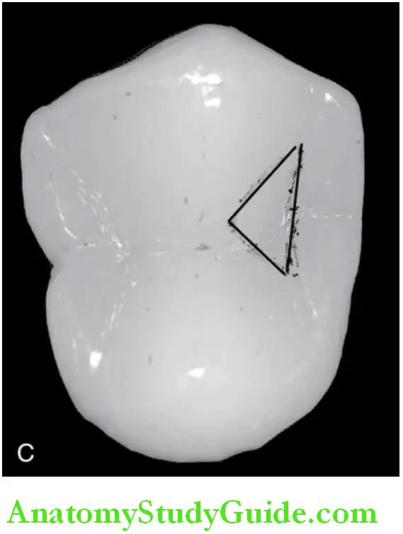
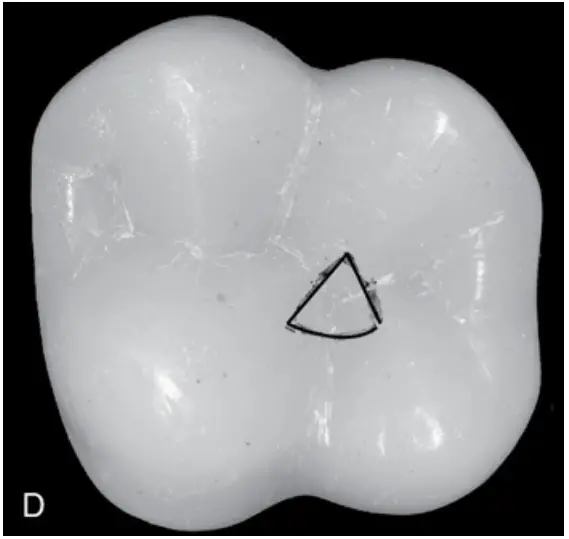
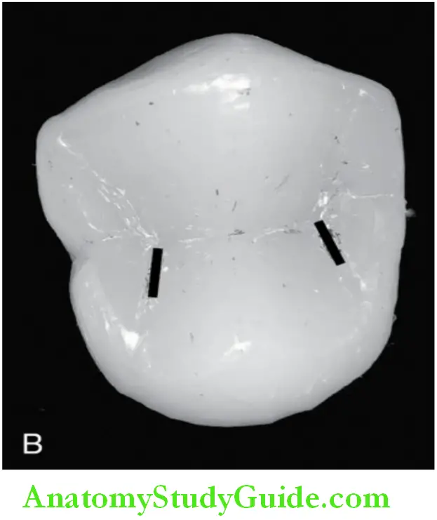
Fossa:
- A fossa is an irregular depression or concavity. It is seen on the lingual surface of the anterior teeth and the occlusal surface of the posterior teeth.
- Lingual fossa: It is seen on the lingual surface of the incisors and canines.
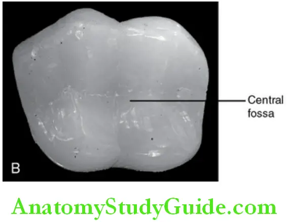
- Central fossa: It is present on the occlusal surface of the molars.
- Triangular fossa: It is found on the occlusal surface of the molars and premolars mesial or distal to the marginal ridges
- Distal fossa: A major fossa located distal to the oblique ridge in the maxillary
Molars
Sulcus:
A sulcus is a long depression or valley on the surface of a tooth running mesiodistally between cusps and ridge.
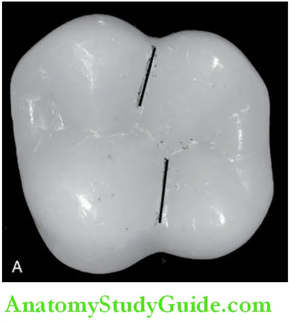
Groove: Groove is a linear depression in the deepest portion of the sulcus.
Developmental groove: A developmental groove is a sharply defined linear depression separating the lobes of major portions of a tooth formed during tooth development.
The major grooves may be named based on the location.
Central groove: A central groove is a groove that runs in a mesiodistal direction on the occlusal surface of a posterior tooth and divides it buccolingually into two halves
Supplementary groove: An accessory groove present on the occlusal surface of posteriors or the lingual surface of the anterior.
Pit:
Pits are small pin-point depressions located at the junction of developmental grooves or at terminals of those grooves. They are the deepest point of the fossa.
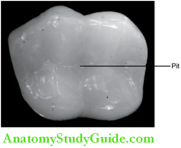
For example, the central pit – the deepest point in the central fossa of molars
Fissure:
A fissure is a narrow cleft or crevice at the depth of any groove formed due to incomplete fusion of enamel during tooth development.
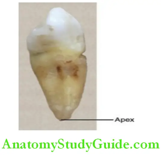
Apex:
The tip at the end of the root is called the apex.
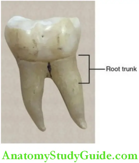
Root trunk:
The root trunk is the part of the root of a multirooted tooth from the cementoenamel junction to the point of splitting of the root.
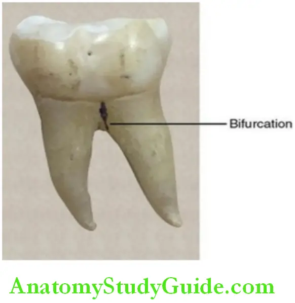
Furcation:
The place on a multirooted tooth where the root trunk divides into separate roots is called the furcation.
- Bifurcation: Seen on a two-rooted tooth
- Trifurcation: Present on a three-rooted tooth
Cervical line:
The junction between the crown and the root is represented by the cervical line.
Line Angles Point Angles And Thirds Of The Crown And Root
The surface of the crown and root are divided into thirds for descriptive purposes for studying the morphological features of the tooth.
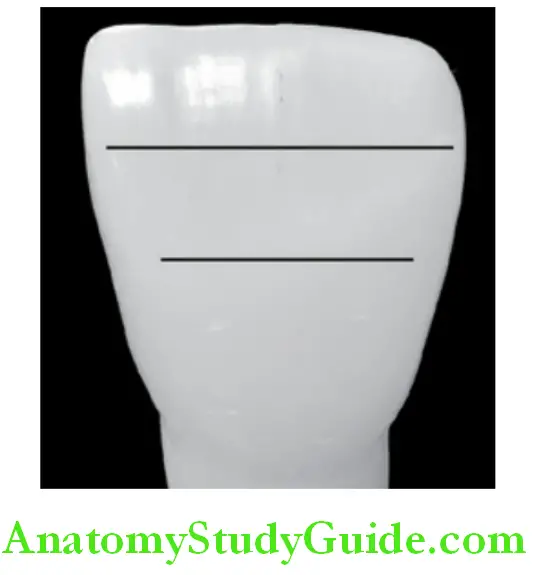
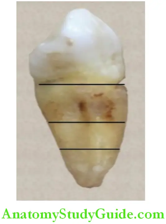
Crown:
The crown is divided into
- Incisal/occlusal third depending on whether the tooth is an anterior or posterior
- Middle third
- Cervical third
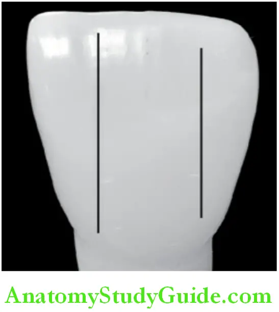
It may be further divided in three directions:
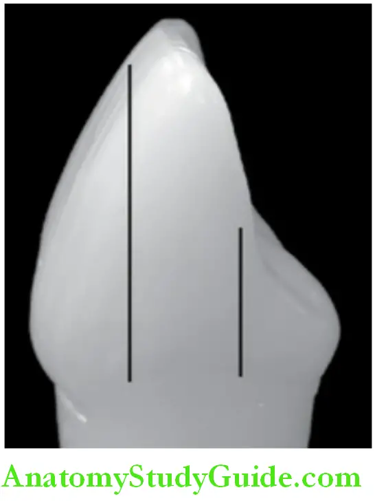
- Mesiodistally into:
- Mesial third
- Middle third
- Distal third
- Labiobuccolingually into:
- Labial or buccal third
- Middle third
- Lingual third; all five surfaces of the crown can thus be divided
Root:
The root can be divided into
- Cervical third
- Middle third
- Apical third
Line angles:
The junction of two surfaces is called a line angle (fig. 16.21). The name of the line angle is derived from the surfaces that join together.
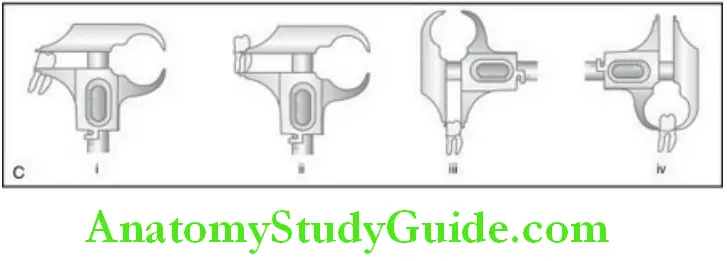
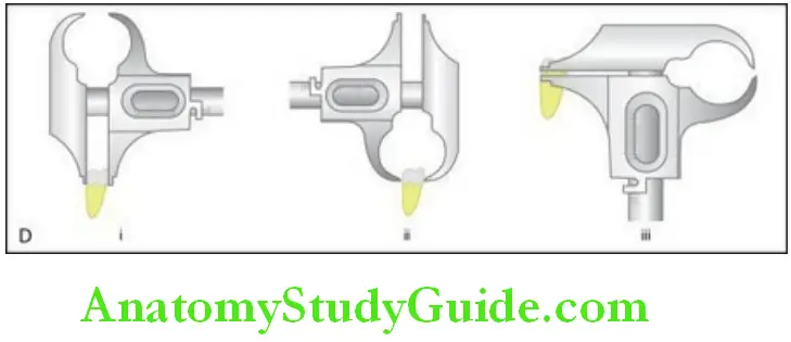
1. Measurements of teeth:
- Length of crown (labial)
- Length of root
- Mesiodistal diameter of the crown
- Mesiodistal diameter of the crown at the cervix.
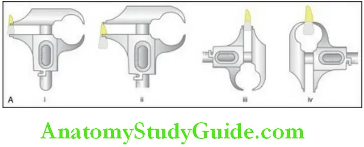
2. Measurements of teeth:
- Labiolingual diameter of the crown
- Lbiolingual diameter of the cervix
- The curvature of the cementoenamel junction of mesial.
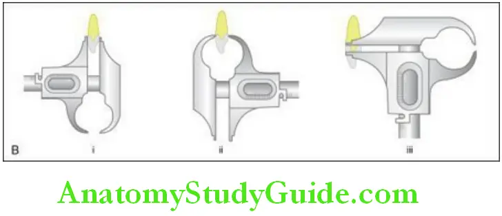
3. Measurements of teeth:
- Length of crown (buccal)
- Length of root
- Mesiodistal diameter of the crown
- Mesiodistal diameter of the crown at the cervix.
4. Measurements of teeth:
- Buccolingual diameter of the crown;
- Buccolingual diameter of the crown at cervix;
- The curvature of the cementoenamel junction of mesial.
For example, nasolabial line angle – is formed by the junction of the labial and mesial surfaces.
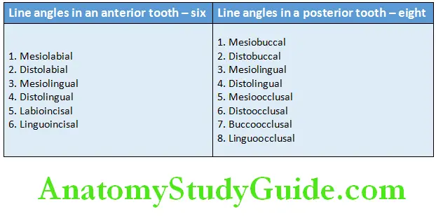
Note: Two opposing surfaces such as the mesial and distal surfaces do not form a line angle.
Point angles:
The junction of three surfaces at a point is called a point angle. The name of the point angle is derived by combining the three surfaces that form it.
For example, mesiobuccoocclusal point angle is formed by the junction of the mesial, buccal, and occlusal surfaces at a point.
Measurement Of Teeth
- The boley gauge is used to measure the record the eight different measurements of a tooth. The method of measuring an anterior and posterior tooth.
- The tissues of a tooth are enamel, dentin, cementum, and pulp. Enamel, dentin, and cementum are the hard tissues and are calcified. Pulp is a soft connective tissue.
- Enamel is the tissue that forms the outermost protective covering of the crown of the tooth. It is highly mineralized and rich in calcium hydroxyapatite. Enamel is laid down by ameloblasts.
- Dentin is a hard tissue that is yellow in colour and is present in between enamel and cementum. It makes up the bulk of the crown. It is less mineralized than enamel. It is laid down by odontoblasts.
- Pulp is the soft connective tissue present in the pulp cavity enclosed by dentin. It is present in the crown and in the root within the pulp chamber and the root canal. The pulp communicates with the tissue at the apex of the tooth via the apical foramen.
- Cementum is the tissue that covers the root of the tooth and is present over the dentin. It varies in thickness from the neck of the tooth to the apex of the root. It is a mineralized tissue which is harder than bone and softer than enamel. It is laid down by cementoblasts.
- Crown: The crown is the part of the tooth covered by enamel.
- Root: The root is the part of the tooth covered by cementum.
- The junction of the crown and root is the cervical/neck area of the tooth. This junction is the cementoenamel junction. The junction is marked by the cervical line.
- Alveolar bone: The part of the jaw that supports the teeth is called the alveolar bone.
- The teeth have surfaces based on the position, location, and uses of the tooth.
- Labial surface: The surface of the tooth facing the lips.
- Buccal surface: The surface of the tooth facing the cheek.
- Lingual surface: The surface of the tooth facing the tongue.
- Palatal surface: The surface of the tooth facing the palate.
- Proximal surfaces: The imaginary vertical line that passes in between the maxillary and mandibular central incisors is considered the midline which is the reference for the mesial and distal surfaces.
- Mesial surface: The surface of the tooth that is closer to the midline of the arch.
- Distal surface: The surface of the tooth that is away from the midline of the arch.
- Incisal and occlusal surfaces: The surfaces of the teeth that come in contact with those in the opposite arch when the jaws are closed are called the incisal or the occlusal surfaces.
- The occlusal surface is applicable to the posteriors, i.e. Premolars and the Molars.
- Lobe: It is one of the primary sections of formation in the development of the crown. The cusps and mamelons are representative of the lobes.
- Mamelons: Mamelons are any one of the three rounded protuberances or tubercles found on the incisal edges of the newly erupted incisor teeth.
- Cusp: A cusp is a pyramidal elevation or mound on the crown portion of the tooth making up a divisional part of the occlusal surface. Cusps are seen in canines and the posterior teeth.
- Tubercle: A tubercle is a smaller elevation on some portion of the crown Produced by an extra formation of enamel.
- Cingulum: Cingulum is the bulge on the cervical third of the lingual surface of the anterior teeth.
- Ridge: A linear elevation on the surface of a tooth and is named according to the location.
- Perikymata: Perikymata are multiple and minute horizontal ridges on the surface of the enamel of newly erupted permanent teeth.
- Fossa: A fossa is an irregular depression or concavity. It is seen on the lingual surface of the anterior teeth and the occlusal surface of the posterior teeth.
- Sulcus: A long depression or valley on the surface of a tooth running Mesiodistally between cusps and ridge.
- Groove: A linear depression in the deepest portion of the sulcus.
- Developmental groove: A sharply defined linear depression separating the lobes of major portions of a tooth formed during tooth development.
- Central groove: It is a groove that runs in a mesiodistal direction on the occlusal surface of a posterior tooth and divides it buccolingually into two halves.
- Pit: Pits are small pin-point depressions located at the junction of developmental grooves or at the terminals of those grooves. They are the deepest point of the fossa.
- Fissure: A fissure is a narrow cleft or crevice at the depth of any groove formed due to incomplete fusion of enamel during tooth development.
- Apex: The tip at the end of the root is called the apex.
- Root trunk: It is the part of the root of a multirooted tooth from the Cementoenamel junction to the point of splitting of the root.
- Furcation: The place on a multirooted tooth where the root trunk divides into separate roots.
- Cervical line: The junction between the crown and the root is represented by the cervical line.
- Line angles: The junction of two surfaces is called a line angle. The name of the line angle is derived from the surfaces that join together.
- Point angles: The junction of three surfaces at a point is called a point angle. The name of the point angle is derived by combining the three surfaces that form it.
Leave a Reply