Question 1. Write a short note on cerebellopontine angle (CP angle) tumors.
Answer:
- The cerebellopontine angle is an area of the lateral cistern containing CSF, arachnoid tissue, cranial nerves, and its associated vessels.
- CP angle tumors constitute the most common posterior fossa tumors and the majority of them are benign.
Various CP angle tumors
cerebellopontine angle Clinical Features
Most common: Progressive unilateral sensory neural hearing loss (SNHL) (retrocochlear) present in 95% of cases and often accompanied by tinnitus which is present in 65% of cases.
Rare presentations include facial numbness or pain, earache or facial weakness, cerebellar ataxia, or symptoms of hydrocephalus (headache, visual disturbance, mental status change, nausea, and vomiting).
cerebellopontine angle Signs
Read And Learn More: General Medicine Question And Answers
Ear: Normal otoscopy
Cranial nerves:
- 5th cranial nerve: The earliest sign is impaired corneal reflex. Motor functions are affected rarely.
- 7th cranial nerve: Sensory first; loss of sensation in the posterior superior aspect of the external auditory canal (EAC) called Hitselberger’s sign. Lower motor neuron facial palsy develops later
- 9th and 10th cranial nerve: Palsy-palatal, pharyngeal, and laryngeal paralysis.
- Eyes: Nystagmus.
- Cerebellar signs present (ipsilateral).
cerebellopontine angle Investigations
- Audiological tests: They show features of retro cochlear hearing loss (high-frequency SNHL), recruitment negative, poor speech discrimination score, and the presence of rollover phenomenon.
- Acoustic reflex: Nearly in 75% of patients the stapedial reflex is lost.
- Caloric test: It is diminished or absent. Normal test finding does not eliminate the diagnosis.
- Plain X-ray: The best view is the periorbital view; a difference of 1 mm in the vertical height of the internal acoustic meatus is significant.
- It cannot detect intermetal tumors.
- MRI: With gadolinium enhancement is the gold standard.
- Treatment
- Surgical excision
- Radiotherapy: Stereotactic radiosurgery (gamma knife) may stop the growth of vestibular schwannoma mainly in small intracanalicular and extra canalicular lesions
Radiosurgery is recommended in bilateral vestibular schwannomas (e.g., Morbus Recklinghausen) but only when the tumors are small.
Annual imaging is recommended for all patients being managed conservatively
for the rest of their life or until vestibular schwannoma growth is seen to a certain limit.
- Few common tumors of CNS.
- Astrocytoma
- Low-grade astrocytoma
- High-grade astrocytoma
- Oligodendrogliomas
- Ependymomas
- Medulloblastoma
- CNS lymphoma (more common in HIV patients)
- Meningiomas
- Schwannomas
- Cerebellopontine angle tumors.
- Vestibular schwannoma/
- acoustic neuroma
- Meningioma
- Cerebellar glioma
- Less likely: Arachnoid cyst, non-acoustic cranial nerve schwannomas, vascular malformations, dermoids, teratomas, and lipomas.
Acoustic Neuroma (Schwannoma)
Question 2. Write a short note on the acoustic neuroma.
Answer:
- It is a benign tumor that arises from Schwann cells of the 8th cranial (vestibular) nerve.
- The majority are sporadic and unilateral.
- Common between the 4th and 6th decade of life, with a slight female preponderance.
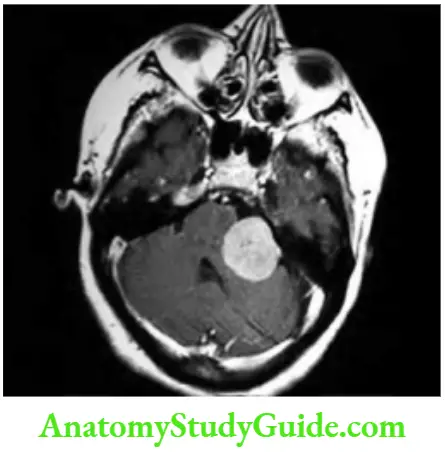
- Site: Commonly arises near the nerve’s entry point into the medulla or in the internal auditory meatus and extends into the cerebellopontine angle. Constitutes 80–90% of tumors at the cerebellopontine angle.
Acoustic Neuroma (Schwannoma) Clinical features: Unilateral progressive hearing impairment/loss, sometimes with tinnitus. Large tumors may manifest signs and symptoms of cerebellar and brainstem involvement.
Acoustic Neuroma (Schwannoma) Investigations: MRI is the investigation of choice.
Acoustic Neuroma (Schwannoma) Management: Total surgical excision is the treatment of choice. Stereotactic radiosurgery (radiotherapy) may be used for some tumors.
Question 3. Write a short note on radiological investigation used in neurology.
Answer:
- About 90% of neurological cases can be diagnosed by history alone, with a lesser contribution from examination and investigation.
- Investigations include assessment of structure (imaging) and function (neurophysiology).
- Neurological imaging is used for the assessment of structure.
- Various techniques include X-rays (plain X-rays, CT, CT angiography, myelography, and angiography), magnetic resonance (MRI, MRA), and ultrasound (Doppler imaging of blood vessels).
- However, it is now possible to use imaging techniques to assess CNS function also.
- Single-photon emission computed tomography scanning can be used to mark CBF by using a lipid-soluble radioactive tracer.
- It is useful in dementia or epilepsy.
- Single photon emission computed tomography (SPECT) is also useful in the diagnosis of movement disorders (e.g., by examining dopamine activity to assess the function of the basal ganglia in patients with suspected parkinsonism).
Magnetic Resonance Imaging
Magnetic resonance imaging is an imaging technique used mainly in the medical field to produce high-quality images of the inside structures of the human.
The principle of MRI is based on the presence of hydrogen atoms in all human tissues.
A hydrogen nucleus/atom is a proton whose electrical charge creates a local electrical field.
Protons in body tissue are aligned to the magnetic axes. When protons are surrounded by sudden strong magnetic impulses/field (as in an MRI machine), the protons are aligned along the field.
Application of a radiofrequency wave at right angles to their alignment and is imaged.
Then the field is suddenly reduced, and the protons resonate and spin, then revert to their normal alignment.
When the magnitude and rate of energy release occur with a return to baseline alignment, images are made at different phases of relaxation.
They are known as T1, T2, T2 STIR, FLAIR, diffusion-weighted imaging (DWI), and other sequences. The recording is done by a coil.
These intensities are used to produce images. The T1-weighted sequence accentuates substances that contain fat and T 2-weighted sequence accentuates substances that contain water.
Acoustic Neuroma Interpretation: In the brain, T1-weighted images reveal the nerve connections of white matter and appear white (hyperintense), the congregations of neurons of gray matter appear gray and cerebrospinal fluid appears dark.
- These are reversed in T 2-weighted imaging.
- Neurological indications for MRI
- MRI is the investigation of choice for the evaluation of all neurological disorders:
- Structural imaging: Produces high-quality soft tissue images.
- Useful in the investigation of disease of the posterior fossa and temporal lobes, inflammatory conditions (e.g., multiple sclerosis), and in investigating epilepsy
- Magnetic resonance angiography (MRA) to study blood vessels in the neck or brain
- Functional MRI: Mainly research tools
- MR spectroscopy: Mainly research tools
Leave a Reply