Hepatic Tuberculosis
- Tuberculosis of the liver occurs as a result of military dissemination from primary complex or from chronic adult pulmonary tuberculosis.
- The diagnosis is possible by liver biopsy. The patients may have unexplained fever, jaundice, hepatomegaly or hepatosplenomegaly. There may be elevated serum alkaline phosphatase levels and hyperglobulinaemia.
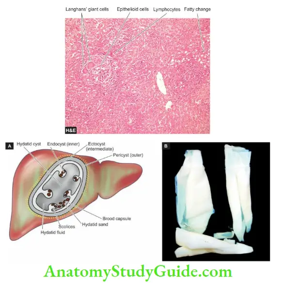
- Morphologic Features Basic lesion is the epithelioid cell granuloma characterised by central caseation necrosis with the destruction of the reticulin framework and peripheral cuff of lymphocytes.
- Ziehl-Neelsen staining for AFB or culture of the organism from the biopsy tissue is confirmatory. Rare lesions consist of tuberculous cholangitis and tuberculous pylephlebitis.
Read And Learn More: Systemic Pathology Notes
Hydatid Disease (Echinococcosis)
- Hydatid disease occurs as a result of infection by the larval cyst stage of the tapeworm, Echinococcus granulosa. The dog is the common definite host, while man, sheep and cattle are the intermediate hosts.
- The dog is infected by eating the viscera of sheep containing hydatid cysts. The infected faeces of the dog contaminate grass and farmland from where the ova are ingested by sheep, pigs and man.
- Thus, man can acquire infection by handling dogs as well as by eating contaminated vegetables.
- The ova ingested by man is liberated from the chitinous wall by gastric juice and pass through the intestinal mucosa from where they are carried to the liver by the portal venous system.
- These are trapped in the hepatic sinusoids where they eventually develop into hydatid cysts. About 70% of hydatid cysts develop in the liver which acts as the first filter for ova.
- However, ova which pass through the liver enter the right side of the heart and are caught in the pulmonary capillary bed and form pulmonary hydatid cysts.
- Some ova which enter the systemic circulation gives rise to hydatid cysts in the brain, spleen, bone and.
- The disease is common in sheep-raising countries such as Australia, New Zealand and South America.
- The uncomplicated hydatid cyst of the liver may be silent or may produce a dull ache in the liver area and some abdominal distension.
- Complications of hydatid cyst include its rupture (for example into the peritoneal cavity, bile ducts and lungs), secondary infection and hydatid allergy due to sensitisation of the host with cyst fluid.
- The diagnosis is made by peripheral blood eosinophilia, radiologic examination and serologic tests such as indirect haemagglutination test and C along skin test.
Morphologic Features: Hydatid cyst grows slowly and may eventually attain a size over 10 cm in diameter in about 5 years. E.
- granulose generally causes unilocular hydatid cysts while E. multilocular results in multilocular or alveolar hydatid disease in the liver.
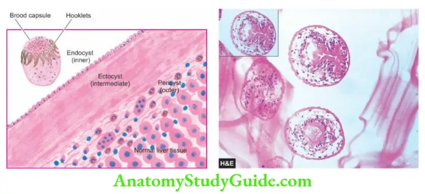
- The cyst wall is composed of 3 distinguishable zones—outer pericyst, intermediate characteristic ectocyst and inner endocyst:
- Pericyst is the outer host inflammatory reaction consisting of fibroblastic proliferation, mononuclear cells, eosinophils and giant cells, eventually developing into dense fibrous capsules which may even calcify.
- Ectocyst is the intermediate layer composed of characteristic acellular, chitinous, laminated hyaline material.
- The endocyst is the inner germinal layer bearing daughter cysts (brood-capsules) and scolices projecting into the lumen.
Hydatid sand: is a grain-like material composed of numerous scolices present in the hydatid fluid.
- Hydatid fluid, in addition, contains antigenic proteins so that its liberation into circulation gives rise to pronounced eosinophilia or may cause anaphylaxis
Other Infections of the Liver
- Cholangitis occurring secondary to obstruction of a major extrahepatic duct causes pyogenic cholangitis.
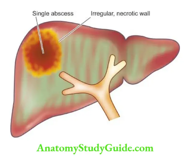
- Pyogenic liver abscesses can occur by ascending infection, portal pyaemia, septicaemia, and direct infection.
- Amoebic liver abscesses are caused by the spread of trophozoites of Entamoeba histolytica from intestinal lesions.
- Tuberculosis of the liver occurs by miliary dissemination from another site, most often chronic adult pulmonary tuberculosis.
- Hydatid cyst has an endocyst, ectocyst and pericyst and it occurs as a result of infection by the tapeworm larvae, Echinococcus granulosa.
Leave a Reply