Visual Pathway Introduction
- The retinal impulses are carried to the visual center in the cerebral cortex by the nervous pathway called the visual pathway or optic pathway.
- In binocular vision, the light rays from the temporal (outer) half of the visual field fall upon the nasal part of the corresponding retina. The rays from the nasal (inner) half of the visual field fall upon the temporal part of the retina.
Read And Learn More: Medical Physiology Notes
Table of Contents
Visual Receptors
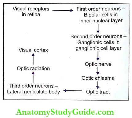
Rods and cones, which are present in the retina of the eye, form the visual receptors. Fibers from the visual receptors synapse with dendrites of bipolar cells of an inner nuclear layer of the retina.
- First-Order Neurons: First-order neurons (primary neurons) are bipolar cells in the retina. Axons from the bipolar cells synapse with dendrites of ganglionic cells.
- Second-Order Neurons: Second-order neurons (secondary neurons) are the ganglionic cells in the ganglionic cell layer of the retina. The axons of the ganglionic cells form the optic nerve. The optic nerve leaves the eye and terminates in the lateral geniculate body.
- Third-Order Neurons: The third-order neurons are in the lateral geniculate body. Fibers arising from here reach the visual cortex.
Connections Of Visual Receptors To Optic Nerve
Two pathways exist between the visual receptors and optic nerve:
- Private pathway
- Diffuse pathway.
1. Private Pathway:
- The individual cones in the fovea centralis are connected to separate bipolar cells. Each bipolar cell is connected to a separate ganglionic cell namely, the midget ganglionic cell.
- Thus, the individual cone is connected to an individual optic nerve fiber. This type of private pathway is responsible for visual acuity and intensity discrimination.
2. Diffuse Pathway: A number of cones and rods are connected with a polysynaptic bipolar cell. The bipolar cells are connected to diffused ganglionic cells. So, there is great overlapping. This type of pathway is present outside the fovea.
Course Of Visual Pathway
The visual pathway consists of six components:
- Optic nerve
- Optic chiasma
- Optic tract
- Lateral geniculate body
- Optic radiation
- Visual cortex.
1. Optic Nerve:
- It is formed by the axons of ganglionic cells. The optic nerve leaves the eye through the optic disk. The fibers from the temporal part of the retina are in the lateral part of the nerve and carry the impulses from the nasal half of the visual field of the same eye.
- The fibers from the nasal part of the retina are in the medial part of the nerve and carry the impulses from the temporal half of the visual field of the same eye.
2. Optic Chiasma: The medial fibers of each optic nerve cross the midline and join the uncrossed lateral fibers of the opposite side to form the optic tract. The area of the crossing of the optic nerve fibers is called optic chiasma.
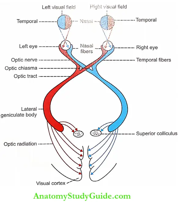
3. Optic Tract:
- It is formed by uncrossed fibers of the optic nerve on the same side and crossed fibers of the optic nerve from the opposite side. All the fibers of the optic tract run backward, outward, and toward the cerebral peduncle. While reaching the peduncle, the fibers pass between the tuber cinereum and the anterior perforated substance.
- Then, the fibers turn around the peduncle to reach the lateral geniculate body in the thalamus. Here, many fibers synapse while few fibers just pass through this and run towards the superior colliculus in the midbrain. Fibers from the fovea do not enter the superior colliculus.
- Some fibers from the fovea of each side pass through an optic tract of the same side and others through an optic tract of the opposite side. Due to the crossing of medial fibers in optic chiasma, the left optic tract carries impulses from the temporal part of the left retina and the nasal part of the right retina, i.e. it is responsible for vision in the nasal half of the left visual field and temporal half of right visual field.
- The right optic tract contains fibers from the nasal half of the left retina and the temporal half of the right retina. It is responsible for vision in the temporal half of the left visual field and the nasal half of the right visual field.
4. Lateral Geniculate Body:
- The majority of the fibers of the optic tract terminate in the lateral geniculate body, which forms the subcortical center for visual sensation. From here, the geniculocalcarine tract or optic radiation arises. This tract is the last relay of the visual pathway.
- Some of the fibers from the optic tract do not synapse in the lateral geniculate body but, pass through it and terminate in one of the following centers:
- The superior colliculus: It is concerned with reflex movements of eyeballs and head in response to optic stimulus
- Pretectal nucleus: It is concerned with light reflexes
- The supraoptic nucleus of the hypothalamus: It is concerned with the retinal control of the pituitary in animals. But in humans, it does not play any important role.
5. Optic Radiation: Fibers from the lateral geniculate body pass through the internal capsule and form optic radiation. The fibers between the lateral geniculate body and the visual cortex are also called geniculocalcarine fibers. Optic radiation ends in the visual cortex.
6. Visual Cortex:
- The primary cortical center for vision is called the visual cortex which is located on the medial surface of the occipital lobe. It forms the walls and lips of a calcarine fissure in the medial surface of the occipital lobe.
- There is definite localization of retinal projections upon the visual cortex, In fact, the point-to-point projection of the retina upon the visual cortex is well established. The peripheral retina! representation occupies the anterior part of the visual cortex. The macular representation occupies the posterior part of the visual cortex near the occipital pole.
Areas of Visual Cortex and their Function: Three areas are present in the visual cortex:
- Primary visual area (area 17) – Concerned with the perception of visual impulses
- Visual association area (area 18) – Concerned with the interpretation of visual impulses
- Occipital eye field (area 19) – Concerned with movement of eyes.
Applied Physiology Effects Of Lesion At Different Levels Of Visual Pathway
The injury to any part of the optic pathway causes a visual defect and the nature of the defect depends upon the location and extent of the injury. The loss of vision in one visual field is known as anopia. Loss of vision in one half of the visual field is called hemianopia. Hemianopia is classified into two types:
- Homonymous hemianopia
- Heteronymous hemianopia.
1. Homonymous hemianopia:
- Homonymous hemianopia means loss of vision in the same halves of both visual fields. Loss of vision in the right half of the visual field of both eyes is known as right homonymous hemianopia.
- Similarly, left homonymous hemianopia means loss of vision in the left half of the visual field of both eyes.
2. Heteronymous hemianopia:
- Heteronymous hemianopia means loss of vision in opposite halves of the visual field. For example, binaural heteronymous hemianopia means loss of vision in the right half of the left visual field and the left half of the right visual field (nasal half of both visual fields).
- Bitemporal heteronymous hemianopia is the loss of sight in the left side of the left visual field and the right side of the right visual field (temporal half of both visual fields).
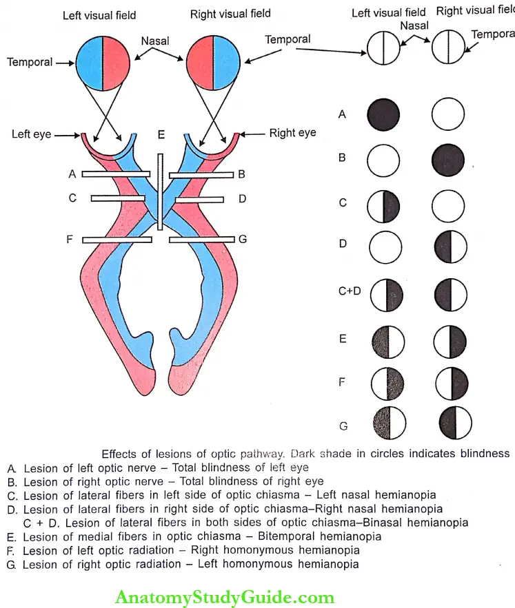
- Effects of Lesion of Optic Nerve: The lesion in one optic nerve will cause total blindness or anopia in the corresponding visual field. The lesion occurs due to increased intracranial pressure.
- Effects of Lesion of Optic Chiasma: The nature of the defect depends upon the fibers involved.
- Pressure on uncrossed lateral fibers by aneurysmal dilatation of the carotid artery causes blindness in the temporal part of the retina of the same side, i.e. the retina cannot receive light stimulus from the objects in the nasal half of the same visual field. So, the hemianopia developed is called left or right nasal hemianopia
- If lateral fibers of both sides are affected, the vision is lost in the nasal half of both visual fields causing binaural hemianopia. It occurs due to dilated third ventricle, which forces the angle of chiasma against carotid arteries. It also occurs due to dilatation of the carotid artery on both sides
- Compression of the nasal, i.e. crossed fibers by pituitary tumor causes bitemporal hemianopia.
- Effects of Lesion of Optic Tract, Lateral Geniculate Body, and Optic Radiation:
- The lesion of the optic tract or lateral geniculate body or optic radiation causes homonymous hemianopia. In the right-sided lesion, there is loss of vision in the right side of both retinas, i.e. in the left side of both visual fields – left homonymous hemianopia.
- In the left-sided lesion, there is loss of vision in the left half of the retina of both eyes and loss of sight on the right half of both visual fields – right homonymous hemianopia.
- Effects of Lesion of Visual Cortex: The lesion of the upper or lower part of the visual cortex leads to inferior or superior homonymous hemianopia.
- Macular sparing: In all the conditions mentioned above, total blindness does not occur because the macular vision is not lost. This phenomenon in which the macular vision is retained (unaffected) in conditions of hemianopia is called macular sparing.
- Macular sparing occurs because of the following reasons:
- The fibers from the macula project into the visual cortex of both sides
- Fibers from the macular region are projected into both the anterior and posterior parts of each visual cortex. Only the bilateral lesion of the visual cortex causes total blindness.
- Macular sparing occurs because of the following reasons:
- Macular sparing: In all the conditions mentioned above, total blindness does not occur because the macular vision is not lost. This phenomenon in which the macular vision is retained (unaffected) in conditions of hemianopia is called macular sparing.

Leave a Reply