Bones Of The Skull
The skull consists of cranium with mandible. The bones of the skull are classified as
Table of Contents
1. Paired, and
2. Unpaired.
1. Paired: These are one on each side.
Read And Learn More: Head Anatomy Notes And Important Questions With Answers
2. Unpaired: They are situated in the midline.
- To recollect the bones, imagine interior of the base of the skull.
- Visualise, that you are sliding your finger from anterior to posterior in the midline.
- It slides on frontal, ethmoid, sphenoid and occipital bone.
- Now slide the finger on the exterior of base of the skull.
- The finger slides on occipital, sphenoid, vomer and mandible.
- Vomer is situated between the two choanae.
Macewen Triangle Boundaries
All unpaired bones are summarized as
Frontal,
Ethmoid,
Sphenoid,
Occipital,
Vomer, and
Mandible.
Pterion
Pterion (Gr. Pteryx-wing)
1. It is H-shaped suture presents on the lateral side of the skull.
2. Formation: It is formed by four bones.
A. Frontal,
B. Parietal,
C. Sphenoid, and
D. Temporal.
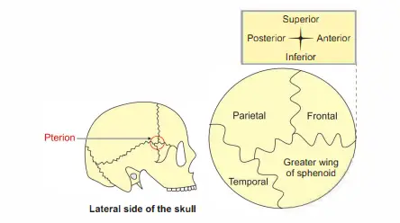
3. Pterion Situation: It is situated 4 cm above the midpoint of zygomatic arch and 2.5 cm behind the frontozygomatic suture.
4. Pterion Relations: Structures deep to pterion.
A. Middle meningeal vessels.
B. Stem of lateral sulcus of cerebral hemisphere (Sylvian point).
Macewen Triangle Boundaries
5. Pterion Applied Anatomy
- Fracture of the pterion can be life-threatening because it overlies the anterior branches of the middle meningeal vessels.
- The vessels lie in the groove on the internal aspect of the lateral wall of the skull.
- The extradural haematoma exerts a local pressure on the corresponding cerebral cortex.
- The motor area lies deep into the haematoma.
- The centre of the pterion is an important landmark for a neurosurgeon to make Burr holes.
- An untreated middle meningeal artery haemorrhage may cause death.
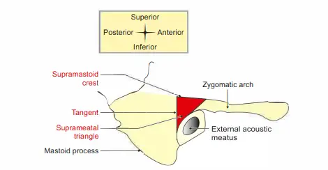
Supramental Triangle (MacEwen’s triangle)
It is a triangular depression, present above and behind the external acoustic meatus.
1. Boundaries: Margins of the supramental triangle are: I …. SETI
2. Above byupramastoid crest.
3. Anteriorly by posterosuperior margin ofxtemal acoustic meatus.
4. Behind by tangential line drawn from the posterior margin of the external acoustic meatus.
Supramental Triangle (MacEwen’s triangle) Applied anatomy
The suprameatal triangle corresponds with cymbal concha of the auricle and the mastoid antrum.
At birth, the mastoid antrum is situated 1 mm deep to the suprameatal triangle.
It increases by 1 mm/year till puberty. In an adult, it is situated 12 mm deep to the suprameatal triangle.
Macewen Triangle Boundaries
Mastoid process (Masts-breast, aid-like)
It is a large projection from the lower part of the mastoid part of the temporal bone.
It forms the lateral wall of the mastoid notch.
1. Situation: Posteroinferior part of the external acoustic meatus.
2. Types of mastoid process: Depending upon the distribution of air cells, it is of the following types:
- Pneumatic: It shows many air cells.
- Sclerotic: It has few or no air cells.
- Mixed: It contains air cells and bone marrow in equal proportion.
3. Mastoid process Development
- It develops in 2nd year of life and is usually better developed in males Cf’ than in females <?
- It is an example of traction epiphysis.
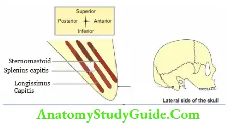
4. Mastoid process Attachments: The following muscles are attached from superficial to deep.
Boundaries Of Suprameatal Triangle
SSC LC
Sternocleidomastoid (spinal root of the accessory nerve),
Splenius capitis (Cl to C6 nerves), and
Longissimus capitis (dorsal rami of cervical nerve).
5. Relations
1. It is grooved on the deep aspect by a digastric notch for the origin of the posterior belly of the digastric.
Medial to the notch is a groove for the occipital artery, facial nerve, and stylomastoid branch of the posterior auricular artery.
Macewen Triangle Boundaries
2. Stylomastoid foramen is present between the mastoid process and the styloid process.
It transmits
1. Facial nerve, and
2. Stylomastoid branch of the posterior auricular artery.
Boundaries Of Suprameatal Triangle – Applied anatomy
The facial nerve is likely to get damaged in the application of forceps during assisted delivery.
At birth, the stylomastoid foramen is more superficial for the following reasons.
- The mastoid process is not developed at birth, and
- The external acoustic canal is short.
Styloid Process
Styloid {stylo-stake (wooden or metal post-pointed at one end), aid-resemblance}
It is a long, slender and pointed bony process. It measures about 2.5 cm, it arises from the inferior surface of the temporal bone.
It is directed downwards and forwards._:
1. Site: It is present on the inferior surface of the petrous part of the temporal bone.
2. Parts of the styloid process
- Tympanohyal {Hyal-hyoid U (shaped like a Greek letter upsilon (u)}: It is covered by a tympanic plate.
- Stylohyal: It is the lower part of the styloid process.
Macewen Triangle Boundaries
3. Styloid Process Relations
- Medially: From anterior to posterior
- Internal carotid artery,
- IX, X and XI cranial nerves, and
- Internal jugular vein.
- Laterally:
- At base: Facial nerve,
- At apex: External carotid artery,
- Overlapped by the parotid gland.
Macewen’s Triangle
4. Styloid Process Attachments
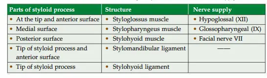
- Styloid process + structures attached to styloid process = styloid apparatus.
5. Styloid Process Development: It is developed from the cartilage of the 2nd pharyngeal arch.
- Its tympanohyal part ossifies before birth.
- The stylohyal part ossifies after birth.
Macewen’s Triangle
Foetal skull Features
- Disproportion between cranial vault and facial skeleton. The vault is very large in proportion to the face.
- The vertical diameter of the orbit equals the sum of the vertical height of the maxilla and mandible.
- The bones of the skull and of the face are loose. They are readily disarticulated in the macerated skull.
- The bones of the vault do not interdigitate in sutures but are separated by linear attachments of fibrous tissue.
- The anterior fontanelle lies between two parietal bones and two halves of frontal bones.
- The posterior fontanelle lies between the apex of the occipital bone and the posterior borders of
the parietal bone. - Stylomastoid foramen is near the lateral surface of the skull.
- External acoustic meatus is wholly cartilaginous
2. Ossification of bones
- Frontal bone: It consists of two halves separated by a median metopic suture.
- Temporal bone: It consists of four parts
- Petromastoid part,
- Squamous part,
- Styloid process, and
- Tympanic plate.
- The mandible is in two halves.
- The occipital bone is in four parts
- Squamous-1
- Basilar-1
- Condylar-2
- Sphenoid is in three pieces
- The central part formed by the body and lesser wings,
- The lateral part formed by the greater wing, and
- Pterygoid process.
Macewen’s Triangle
3. The following structures are absent in the foetal skull.
- Glabella, superciliary arch and mastoid process.
- The bony part of the external acoustic meatus.
- There is no diploe in the bones of the cranial vault.
- The tympanic part is present as C C-shaped ring.
- The maxillary air sinus is a narrow slit.
4. The following structures are of adult size in foetal skull
- Tympanic membrane
- The bones of the middle ear (malleus, incus and stapes).
- The middle ear, internal ear and mastoid antrum. These are enclosed in the petromastoid part of the temporal bone.
5. Applied anatomy: As the stylomastoid foramen is near the lateral surface of the skull, the facial nerve is unprotected and vulnerable.
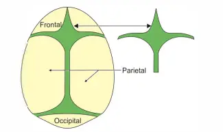
Fontanelle (fonticuli)
Fontanelle (fonticuli) Introduction: They are membranous gaps present at the 4 angles of the parietal bone.
1. Fontanelle Features: They are paired and unpaired.
2. Fontanelle Unpaired
- Anterior fontanelle, and
- Posterior fontanelle.
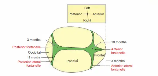
3. Fontanelle Paired
- Anterolateral fontanelle, and
- Posterolateral fontanelle.
Macewen’s Triangle
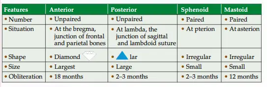
4. Fontanelle (fonticuli) – Applied anatomy
- It helps to determine the age of the child. If it persists after 2 years, it indicates
- Disturbance in calcium metabolism or/ and
- Deficiency of vitamin D.
- Abnormal bulging indicates increased intracranial tension.
- Abnormally depressed fontanelle indicates dehydration.
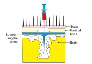
- The superior sagittal sinus can be punctured at the anterior fontanelle to
- Collect the venous blood for investigation.
- Start the infusion of fluid.
- Transfuse blood.
- Nowadays, puncture of the superior sagittal sinus is not used for the above purposes.
- Central venous catheterization is used for the above purposes.
- Cerebrospinal fluid can be obtained by introducing a needle from its lateral angle in a downward and lateral direction.
- It allows malleability of the foetal head (moulding of skull bones) during its passage through the birth canal at the time of delivery.
- It helps in the diagnosis of the presentation, position and attitude of the foetal head.
Emissary’s veins
Emissary veins (emissary-drain, to send, messenger, ambassador)
Emissary veins Introduction: These veins connect the intracranial venous sinuses to extracranial veins
- Peculiarities: They lack
- Valves,
- Smooth muscles, and
- Compressibility.
- Function: They maintain intracranial pressure.
Suprameatal Triangle Boundaries
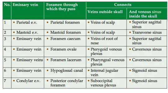
Emissary veins Applied anatomy: Infection outside the cranium spreads to the venous sinuses through the emissary vein.
Enumerate structures within the parotid salivary gland.
They are grouped as
- Arteries: External carotid artery and its two terminal branches
Maxillary artery, and
Superficial temporal artery. - Veins: Formation of the retromandibular vein and its tributaries. Veins forming retromandibular vein
Maxillary vein, and
Superficial temporal vein
Tributaries of retromandibular vein
Anterior division of retromandibular vein
Posterior division of retromandibular vein - Nerves
Terminal branches of the facial nerve
Temporal
Zygomatic
Buccal which subdivides into
Upper buccal, and
Lower buccal
Mandibular
Cervical
Auriculotemporal nerve branch of the posterior division of mandibular nerve. - Parotid duct
Suprameatal Triangle Boundaries
Foramina of middle cranial fossa
- Optic canal
Optic nerve (kind cranial nerve) with meninges,
Ophthalmic artery with sympathetic plexus, and
The central artery of the retina.
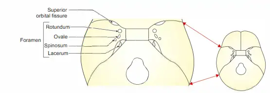
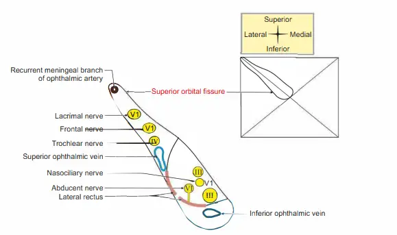
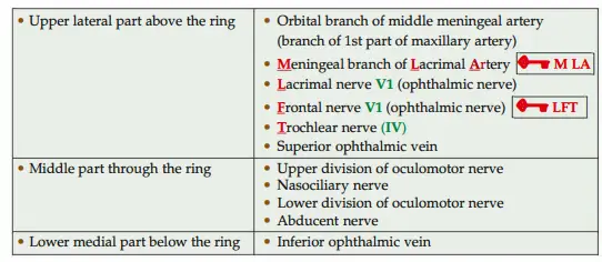
- Superior orbital fissure
Outside tendinous ring (from above downwards)
Vessels
Meningeal branch of the lacrimal artery
Anastomosing branch of middle meningeal artery1
Superior ophthalmic vein
Nerves I …. LFTI
Lacrimal branch of the ophthalmic division of trigeminal nerve (Vl)
Frontal branch of ophthalmic division of trigeminal nerve (Vl)
Trochlear nerve (IV)
In the tendinous ring
Upper-division of oculomotor nerve (III)
Nasociliary branch of the ophthalmic division of trigeminal nerve (Vl)
Lower division of oculomotor nerve (III)
Abducent nerve (VI)
Below the tendinous ring
Inferior ophthalmic vein
Foramen ovale (Y’>f I …. MALE
The trunk of the Mandibular nerve, 3rd division of the trigeminal nerve, the Accessory meningeal artery (branch of the maxillary artery),
Lesser petrosal nerve (branch of tympanic plexus), and Emissary vein.
It communicates cavernous sinus with
Pterygoid venous plexus, and
The anterior division of the middle meningeal sinus. - Foramen spinosum
Middle meningeal artery (branch of the maxillary artery)
Middle meningeal vein
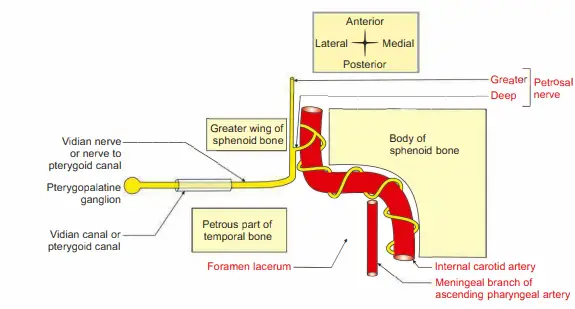
Meningeal branch of mandibular nerve - Foramen lacerum
No structures pass through and through except the Meningeal branch of
Ascending Pharyngeal artery.
The internal carotid artery passes from lateral to medial.
The nerve to the pterygoid canal (Vidian nerve) is formed by deep petrosal (sympathetic plexus around internal carotid artery) and greater petrosal nerves (branch of the facial nerve).
Superior orbital fissure
1. Superior Orbital Fissure Location: It is present in the superior wall of the orbit.
2. Superior Orbital Fissure Shape: Retort shaped.
3. Superior Orbital Fissure Features
It is the gap present between the greater and lesser wings of the sphenoid bone.
It is completed laterally by the frontal bone uniting the two wings.
Note: The dimensions of various nerves
Optic nerve: Thickest sensory cranial nerve. It contains 1 million neurons.
The lower division of the oculomotor nerve is thicker as compared to the upper division. It has more branches.
Trochlear and abducent nerves are less thick than the oculomotor nerve.
The nasociliary is a thin nerve.
Suprameatal Triangle Boundaries
4. Superior Orbital Fissure Contents
5. Superior Orbital Fissure Applied Anatomy: Fracture causing craniofacial disjunction may damage the 3rd,4th and 6th cranial nerves in the superior orbital fissure. I
Inferior orbital fissure
- Location: It is located between the upper margin of the posterior wall of the maxilla and the greater wing of the sphenoid bone.
- Communication: It communicates with
Orbit superiorly,
Infratemporal fossa laterally, and
Pterygopalatine fossa medially.
Suprameatal Triangle Boundaries
- Contents
Maxillary nerve which continues as an infraorbital nerve once it enters the orbit.
Zygomatic nerve, a branch of the maxillary nerve.
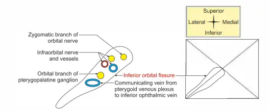
- Infratemporal artery, is a branch of the maxillary artery.
Infratemporal vein, a tributary of maxillary vein.
Connecting channels between the inferior ophthalmic vein and the pterygoid plexus of veins. - Applied anatomy
The infraorbital nerve is invariably damaged in depressed fractures of the zygomatic bone or in fractures of the orbital floor.
Malignant tumours of the maxillary sinus may cause pressure effects on the nerve.
There may be pain or numbness of the cheek, lower eyelid, incisor teeth and adjacent gums.
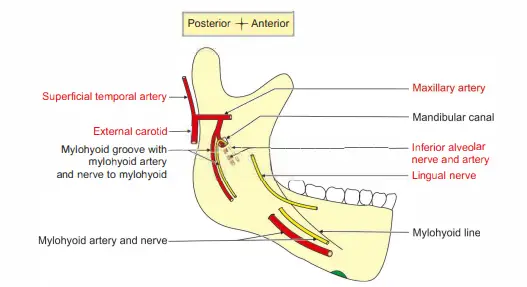
Arteries and nerves related to the ramus of the mandible
Following are the nerves and vessels related to the mandible.
1. Masseteric nerves and vessels pass through the mandibular notch.
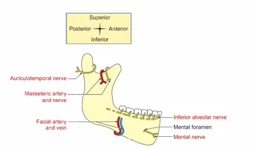
2. Inferior alveolar nerves and vessels enter the mandibular canal through the mandibular foramen.
3. Mylohyoid nerves and vessels run forward in the mylohyoid groove.
4. Mental nerves and vessels emerge through the mental foramen.
5. The facial artery is closely related to the mandible.
6. The maxillary artery and some of its branches are related between the lower border of the lateral pterygoid and medial pterygoid.
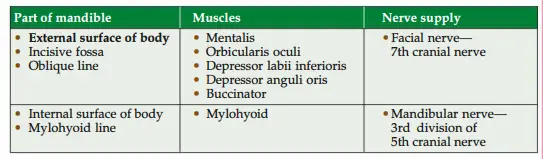
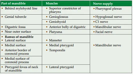
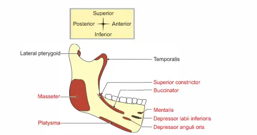
Name the muscles attached to the mandible
Spine of sphenoid
Spine of sphenoid Introduction: It is an irregular downward projection arising from the inferior surface of the greater wing of the sphenoid bone.
1. Attachment: It gives attachment to the sphenomandibular ligament.
2. Relations
Medially: Chorda tympani nerve, a branch of the facial nerve-7th cranial nerve.
Laterally: Auriculotemporal nerve, a branch of the posterior division of the mandibular nerve.
Suprameatal Triangle Boundaries
3. Applied anatomy: The damage to the spine of the sphenoid stops the secretion of all major salivary glands.
This is because of its close association with chorda tympani and the auriculotemporal nerve.
Lateral pterygoid plate
Lateral Pterygoid Plate Introduction: It is a quadrilateral, thin plate arising from the body of the sphenoid bone.
1. Surfaces: It has medial and lateral surfaces.
2. Features
The lateral surface forms the medial boundary of the infratemporal fossa.
The medial surface forms the lateral wall of the pterygoid fossa.
The anterior border of the lateral pterygoid plate forms the posterior boundary of -o
4. Ossification: The pterygoid plate develops in the membrane.
3. Attachments
Lateral surface-lower head of lateral pterygoid muscle.
Medial surface-origin to medial pterygoid muscle.
Posterior border-pterygospinous ligament.
Jugular foramen
(Jugular-pertaining to neck)
1. Jugular Foramen Location: It is situated between the jugular process of the occipital bone and the petrous part of the temporal bone.
2. Jugular Foramen Features
The anterior wall is hollowed and forms the jugular fossa. It lodges the superior bulb of the internal jugular vein.
It presents three canaliculi
The mastoid canaliculus transmits the auricular branch of the vagus nerve.
The cochlear canaliculus transmits the aqueduct of the cochlea.
The tympanic canaliculus transmits the tympanic branch of the glossopharyngeal nerve.
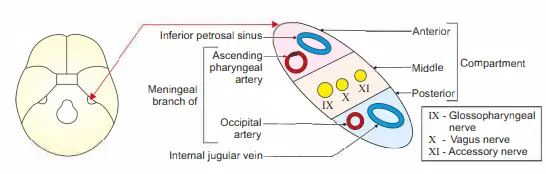
3. Jugular Foramen Contents: It is divided into three parts. The contents are
Vessels in front
Inferior petrosal sinus.
The meningeal artery, is a branch of the ascending pharyngeal artery.
Vessels behind
Internal jugular vein, a direct continuation of sigmoid sinus
Meningeal artery, a branch of the occipital artery
Three nerves in between
Glossopharyngeal nerve (IX),
Vagus nerve (X), and
Accessory nerve (XI).
Anterior Longitudinal Ligament
1. Anterior Longitudinal Ligament Attachment: It is attached to upper and lower borders of anterior surfaces of bodies of all vertebrae.
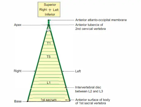
2. Extent: It extends from the anterior surface of the upper sacral vertebra to the anterior tubercle of the axis (second cervical vertebra). Anterior ligament to anterior tubercle of axis.
Suprameatal Triangle Boundaries
3. Variation in thickness: It is ..A. lar in shape. The base is at the upper sacral vertebra and the apex is at the axis.
4. Termination: It continues as anterior atlanto-occipital membrane.
Posterior longitudinal ligament
1. It is attached to the upper and lower borders of the posterior surface of the bodies of all vertebrae.
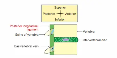
2. Extent: It extends from the body of 1st sacral to the lower border of the body of the second cervical vertebra.
3. Termination: It continues as membrane tectoria (tectum-roof) above the second cervical vertebra.
4. Relations
Anterior: Basivertebral venous plexus,
Posterior: Spinal cord with meninges.
5. Variation in thickness: The upper part is broad and has uniform width, lower part is narrow.
6. Types of fibres
Superficial fibres: They join 3 to 4 vertebrae.
Deep fibres: They merge with annulus fibrosus.
7. Applied anatomy: The posterior longitudinal ligament will be tom in excessive flexion of the neck leading to prolapse of the intervertebral disc.
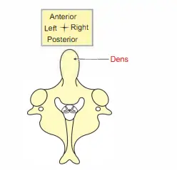
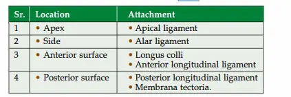
Dens (odontoid process)
Dens (odontoid process) Introduction: It is a strong tooth-like process projecting upwards from the body of the axis (2nd cervical) vertebra.
1. Evolution: It represents the body of the 1st cervical vertebra.
2. Articulations
Anteriorly with the anterior arch of the atlas.
Posteriorly with transverse ligament of atlas
3. Dens (odontoid process) Attachments:
Foramen lacerum
(lacerum-irregular)
1. Foramen Lacerum Site: Middle cranial fossa.
2. Boundaries
Medially-body of sphenoid.
Laterally-greater wing of sphenoid.
Posteriorly-apex of petrous part of temporal bone
3. Communications
Anteriorly, with pterygopalatine fossa.
Posteriorly, with the carotid canal.
4. Foramen Lacerum Contents
No structures pass through and through except (Meningeal branch of Ascending Pharyngeal artery).
The structure crossing from lateral to medial is internal carotid artery.
The structure formed is nerve of pterygoid canal (Vidian’s nerve).
Boundaries Of Suprameatal Triangle
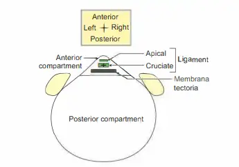
Foramen Magnum
(Magnum-great)
Foramen Magnum Introduction: It is the largest foramen of the skull.
1.Foramen Magnum Site: Posterior cranial fossa in occipital bone.
2. Communication: It connects the posterior cranial fossa to the vertebral canal.
3. Shape: Oval-., it is wider in the posterior half.
4. Structures passing (depending upon importance) (Figs 1.20A and B)
Most important structures: The lowest part of medulla oblongata with meninges.
Important structures pass through subarachnoid space.
Two vertebral arteries (branches of 1st part of subclavian artery) with sympathetic plexus
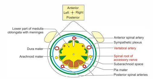
- One anterior spinal artery (branch of vertebral artery).
- Two posterior spinal arteries (branch of vertebral artery).
- Spinal roots of accessory nerves (Xlth cranial nerve).
- Less important
- Apical ligament of dens
- Vertical band of cruciate ligament
- Ligamentum denticulatum.
Maxilla
It is a paired bone of the face forming the upper jaw.
1. Maxilla Classification: It is a pneumatic bone.
2. Maxilla Development: It is developed in the membrane.
3.Maxilla Side determination
It has a frontal process pointing medially and upwards.
The alveolar process is thick, arched and projects downwards.
4. Maxilla Features: It has a body which is a hollow pyramid and has the following surfaces.
A. Anterior surface: It presents
Incisive fossa: A fossa above the incisor teeth. It gives attachment to the following muscles.
Depressor septi from the fossa.
Orbicularis oris below the fossa.
Nasalis lateral to fossa.
Canine fossa: It is lateral to incisive fossa. It gives origin to levator anguli oris
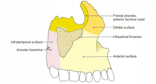
- Infraorbital Foramen: It lies above the canine fossa. It transmits the Infraorbital artery, a branch of the maxillary artery.
- Infraorbital nerve, is a branch of the maxillary nerve.
- Origin of levator labii superioris.
- Posterior (infratemporal) surface: It has foramina for posterior superior alveolar vessels and nerves.
- It presents maxillary tuberosity: It has articular and non-articular surfaces.
- The articular surface articulates with the pyramidal processes of the palatine bone.
- The non-articular surface gives attachment to the superficial head of the medial pterygoid.
- It forms the anterior boundary of the pterygopalatine fossa.
- Superior (orbital) surface: It forms the floor of the orbit. It is ..A. tar. It has Anterior, posterior and medial borders.
- It presents a lacrimal notch which is converted into the nasolacrimal canal.
- The surface presents an infraorbital groove and canal for the infraorbital vessels and nerve.
- The inferior oblique muscle of the eyeball arises from the surface.
Its posterior border forms the inferior border of the inferior orbital fissure. It transmits
- Infraorbital vessels,
Maxillary nerve,
Zygomatic nerve,
Sympathetic plexus, and
Veins connecting ophthalmic vein to pterygoid plexus. - Medial surface
It forms the lateral wall of the nose. It presents
Maxillary hiatus: It leads to maxillary air sinus.
Nasolacrimal groove, - Concha crest,
Inferior meatus of the nasal cavity, and
Atrium of the middle meatus. - Processes
Frontal process: It has - Two surfaces: Lateral and medial
Lateral surface presents
Anterior lacrimal crest that gives attachment to Lacrimal fascia, and Medial palpebral ligament.
The anterior smooth area that gives attachment to orbicularis oculi and levator labii superioris alaeque nasi.
The medial surface forms a lateral wall of the nasal cavity. - Two borders: Anterior and posterior
One end
Zygomatic process: A short process projecting laterally articulating with
the zygomatic process of the temporal bone. - Alveolar process: It presents
Sockets for the teeth.
Origin of the buccinator muscle.
Palatine process: It is strong, thick and plate-like. It articulates with its fellow on the opposite side and forms the hard palate. - Inferior surface
It has Pits for palatine glands.
Groove for greater palatine vessels and nerves.
Incisive canal which transmits terminal branches of greater palatine vessels and nasopalatine nerve.
Anterior and posterior incisive foramina.
Nasal crest present on the medial border.
The incisor crest is the anterior part of the nasal crest.
The anterior nasal spine is the anterior end of the incisor crest. - Processes: It has four processes.
A process above-frontal process:
A process below and medially palatine
A process laterally and above-zygomatic
A process below and anteriorly-alveolar. - Ossification: It ossifies in the membrane from a single centre which appears on the 6th week of intrauterine life.
The centre appears in the canine fossa.
Two more centres appear in the 7th week of intrauterine life.
5. Maxillary Sinus – Applied anatomy
The pus in the maxillary sinus is not drained naturally because the antrum of Highmore is at a higher level.
In chronic maxillary sinusitis, the pus is drained by antral puncture or antrostomy.
A trocar and cannula are passed through the nasal cavity in an upward and backward direction.
It passes below the inferior nasal concha.
A hole is produced in the lateral wall of nasal cavity.
Note: Nowadays, it has been taken over by the endoscopic method of drainage.
Caldwell-Luc operation: For adequate drainage in chronic sinusitis, a part of the medial wall of the sinus below the inferior nasal concha is removed to avoid injury to the infraorbital nerve.
Maxillary Tumours can produce a bulging in the following adjacent structures.
- Superiorly in the floor of the orbit,
- Inferiorly in the roof of the oral cavity,
- Anteriorly in the face,
- Posteriorly in the infratemporal fossa, and
- Medially in the lateral wall of the nose.
There may be unilateral or bilateral fractures of the maxilla.
Unilateral fracture involves the alveolar process.
Bilateral fractures are subclassified depending upon the involvement of zygomatic bone, orbit, and cranium.
They are classified as
- LeFort I,
- LeFort II, and
- LeFort III.
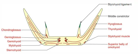
Hyoid bone
(U-shaped)
Hyoid Bone Introduction: It is an unpaired midline bone of the neck. It is suspended by muscles and hence very much mobile.
1. Hyoid Bone Classification: It is irregular bone.
2. Hyoid Bone Development
- Smaller come and Superior parts of the body develop from the second pharyngeal arch.
- The lower part of the body and greater come develop from the third pharyngeal arch.
3. Hyoid Bone Features: It has a body, greater and lesser comma.
- Body: It has two borders and two surfaces.
a. Surfaces - Anterior surface: A median ridge divides into lateral halves. (The content of the brackets indicates the nerve supplying the muscles.)
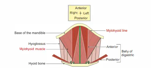
- Geniohyoid (Cl), mylohyoid (V3) and hyoglossus (XII) are attached to the anterior surface.
- The investing layer of deep cervical fascia is attached deep to the mylohyoid.
Posterior surface: It is related to
- Bursa,
- Thyrohyoid membrane, and
- Epiglottis
Hyoid Bone Borders
Upper border: It provides attachment to the following structures from anterior to posterior
- Genioglossus muscle (XII)
- Hyoepiglottic ligament.
- Thyrohyoid membrane.
Lower border: Stemohyoid (ansa cervicalis) and omohyoid (ansa cervicalis) are attached to the lower border.
Greater cornu: It has two borders and two surfaces.
Surfaces: The following muscles are attached to the anterior surface (from medial to lateral)
- Middle constrictor (pharyngeal plexus)
- Hyoglossus (XII)
- Stylohyoid (VII)
- Intermediate loop of digastric.
Borders: They give attachments to the following structures.
- Medial-thyrohyoid membrane
- Lateral-thyrohyoid muscle.
Lesser cornu: It has the following attachments.
- Stylohyoid ligament
- Middle constrictor of the pharynx.
4. Joints: Synovial joint is formed between lesser and greater comua of hyoid bone and gets obliterated in the later part of life.
5. Movements
- The suprahyoid muscle (mylohyoid, stylohyoid and geniohyoid)-elevators.
- The infrahyoid muscles (stemohyoid and omohyoid)-depressor.
6. Ossification: It ossifies from 6 centres. They are as follows
- Greater come (before birth)-2
- Body (after birth)-2, and
- Lesser comu (at puberty).
7. Applied anatomy
A postmortem finding of the fracture of the greater comic is indicative of homicidal throttling of the hyoid bone.
This is true in the case of
- Sudden and
- Unnatural death with other signs of suffocation.
The hyoid bone moves with deglutination.
Leave a Reply