Stomach And Spleen
Stomach
Question 1. Describe the stomach under the following headings:
Table of Contents
- stomach Introduction
- stomach External features
- stomach Relations
- stomach Arterial supply
- stomach Venous drainage
- stomach Lymphatic drainage
- stomach Nerve supply and
- stomach Applied anatomy.
Answer:
1. Stomach Introduction:
- The stomach is the widest and most distensible part of the gut.
- It lies obliquely in the upper, left part of the abdomen occupying epigastric, umbilical and left hypochondriac regions.
Its main functions are:
- Storage of food (capacity 1000–1500 ml).
- Formation of chyme.
- Secretion of HCl and Castle’s intrinsic factor.
2. Stomach External features:
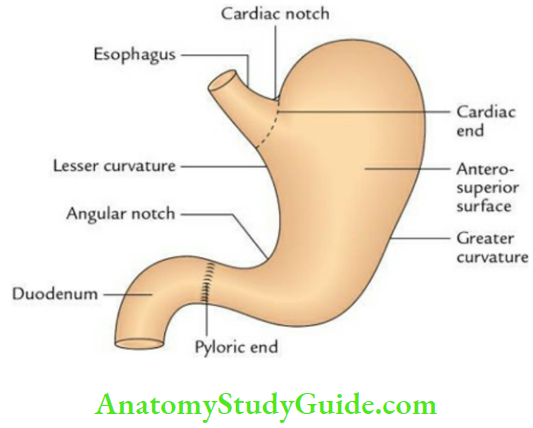
- Two orifices: Cardiac and pyloric
- Two curvatures: Greater and lesser
- Two surfaces: Anterosuperior and posteroinferior
- Four parts: Cardiac, fundus, body, and pyloric.
3. Stomach Relations:
- Anterior:
- Liver
- Diaphragm
- Anterior abdominal wall
- Posterior:
- Diaphragm (left crus)
- Left kidney
- Left suprarenal gland
- Splenic artery
- Pancreas
- Transverse mesocolon
- Left colic flexure (splenic flexure of colon)
- Spleen
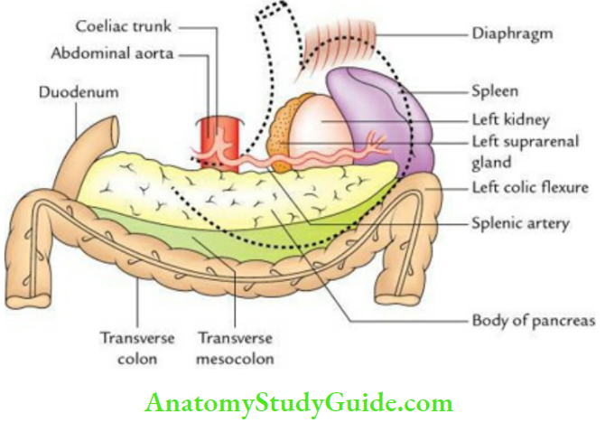
Note: All the structures forming the stomach bed, are separated from the stomach by a lesser sac except the spleen, which is separated from the stomach by a greater sac.
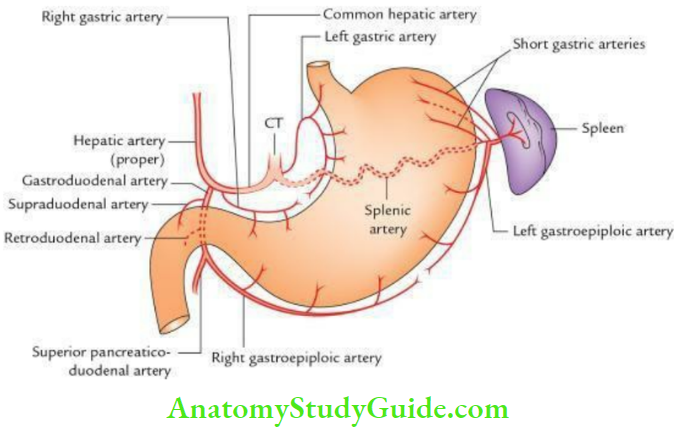
4. Stomach Arterial supply: It is provided by
- Left gastric artery, a branch of the celiac trunk
- Right gastric artery, a branch of common hepatic artery
- Left gastroepiploic artery, a branch of the splenic artery
- Right gastroepiploic artery, a branch of the gastroduodenal artery.
- Short gastric arteries, branches of splenic artery
5. Stomach Venous drainage: It is done by
- Left and right gastric veins into the portal vein.
- Short gastric and left gastroepiploic vein into the splenic vein.
- Right gastroepiploic vein into a superior mesenteric vein.
6. Stomach Lymphatic drainage:
The lymph from the stomach is drained as follows:
- From the upper 1/3rd of the area near the greater curvature (pancreaticolienal territory):
- into pancreaticolienal (pancreaticosplenic) lymph nodes
- From lower 2/3rd of the area near the greater curvature (inferior gastric territory): into right gastroepiploic lymph nodes.
- From right 2/3rd of the area near the lesser curvature (superior gastric territory): into left gastric lymph nodes.
- From pyloric canal: into pyloric right gastric and hepatic lymph nodes.
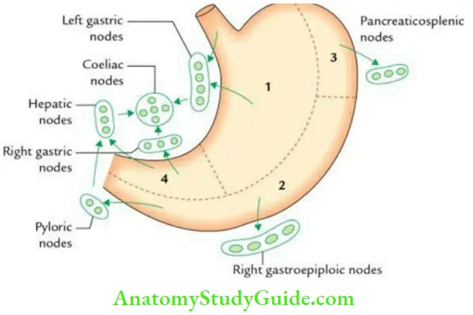
Note: Ultimately, lymph from all these regional nodes drains into celiac group of lymph nodes.
7. Stomach Nerve supply: It is provided by
- Sympathetic nerve fibers, derived from T6 to T9 spinal segments. They carry pain sensations and constrict the pyloric sphincter.
- Parasympathetic nerve fibers, are derived from vagus nerves. They increase gastric motility and relax the pyloric sphincter.
8. Stomach Applied anatomy:
- Gastritis: It is the inflammation of the mucous membrane of the stomach caused by HCl.
- Gastric carcinoma: It commonly occurs in the pyloric antrum along the greater curvature.
- Gastric ulcers: They commonly occur along the lesser curvature because during swallowing, the liquids or bolus of food passes along this curvature in the gastric canal (ofMagenstrasse).
- Gastric pain: It is referred in the epigastrium because the stomach is supplied by T6 to T10 spinal segments.
Read And Learn More: Anatomy Question And Answers
Question 2. Write a short note on your stomach bed.
Answer:
It refers to the structures upon which the stomach lies i.e. rests.
For examples:
- Pancreas
- Spleen
- Left kidney
- Left suprarenal gland
- Transverse colon
- Transverse mesocolon
- Diaphragm
All these structures form posterior relations of the stomach
Question 3. Enumerate the histological features of the stomach in general.
Answer:
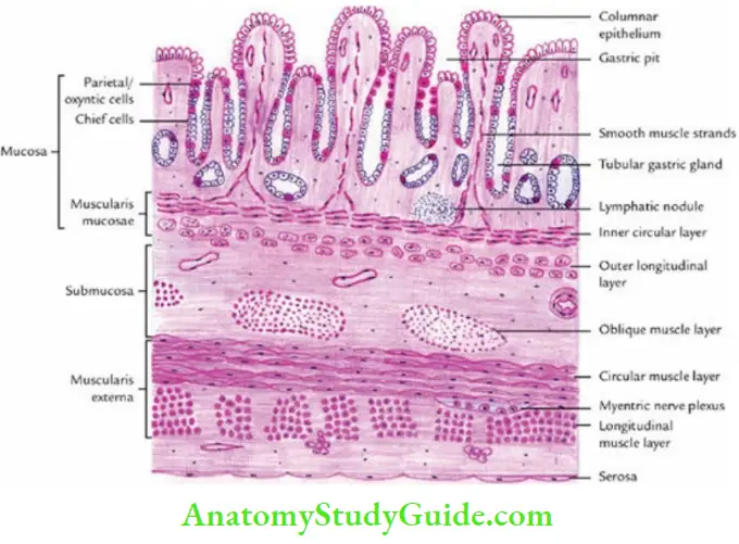
The key histological features of the stomach in general are:
- Lumen is lined by simple columnar epithelium.
- The mucous membrane contains gastric glands, which open on the surface in gastric pits.
- Muscularis external is thick and consists of 3 layers: instead of two viz, outer longitudinal, middle circular, and inner oblique.
The three parts/regions, viz. cardiac, fundus, and pylorus, present different histological structures.
Question 4. Write a short note on histological features (microscopic anatomy) of the cardiac part of the stomach.
Answer:
A histological section through the cardiac part often passes through the cardio esophageal junction. Hence, the present account presents the histological features of cardio esophageal junction.
Mucous membrane:
- Epithelium:
- The stratified squamous epithelium of the esophagus abruptly changes into the simple columnar epithelium of cardiac part of the stomach.
- Cells of columnar epithelium look alike and have basal oval nuclei.
- Absence of goblet cells in the epithelium.
- Columnar epithelium, lining the surface dips from it to form gastric pits.
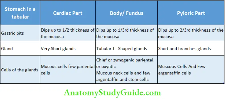
- Lamina propria: Lamina propria contains gastric glands, which never extend through the muscularis mucosae. This gland dips up to ½ the thickness of mucosa.
- Muscularis: Muscularis mucosae is thin and consists of an inner circular layer and an outer longitudinal layer.
- Submucosa: Submucosa shows some mucous secreting acini of the esophagus.
- Muscularis: Muscularis externa is made up of inner circular and outer longitudinal layers.
- Serosa: Serosa is the outermost covering and is lined by squamous cells.
Question 5. Write a short note on histological features (microscopic anatomy) of the fundus of stomach
Answer:
A section through the fundus/body of the stomach presents the following microscopic features:
1 . Mucosa:
It is thick and thrown into prominent folds called rugae.
- Lining epithelium: It is lined by simple columnar epithelium, which invaginates into lamina propria to form gastric pits.
- Laminae propria: It contains a large number of straight tubular glands, which open into gastric pits. These glands are oriented perpendicular to the surface.
- The deeper 2/3rd of each gland is a secretory portion, while the upper 1/3rd is conducting portion.
- Glands are lined by 3 main types of cells: viz chief, parietal and mucous.
- Chief/peptic/zymogenic cells: They are pyramidal in shape and occur in clusters in the basal third of the gland. They secrete pepsin and lipase.
- Parietal/oxyntic cells: They are found along the whole length of the gland but more in the middle part. They give characteristic fried egg appearance and secrete HCl and intrinsic factors.
- Mucous cells: They line the proximal parts (necks) of the gland. They secrete mucous.
- Muscularis mucosae: It is thin and made up of inner circular and outer longitudinal layers.
Note: Other cell types present in the gastric glands are: argentaffin cells in the basal part which secrete serotonin, and undifferentiated stem cells which replace the damaged cells.
2. Submucosa: It is made up of loose tissue and contains nerves and vessels.
3. Muscularis externa: It is thick and consists of inner oblique, middle circular, and outer longitudinal layers of smooth muscle fibers.
4. Serosa: It is a thin outer coat made up of mesothelium i.e. lines by squamous cells.
Question 6. Write a short note on histological features of the pyloric part of the stomach.
Answer:
A section through pylorus presents the following features:
1 . Mucosa:
- Lining epithelium: It is lined up by tall columnar epithelium. Surface epithelium dips down to form deep gastric pits, which extend up to 1⁄2 to 2⁄3rd of the thickness of the mucous membrane.
- Lamina propria: It contains short and branched pyloric glands, which open into gastric pits. Since gastric pits are deeper, the ratio of pit length to gland length is 1:1.
- Muscularis mucosae: It is made up of inner circular and outer longitudinal layers.
2. Submucosa: It is made up of loose connective tissue and contains nerves and vessels.
3. Muscularis externa: It is mainly made up of thickened circular layer (which forms a pyloric sphincter).
4. Serosa: It is made up of mesothelium lined by squamous cells.
Question 7. Give the differences in histological features of the cardiac, body/fundus, and pyloric parts of the stomach in a tabular form.
Answer:
Histological features of the cardiac, body/fundus, and pyloric parts of the stomach in a tabular form:
Spleen
Question 8. Describe the spleen under the following headings:
- Spleen Introduction
- Spleen External features
- Spleen Relations
- Spleen Blood supply
- Spleen Histology and
- Spleen Applied anatomy.
Answer:
1. Spleen Introduction:
The spleen is the highly vascular largest lymphoid organ (also called hemolymphoid organ) in the body, located in the left hypochondrium. It lies obliquely along the long axis of the 10th rib and is directed downwards, forwards, and laterally. It filters blood from antigens and microorganisms and removes old and abnormal RBCs.
Its shape, size, and weight are as follows:
- Shape: Wedge–shaped.
- Size: 1 inch thick; 3 inches broad; 5 inches long.
- Weight: 7 ounces (1 ounce = 30 gm).
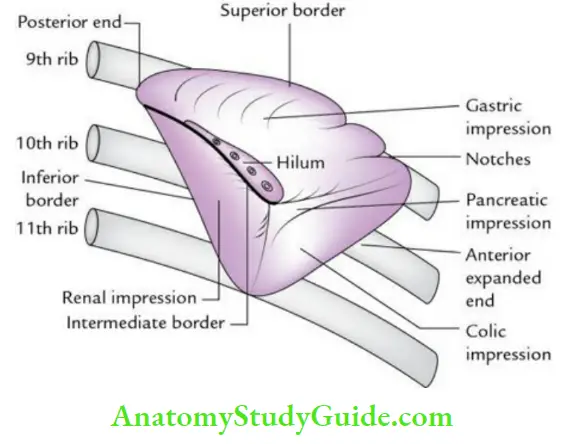
2. External features:
Two ends:
- Anterior and Posterior: The superficial anterior end is expanded and looks like a border. It reaches up to the midaxillary line but does not cross it.
- The Posterior end is deep and extends into the epigastric region.
Two surfaces:
- The diaphragmatic surface is convex and smooth.
- The visceral surface is concave and irregular. It presents gastric, renal, colic, and pancreatic impressions.
Three borders:
- The superior border is characteristically notched near its anterior end.
- The inferior border is rounded.
- The intermediate border is also rounded and directed to the right.
Hilum:
- It is located on the anteromedial part of the gastric impression along the long axis of the spleen.
- Just above the intermedial border.
3. Spleen Relations:
Visceral surface It is related to
- Stomach
- Left kidney
- Left colic flexure
- Tail of pancreas
These viscera produce impressions on this surface as follows:
- Gastric impression: Between the superior and intermediate borders.
- Renal impression: Between the intermediate and inferior borders.
- Colic impression: Near the anterior end.
- Pancreatic impression: Between the hilum and the colic impression.
Diaphragmatic surface: It is related to
- Diaphragm.
- Costodiaphragmatic recess.
- Left lung.
- 9th, 10th, and 11th ribs (of the left side).
Peritoneal relations:
The spleen is surrounded by the peritoneum and reflected at the hilum to form lienorenal and gastrosplenic ligaments, which together constitute the splenic pedicle.
- Lienorenal ligament: Lienorenal ligament contains the tail of the pancreas, splenic vessels, and pancreaticosplenic lymph nodes.
- Gastrosplenic ligament: The gastrosplenic ligament contains left gastroepiploic vessels and short gastric vessels.
4. Spleen Blood supply:
- Arterial supply: By the splenic artery, a branch of the celiac trunk.
- Venous drainage: By the splenic vein that joins the superior mesenteric vein to form the portal vein.
5. Spleen Histology:
The histological section through the spleen presents the following features:
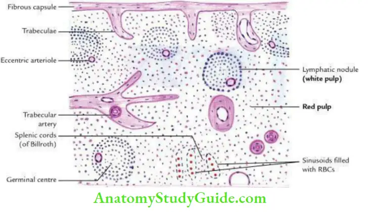
- Fibrous capsule: It consists of collagen fibers, elastic fibers, and scattered muscle bundles. It sends trabeculae inside the splenic parenchyma.
- Parenchyma: The parenchyma of the spleen is not differentiated into the cortex and medulla and presents uniform features.
The splenic parenchyma (also called splenic pulp) is divided into two types depending on the type of blood cells:
- White pulp and
- Red pulp.
The Splenic Parachyma are:
- White pulp:
- It consists of discrete lymphoid nodules having eccentric arteriole (Malpighian corpuscles) – a striking/characteristic feature of the parenchyma of the spleen.
- The germinal center may be seen. It is surrounded by red pulp.
- Red pulp:
- It consists of a diffuse delicate meshwork of reticular cells and reticular fibers.
- The spaces of the network are filled with Lymphocytes, Macrophages, and RBCs.
- These cells are arranged in the form of anastomosing cords called cords of Billroth.
- The spaces between Billroth’s cords are occupied by sinusoids. \ The red pulp appears red because it contains numerous sinusoids filled with RBCs.
Note: Splenic nodules with eccentric arteriole are actually the periarterial aggregations of lymphatic tissue. The splenic nodules are also called Malpighian corpuscles.
Spleen Applied anatomy
Palpation of the spleen:
- A normal spleen is not palpable. An enlarged spleen can be palpated underneath the left costal margin, during deep inspiration, in the left lateral position.
- Note that the spleen becomes palpable only when it is enlarged twice of its normal size.
Splenomegaly (enlargement of the spleen):
The enlarged spleen (if massive) projects toward the right iliac fossa in the direction of the axis of the 10th rib.
Laceration of the spleen:
- It is often caused by a fractured rib.
- The blood collects underneath the left dome of the diaphragm, leading to its irritation. As a result, the pain is referred to the tip of the left shoulder (Kehr’s sign).
Leave a Reply