Autonomic Nervous System
- ANS controls and coordinates the activities of visceral organs by controlling visceral sensation and visceral movements. ANS is formed of 2 systems: The sympathetic and parasympathetic systems.
- The sympathetic system is formed of thoracic-Iumbar outflow, sympathetic cords, and post-ganglionic nerve fibers while the parasympathetic system is formed of craniosacral outflow, parasympathetic ganglia, and postganglionic nerve fibers.
- The sympathetic and para-sympathetic nervous systems are often antagonistic in their actions. The homeostasis of body fluids is maintained by a balance between these two systems. Sympathetic nerve fibers are adrenergic while parasympathetic nerve fibers are cholinergic in their mode of action.
- Auerbach’s plexus is that part of the autonomic nervous system (mostly from the vagus nerve) lying between the two main muscle layers of the gut and controlling its peristaltic movements.
Cerebrospinal Fluid (Csf)
- The entire nervous system (CNS) contains 80-150 ml of cerebrospinal fluid (CSF).
- It is a clear, colorless, slightly alkaline (7.33) pH fluid of 1.005 specific gravity that occurs in the ventricles of the brain, the central canal of the spinal cord, and subarachnoid space.
- It is filtered out of blood in the choroid plexus and passes back into it. The fluid contains proteins, amino acids, glucose, respiratory gases, urea, Na, K, Ca, Cl, HC03, etc.
- CSF supplies food and oxygen to different parts of the CNS.
- It picks up C02, urea, and other waste products from CNS.
- Protection of CNS from shock,
- Maintenance of a constant internal pressure,
- Keeping CNS moist.
- Reflex actions
- These are spontaneous, automatic, and mechanical responses without involving the will and are controlled by the spinal cord. The path followed in a reflex action is called reflex are. Reflex actions enable the body to give quick responses to harmful stimuli and prevent the overloading of the brain.
- Reflexes are The entire impulse circuit of a reflex response- receptors → CNS→ effectors – is called a reflex arc. It is the basic functional unit of the nervous system.
- Reflexes are of two types: unconditioned and conditioned reflexes. Unconditioned reflexes are inbom and need no training while conditioned reflexes are developed only after training. Conditioned reflex was first demonstrated by Pavlov (1929) in a dog experiment.
- Pavlov is called the ‘Father of conditioned Reflexes.
- Monosynaptic and polysynaptic reflexes. Reflexes in which sensory impulses are directly transferred from sensory neurons to motor neurons are called monosynaptic.
- Such reflexes are uncommon. Usually, a number of small neurons, called association or internuncial neurons or interneurons, present in the grey matter of CNS, serve to transfer a reflex impulse from sensory neurons to the motor neurons.
- Such reflexes are, therefore, called polysynaptic. Interneurons can carry impulses of reflex responses to the effectors located at considerable distances from receptors.
- The reflex action involving the spinal cord and a spinal nerve is called spinal reflex or reflex arc. The reflex action controlled by the brain is called cerebral reflex action.
- Righting Reflex. Adjustment of the position of the head to the earth’s surface, then the position of the trunk to the head, and the position of the body involving the spinal cord, medulla oblongata, and midbrain is termed a righting reflex.
- It was discovered by Magnus.
- Antigravity reflex. It maintains the equilibrium against the force of gravity. It was first reported by Sherrington.
- Transmission Of The Nerve Impulse. When a stimulus is given, there is n response oiler n Irnctlon of it second. A wave of excitation is conducted front the point of stimulation by electrical, chemical, physical, or electromagnetic changes. Transmission excitation Is called conductivity. Electrical currents in the nerve conduction. The electrical events Involve the following changes in the electrical potential of nerve fiber.
- Resting potential stale. The nerve cells remain bathed in a fluid called interstitial fluid. In this fluid remain dissolved sodium (Na++ ) and potassium (K1) ions. During the resting phase, the neurilemma is comparatively 30 times more permeable to potassium ions (K+ than to sodium ions (Na+).
- As a result Na+ ions arc present in high concentration outside the membrane and in low concentration on the inner side. Thus outer surface shows 1+vcT electric charge and the inner surface (-vc) charge due to organic anions. The membrane at this stage is said to be in a polarised state and the resting potential remains at a value of 80 mV. Points
- Depolarization and action potential. When a stimulus of any kind is applied to the nerve, it disturbs the setup. There is a marked change in the potential, it is called an action potential.
- The polarity of the membrane gets reversed after excitation because the Na+ ions move inward and K+ ions move outward. Thus the membrane is said to be depolarised. It is termed the active phase during which the inner surface is positively charged and the outer surface is not exactly charged.
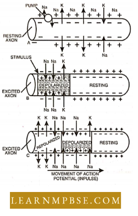
Table of Contents
- Conduction of impulse. The electrochemical changes are conducted up to synapses as the electric wave of change of potential progresses forward along the fiber.
- Repolarization. In the recovery phase, the axon again restores the original +ve concentration by the outside movement of Na+ ions from the inner side of the membrane. The process of change requires some period during which the nerve cannot be stimulated again. It is of the duration 1/1000 second and is called the refractory period.
- At the synapse, the conduction of impulse is due to the release of neurotransmitter substances such as acetylcholine and norepinephrine later on inactivated by acetylcholinesterase and mono-amine oxidase respectively. Accordingly, the nerve fibers are called cholinergic and adrenergic nerve fibers.
- A membrane’s excitability is a function of two properties: capacitance the concentration of oppositely charged ions across the membrane, and conductance, the permeability of the membrane to ionic currents.
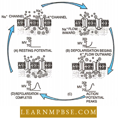
- When an axon membrane shows a steady resting potential, there are 30 times more potassium ions inside the axon than outside and 10 times more sodium ions outside the axon than inside. In this state, the axon membrane is 10 times more permeable to K+ than it is to Na+.
- In the resting state, the axon membrane is polarized, with more positively charged ions outside than inside. This unequal distribution of ions is due to :
- the selective permeability of the membrane, which forms an almost impenetrable barrier to Na+, and
- the action of the Nn+ K+ pump which pumps three Na+ out of the neuron for every two K+ brought in.
- Typically, it lakes many simultaneous graded electronic potentials to initiate an action potential.
- An action potential is initiated each time the membrane potential is raised to threshold potential. Because action potentials never occur below this potential and upon occurring arc always the same size, action potentials arc called all or none events.
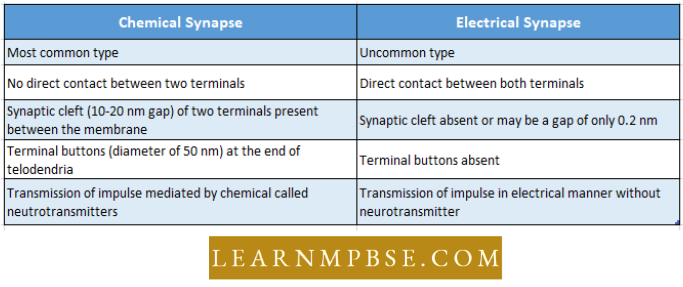
Synapse
- It is the joint of two neutrons – between the axon of one neuron and the dendron of another neuron.
- Axon is termed a presynaptic terminal while, dendron is a post-synaptic terminal,
- Synapses are of the following two types: chemical synapse and electrical synapse.
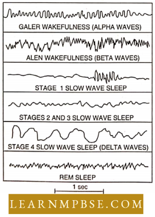
Electroencephalogram
- EEG is a tool to study the different states of consciousness of the mind. But, action potential has little role in it. EEG is the summed-up post-synaptic potential mainly in the large neuron whose processes are perpendicular to the cortical surface.
- Analysis of waves is complicated because the postsynaptic potentials are both excita¬tory’ and inhibitory types, some are near the surface and others are deeper.
- The frequency of waves may vary from 1 to 30 Hz and their amplitudes also vary.
- Alpha waves. Frequency ranges from 8-13 Hz, a pattern seen in adults in awake, relaxed with closed eyes. •
- Beta waves. A frequency of 18 – 20 Hz, shows an attentive, alert, and actively thinking state of mind, the opening of the eye in a dark room.
- Delta wave. The very low-frequency waves of 0.5 – 2 Hz but of higher voltage (amplitude) characterize deep sleep. Similar waves are also found in cases of brain injury.
- Theta waves. The low-frequency waves of 5 0 8 Hz, are prominent in children between 2 to 5 years.
Memory Tips
- Spike Potential. It is n rapid depolarisation of nerve membrane, changing negative testing potential of 80 mV to + 60 m V (spike potential) before falling to action potential of + 20 m V,
- Conduction of nerve impulse. More in myelinated nerve fibers than non-myeli- nated nerve fibers. Invertebrates have mostly non-myelinated nerve fibers.
- In Giant Squid (thickened nerve fibers) the impulse velocity is 20 m/sec (72 km.hr), in fishes 2-35 m./sec (7.2-126 km/.hr,) amphibians 20-40 m/sec (72-144 km/ hr.) in reptiles 15-35 m/sec (54-126 km.hr) while in mammals it is 100-148 m/sec (360-532 km,/hr).
- Excitatory Postsynaptic Potential (E.P.S.P.) It is the potential that is passed from one neuron to another through a synaptic junction.
- Neurotransmitters. Chemical substances help in synaptic neurotransmission.
- There are about 40 types divided into 2 For example tries :
- Neurotransmitters are the chemicals stored in the synaptic vesicle meant for transmitting nerve impulses across the synapse.
- Rapidly acting small-sized neurotransmitters, For Example, ACh, Epinephrine, Norepinephrine, Dopamine, Histamine, and Gaba-Amino butyric acid (GABA). GABA is an amino-acid derivative and plays a role in pre-synaptic inhibition.
- Slowly acting neuropeptides For example. TSH-RH, LH-RH, somatostatin, ACTH, TSH, MSH, vasopressin, Gastrin, Insulin, Angiotensin-11, Bradykinin, etc.
- On the repeated transmission of nerve impulses through a synapse, there occurs a temporary suspension of impulse transmission at the synapse. This is called synap¬tic fatigue. It results from an exhaustion of the neurotransmitter in the synaptic vesicles of the axon terminal.
Leave a Reply