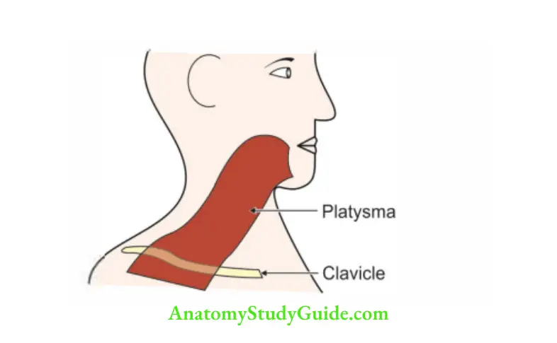
Platysma
It is a broad, flat muscle, remnant of pannculus camosus.
It lies superficial to deep fascia of neck.
Table of Contents
1 Proximal attachments are fascia over the
- Pectoralis major, and
- Deltoid.
Read And Learn More: Neck Anatomy Notes And Important Questions With Answers
2 Platysma Distal attachments: They are divided into anterior and posterior fibres
- Anterior fibres are inserted to the base of mandible.
- Posterior fibres to the skin of the lower part of face and lip.
3 Platysma Action: Depresses the skin It pulls the angle of the mouth downwards as in horror
4 Platysma Development: It is developed from the mesoderm of 2nd pharyngeal arch hence supplied by cervical branch of facial nerve
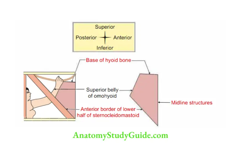
Muscular Triangle
1 Muscular Triangle Boundary
- Anteriorly: Anterior median line of the neck from the hyoid bone to the sternum.
- Posterosuperiorly: Superior belly of the omohyoid muscle.
- Posteroinferiorly: Anterior border of the stemocleidomastoid muscle.
2 Muscular Triangle Contents: Infrahyoid muscles are the chief contents of the triangle These are
- Stemohyoid,
- Stemothyroid,
- Thyrohyoid, and
- Omohyoid.
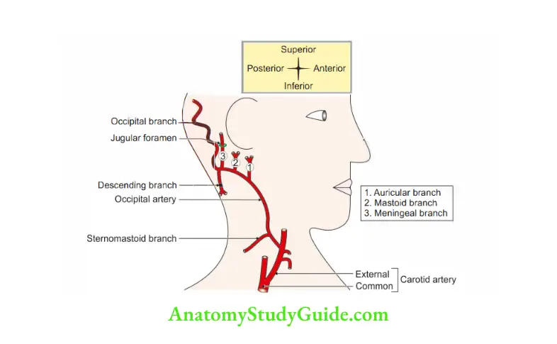
Occipital Artery
Occipital Artery Introduction: It is the artery supplying the posterior aspect of scalp It gives muscular branches to the sternocleidomastoid and stylohyoid
1 Occipital Artery Origin: It is the 1st dorsalbranch of external carotid artery arising opposite facial arteryemergesI greater atoccipitalthe apex nerve of posterior triangle of neck It goes along with greater occipital nerve.
2 Occipital Artery Course and relations
- Courses deep to the lower border of posterior belly of digastric.
- Grooves thebase of skull at the occipitomastoid suture.
- Lies deep to the digastric notch.
3 Occipital Artery Peculiarities
- At its origin, it is crossed superficially by hypoglossal nerve.
- Its upper branch acts as a guide to the accessory nerve.
- It has a tortuous course in the superficial fascia of scalp.
4 Occipital Artery Branches
- Muscular branch to stylohyoid and sternocleidomastoid.
- Meningeal branch.
- Bonybranch to mastoid process.
Question 1: Describe digastric triangle under the following headings:
1 Digastric Triangle Boundaries,
2 Digastric Triangle Roof,
3 Digastric Triangle Floor,
4 Digastric Triangle Contents, and
5 Digastric Triangle Applied anatomy
Answer: 1 Digastric Triangle Boundaries
- Anteroinferior: Anterior belly of digastric
- Posteroinferior: Posterior belly of digastric and stylohyoid
- Superior (base) is by
- Base of the mandible, and
- Line joining the angle of the mandible to the mastoid process
2 Digastric Triangle Roof
Skin
Superficial fascia containing
- Cutaneous vein (tributaries of external jugular vein).
- Cutaneous branches of great auricular nerve.
- Cervical branch of facial nerve.
Deep fascia encloses submandibular salivary gland.
3 Digastric Triangle Floor: From anterior to posterior
- Hyoglossus,
- Mylohyoid, and
- Middle constrictor of the pharynx
4 Digastric Triangle Contents: They are grouped as structures in the
The anterior part of the triangle
Structures superficial to mylohyoid are superficial part of submandibular gland
and related structures The relations can be visualized
- Submandibular gland hugs the mylohyoid by laying its major superficial part anteriorly and small deep part posteriorly.
- Semiflex your left wrist in such a way that the 4 fingers are placed anteriorly and thumb posteriorly Tip of fingers and thumb referred as anterior ends of the superficial part of submandibular gland.
- The number of fingers indicates size of the gland.
- Four fingers of left hand refer the larger superficial part .
- Index finger of left hand represents the facial vein.
- Middle finger of left hand represents the mylohyoid vessels and nerve.
- Ring finger of left hand represents submental vessels and nerve.
- Little finger of left hand represents submandibular lymph node, and
- Thumb of left hand refers smaller deep part Position of the wrist indicates that superficial part of gland is continuous with deep part of the submandibular gland.
Posterior part of triangle
5 Digastric Triangle Applied anatomy
Ludwig’s angina: It is an triangular swelling due to the infection of the submandibular region It is bounded
- Posteriorly by the hyoid bone, and
- Anterolaterally on each side by the two halves of base of mandible This is so because the investing layer of deep cervical fascia is attached to these bones
Collection of pus may push the tongue upwards
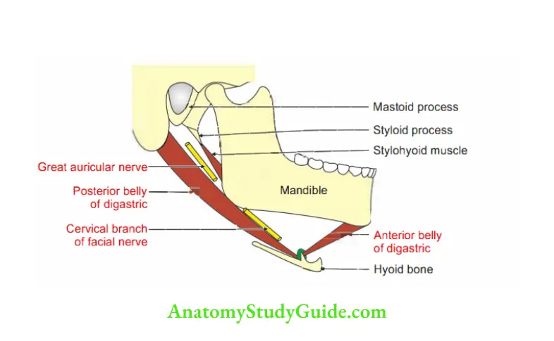
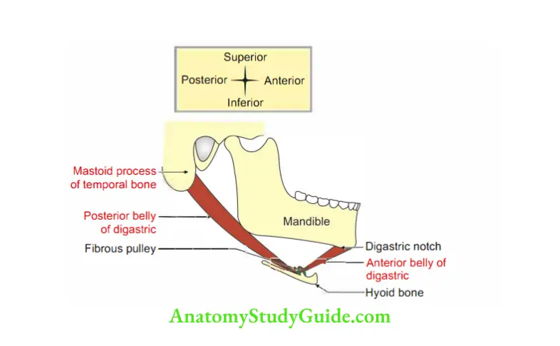
Digastric Muscle
Digastric Muscle Introduction: It is suprahyoid muscle of the neck.
1 Digastric Muscle Origin: It has two bellies.
- Anterior belly arises from digastric fossa of mandible.
- Posterior belly arises from digastric notch, present medial to mastoid process.
2 Digastric Muscle Insertion: Both heads meet at the intermediate tendon which is held by a fibrous pulley attached to the hyoid bone.
3 Digastric Muscle Nerve supply
- An Anterior belly is supplied by nerve to mylohyoid, a branch of inferior alveolar nerve, branch of anterior division of mandibular nerve.
- Posterior belly is supplied by facial nerve.
4 Digastric Muscle Action
- It depresses mandible when mouth is opened widely.
- It elevates the hyoid bone.
5 Digastric Muscle Development
- Anterior belly develops from mesenchyme of the first pharyngeal arch.
- Posterior belly develops from mesenchyme of the second pharyngeal arch.
Question 2: Describe Carotid Triangle under the following headings:
1 Carotid Triangle Boundaries,
2 Carotid Triangle Roof,
3 Carotid Triangle Floor,
4 Carotid Triangle Contents, and
5 Carotid Triangle Applied anatomy
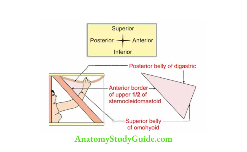
Answer:1 Carotid Triangle Boundaries
Superiorly: Posterior belly of the digastric muscle and the stylohyoid.
Anteroinferiorly: Superior belly of the omohyoid.
Posteriorly: Anterior border of upper half of the stemomastoid muscle.
2 Carotid Triangle Roof
Skin
Superficial fascia It contains
- Platysma muscle,
- Cervical branch of the facial nerve, and
- Transverse cutaneous nerve of the neck
Investing layer of deep cervical fascia
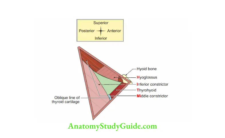
3 Carotid Triangle Floor: It is formed by I Mi I
- Hyoglossus
- Inferior constrictors of pharynx
- Throhyoid muscle
- Middle constrictor of pharynx
4 Carotid Triangle Contents
Carotid sheath
Contents
Common carotid artery and its two terminal branches
- Internal carotid artery
- External carotid artery
Internal jugular vein
Vagus nerve It is present posteromedially
Relations of carotid sheath
- Anterior wall: Ansa cervicalis
- Posterior wall: Sympathetic trunk
Carotid body and carotid sinus
Deep cervical lymph nodes
Vessels and nerves, I 3, 2-5 arteries, 3 veins and 2 ,i4il ranches of external carotid artery
- Ascending pharyngeal artery,
- Superior thyroid artery,
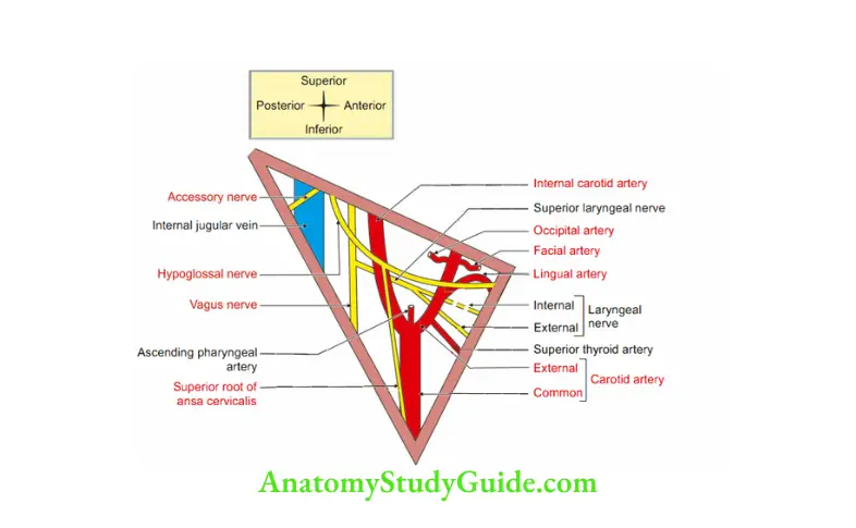
- Lingual artery,
- Facial artery, and
- Occipital artery
3 tributaries of internal jugular vein
- Pharyngeal vein,
- Lingual vein,
- Common facial vein
2 nerves
- Spinal accessory nerve: It is present at the posterosuperior angle of the triangle It passes superficial to triangle .
- Hypoglossal nerve: It always crosses the loop of lingual artery
5 Carotid Triangle Applied anatomy
- A strong carotid pulse is palpable in the carotid triangle inferior to the level of -o
Adam’s apple by gently pressing the common carotid artery against the C’underlying anterior tubercle of 6th cervical vertebra. - In the elderly, atheromatous plaques may be dislodged by palpations on le tide Hence,pulsation should be felt on the right side since a stroke induced in the right cerebralhemisphere is less devastating.
External Carotid Artery
1 External Carotid Artery Origin: It is one of the terminal branches of the common carotid artery given at the level of superior comu of thyroid cartilage.
2 External Carotid Artery Extent: From the upper border of thyroid cartilage to the neck of mandible.
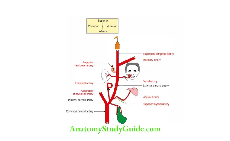
3 External Carotid Artery Branches: All the branches of external carotid artery lie above the level of angle of jaw, and hence, supply the face rather than neck.
The exception is superior thyroid artery, (which falls down on the job and is afraid of
heights) and reaches down to grab the thyroid
1. Medial: Ascending pharyngeal
2. Dorsal:
- Occipital, and
- Posterior auricular
3. Ventral:
- Superior thyroid,
- Lingual, and
- Facial
4. Terminal:
- Superficial temporal, and
- Maxillary
4 External Carotid Artery Course: It lies anterior and medial to internal carotid artery at its origin It passes deep to the posterior belly of digastric and stylohyoid muscle, enters the parotid gland and divides into terminal branches.
5 External Carotid Artery Relations
1. Superficial
- In the carotid triangle, it is overlapped by sternomastoid and crossed by hypglossal nerve, lingual nerve and facial vein.
- In th digastric triangle, it is related toposteriorbelly ofdigastricand stylohyoid muscle.
- In parotid gland, it is overlapped by retromandibular vein.
2. Deep
- Constrictor muscles of pharynx.
- Superior larynl nerve and its tw branches: Internl and externl laryngeal nerves.
- Internal carotid artery.
Lingual Artery
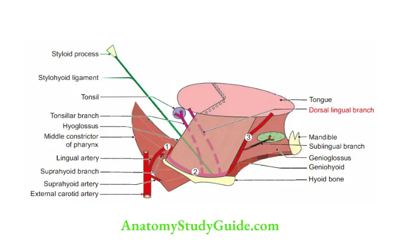
1 Lingual Artery Origin: It is the 2nd ventral branch of external carotid artery, arises opposite to the tip of greater cornu of hyoid bone.
2 Lingual Artery Course and relations: It is divided into three parts by hyoglossus muscle.
- 1st part (lateral to hyoglossus) extends from origin (external carotid artery) to the tip of greater cornu of hyoid bone It forms upward loop to avoid rupture during the movements of hyoid bone.
- 2nd part lies deep to the hyoglossus muscle and on the upper border of greater cornu of hyoid bone It lies superficial to middle constrictor.
- 3rd part runs along anterior border of hyoglossus muscle It is also called deep lingual artery
3 Lingual Artery Branches.
- 1st part: Suprahyoid artery
- 2nd part: Dorsal lingual artery
- 3rd part (deep lingual artery): Sublingual artery
4 Lingual Artery Applied anatomy
- In surgical removal of tongue, the 1st part of the artery is ligated before it gives
any branch to tongue or tonsil. - Bleeding from the lingual artery is arrested by pulling the tongue out.
Question 3: Enumerate the branches of Facial Artery in Neck
Answer: 1 Ascending palatine
2 Tonsillar branch
3 Submental
4 Glandular
Question: Why is Facial Artery Tortuous?
Answer: 1 Facial artery has cervical and facial parts In both segments, the artery is tortuous
2 The cervical part of facial artery is tortuous to adapt to the movements of pharynx during deglutition
3 The facial part is tortuous to adapt to the movements of mandible, lips and cheek
Facial artery
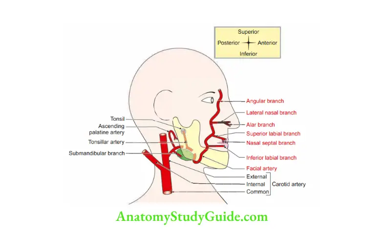
It is the chief artery of the face
1 Facial Artery Tortuous Peculiarity: The artery is tortuous to avoid the rupture during the movements of pharynx, the contraction of the muscles of face and the movements of temporomandibular joint.
2 Facial Artery Tortuous Origin: It is the 3rd ventral branch of external carotid artery arising above the level of the tip of greater cornu of hyoid bone.
3 Facial Artery Tortuous Course: It is divided into cervical and facial.
- It runs superficial to superior constrictor of the pharynx and dep to posterior belly of gigastric and to the ramus of mandible, at the anterior border of the masseter muscle It grooves the submandibular gland.
- It enters the face by winding round the base of the mandible and pierces deep cervical fascia at the junction of ramus and body of mandible.
- It crosses the masseter at anteroinferior angle It runs upwards half an inch lateral to angle of mouth and ascends by the side of nose and anastomoses with ophthalmic artery, branch of internal carotid artery.
4 Facial Artery Tortuous Branches
A Cervical part
- Ascending palatine,
- Ionsillar,
- Submental, and
- Submandibular
Facial part
- Inferior labial,
- Superior labial, and
- Lateral nasal
5 Applied anatomy: The wounds of face bleed profusely but heal quickly because
of rich blood supply and profuse anastomosis
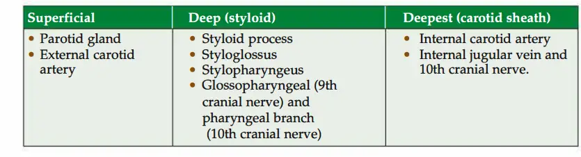
Question 4: Structures passing between external and internal carotid arteries
Answer :1 Muscles
- Styloglossus
- Stylopharyngeus
2 Nerves
- Glossopharyngeal nerve
- Pharyngeal branch of the vagus nerve
3 Bone: Styloid process of temporal bone
4 Gland: Part of the parotid gland
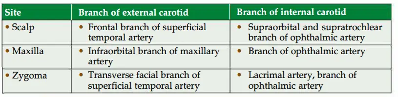
Sites of anastomosis of external and internal carotid arteries
Ansa cervicalis (ansa hypoglossi)
(Ansa-loop, cervicalis-cervical)
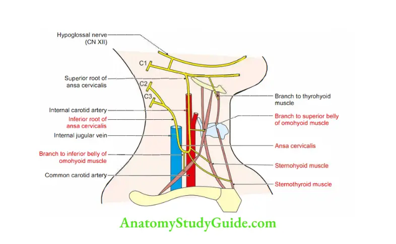
Ansa cervicalis Introduction: A loop formed by ventral rami of cervical nerves.
1 Ansa cervicalis Formation: It is formed by ventral rami of 1st, 2nd and 3rd cervical nerves.
2 Ansa cervicalis Roots
- Superior root (descending hyoglossi or anterior root) is formed by ventral ramus of Cl.
- Inferior root (descending cervicalis or posterior root) is formed by ventral rami of C2 and C3.
3. Ansa cervicalis Relations: It lies on the anterior wall of carotid sheath.
4 Ansa cervicalis Distribution
Superior root: Superior belly of omohyoid
Inferior root
- Sternohyoid,
- Sternothyroid, and
- Inferior belly of omohyoid.
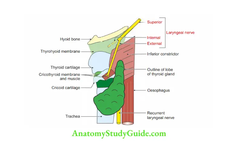
Superior Laryngeal Nerve
1 Superior Laryngeal Nerve Origin: It arises from inferior ganglion of the vagus
2 Superior Laryngeal Nerve Course:
- It passes downwards and forwards on the superior constrictor of pharynx Here,it is deep to internal carotid artery
- It reaches middle constrictor of pharynx and divides into branches on thyrohyoid membrane
3. Superior Laryngeal Nerve Branches
1. Internal laryngeal nerve
- It is the larger terminal branch of superior laryngeal nerve given in the carotid sheath
- It pierces the thyrohyoid membrane and passes deep to it It passes along with
superior laryngeal vessels It is sensory to the mucous membrane of - Posterior one-third of tongue, and
- Larynx above the vocal fold It includes
- Laryngeal mucous membrane of the laryngeal vestibule,
- Middle laryngeal cavity, and
- Superior surface of the vocal folds
2. External laryngeal nerve
- It is the smaller terminal branch of superior laryngeal nerve given in the carotid sheath.
- It passes deep to superior thyroid artery.
- It is motor branch to cricothyroid, the only external muscle of larynx.
4 Superior Laryngeal Nerve Relations: Superior laryngeal nerve is accompanied by superior thyroid artery The
external laryngeal nerve has intimate and important relations with branches of
superior thyroid artery supplying lateral lobe of thyroid gland.
External laryngeal nerve and branches of superior thyroid artery are very close when they are away
from lateral lobe of thyroid gland and go apart when they reach the gland
5 Superior Laryngeal Nerve Development: It is nerve of the IVth pharyngeal arch.
6 Superior Laryngeal Nerve Applied anatomy
- During thyroidectomy, superior thyroid artery is ligated near the lateral lobe of gland to avoid the damage to superior laryngeal nerve.
- Damage to the internal laryngeal nerve causes anaesthesia of the superior laryngeal mucosa It results into lossof protective mechanism of the larynx Thus, the foreign bodies can easily enter the larynx.
- Injury to the external laryngeal nerve results in paralysis of cricothyroid muscle It is unable to vary the length and tension of vocal cords It manifests as monotonous voice It goes unnoticed in persons who do not use wide range of tone in their speech It is critical to singers and public speakers.
- Superior laryngeal nerve block is used with end tracheal intubation in the conscious patients.
Question 5: What is the effect of pressure damage to internal laryngeal nerve, external laryngeal nerve and recurrent laryngeal nerve?
Answer :1 Pressure on internal laryngeal nerve causes loss of sensation of larynx above the
vocal cords on the affected side.
2 Pressure on external laryngeal nerve causes paralysis of cricothyroid muscle It leads to weakness of phonation due to the loss of the tightening effect of the cricothyroid muscle.
3 Pressure on the recurrent laryngeal nerve causes change in the voice.
- Unilateral pressure does not cause complete loss of speech.
- Bilateral pressure causes remarkable loss of speech.
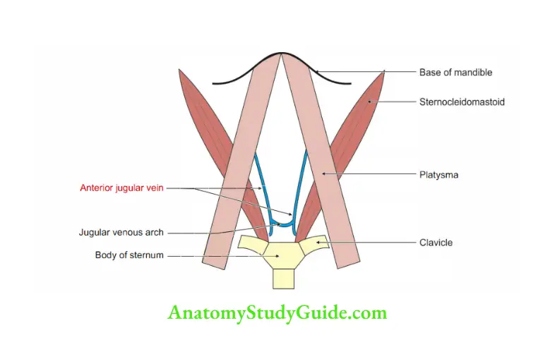
Anterior Jugular Vein
1 Anterior Jugular Vein Origin: Begins in the submental region.
2 Anterior Jugular Vein Course: It runs in the superficial fascia 1cm lateral to median plane in the anterior triangle of neck It enters the suprasternal space by piercing investing layer of deep cervical fascia.
It joins with its fellow of opposite side by transverse channel, the jugular venous arch and then runs deep to sternocleidomastoid
3 Anterior Jugular Vein Termination: It opens into external jugular vein.
4 Anterior Jugular Vein Applied anatomy: It is one of the contents of suprastemal space Injury to the vein results into air embolism.
Leave a Reply