Cell division
Mitosis (Mitos—thread, schisis—cleavage)
It is cell division where threads appear during the cleavage of somatic cells.
Table of Contents
It results in the distribution of identical copies of the parent cell into two daughter cells.
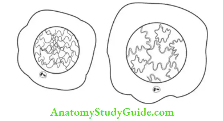
1. Interphase :
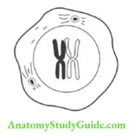
Cells do not actively divide but elongate. With the onset of mitosis, chromosomes undergo coiling condensation. A chromosome contains two parallel subunits called chromatids connected by a centromere.
1. Before mitosis: Chromosome number is diploid and DNA is tetraploid.
Read And Learn More: General Histology Question And Answers
2. After mitosis:
- Chromosomes remain diploid, and
- DNA material becomes diploid in amount.
2. Phases
1. Prophase (Pro—before): It is the 1st stage of cell division.
- Chromosomes are recognized as a structure of chromatids and centromere.
- Chromatids are shortened.
- Centrioles separate and start migrating to each pole.
- Spindles and asters are formed, together called as disasters.
- Nucleolus and nuclear membrane disappear.
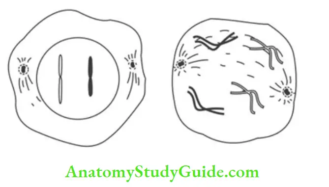
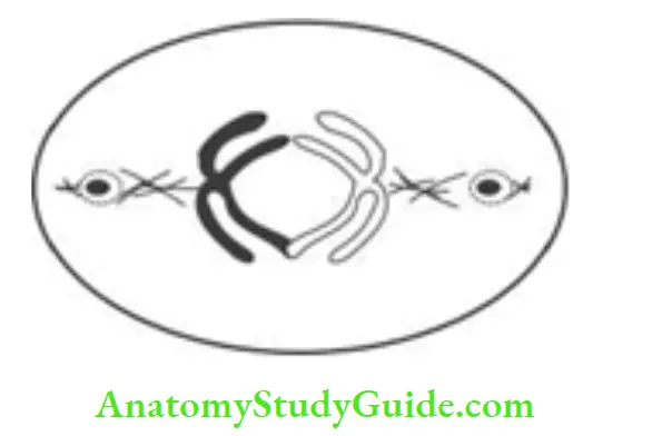
2. Metaphase (meta—after): It is the cell division after prophase.
- Chromosomes migrate toward equators.
- The spindle occupies a central position.
- Centromeres are attached to microtubules.
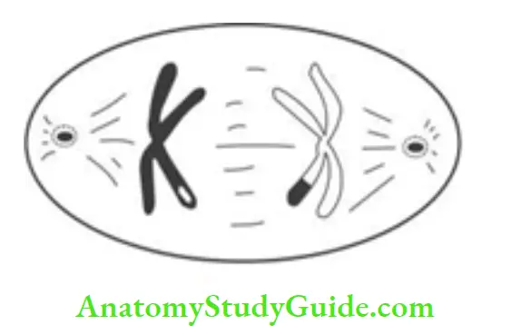
3. Anaphase (ana—apart): It is the cell division where the chromosomes move
- Cytoplasmic division starts with the appearance of a cleavage furrow.
- Chromosomes divide and split at the centromere (the centromere is a double structure).
- Such chromosomes start migrating to each end.
3. Applied anatomy: Radiation causes a change in the number of chromosomes and forms abnormal chromosomes.
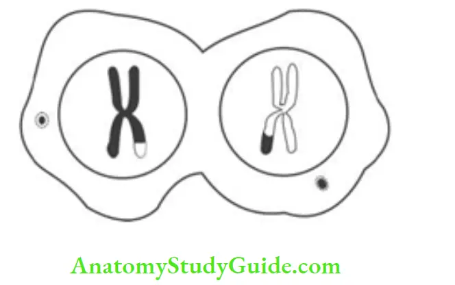
4. Telophase (telo—end): It is the end phase of cell division.
- The nuclear membrane reappears.
- Nucleoli reappear.
- Chromosomes are grouped at each end with diploid numbers.
Meiosis
1. Meiosis:
It is the cell division occurring only in the germ cells. There is an exchange of genetic material. The daughter cells are not identical. It occurs just before the formation of the gamete. The number of chromosomes is reduced to half.
2. Peculiarities:
1. Pairing of homologous chromosomes occurs only in meiosis and not in mitosis.
2. After fertility, the diploid number is restored.
3. The genetic exchange occurs in homologous chromosomal pairs.
- In interphase, there is a replication of DNA.
- DNA is tetraploid and chromosome is diploid.
Meiosis I:
It is a reduction division. At the end of meiosis I, each cell contains a haploid number of chromosomes.
Meiosis II:
The haploid number of chromosomes is maintained. It resembles mitosis but differs in two respects.
- There is no DNA replication prior to this.
- 2nd meiotic division follows meiosis I without interphase.
3. Site:
Meiosis occurs in the genn cells, i.e. spermatogonium in case of male ♂ and oogonium in case of female ♀.
4. Stages:
Meiosis includes 1st and 2nd meiotic division.
1. 1st meiotic division: It consists of
1. Prophase: It is long and complex process. It has various stages.
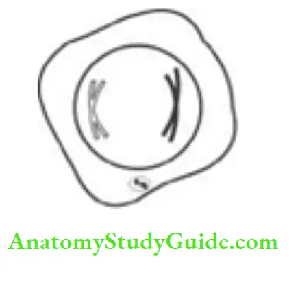
Leptotene (laptops—slender, tene—ribbon):
The chromosomes appear as individual threads, one end of which is attached to the nuclear membrane.
They appear beaded due to the presence of a centromere.
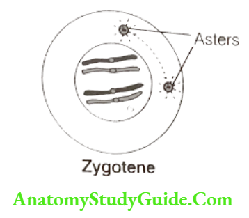
Zygotene:
- There is the pairing of homologous chromosomes. Point-to-point pairing occurs.
- Each pair is bivalent.
- There is limited pairing in X and Y chromosomes.
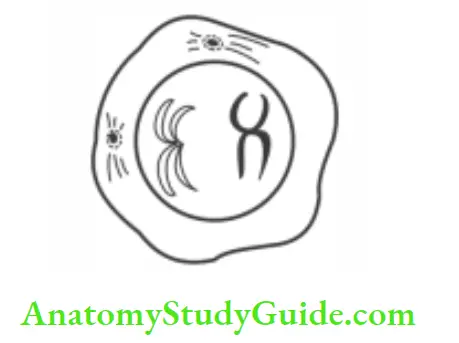
Pachytene (pachy—thick):
- Coiling condensation takes place.
- Centromere and two chromatids become prominent in chromosomes.
- Each bivalent pair consists of four chromatids and forms tetrad.
- Crossing over: It is an important stage. The two central chromatids (1 belonging to each chromosome bivalent) become coiled over other so that they cross at a number point.
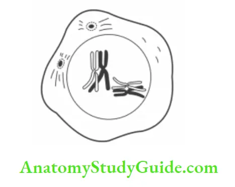
Diplotene (diplotene—duplication):
This stage is characterized by longitudinal separation of members of bivalent, without split in the centromere.
Crossing over is complete. In females ♀, the growth of the primary oocyte is arrested in diplotene and prolongs up to ovulation.
Diakinesis:
- The chiasmata start resolving.
- The chromosomes further become condensed and get coiled up.
- They start migrating toward equator.
- At the end of prophase, nucleoli and nuclear membrane disappear.
2. Metaphase:
- It resembles mitotic metaphase.
- Chromosomes are attached by a centromere. The spindle is formed.
- Homologous pairs lie parallel to the equator with one member on each side of the equator.
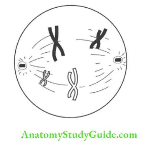
3. Anaphase :
The whole chromosome with two chromatids goes to one pole while another chromosome of homologous pair goes to other side. Such migration of bivalent pairs is at random.
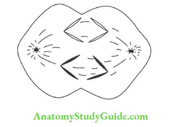
4. Telophase :
The chromosome in each cell is reduced to a haploid number.
2. The 2nd meiotic division is followed by a short interphase:
It differs from usual interphase in that there is no duplication of DNA. It is similar to mitosis. However, the daughter cells are not identical in genetic content.
5. Mitosis Cell Division Significance:
1. 1st meiotic division provides:
- Genetic variability through the process of crossing over.
- Random distribution of homologous chromosomes to daughter cells.
2. 2nd meiotic division provides:
- Germ cells with haploid number of the chromosome, and
- Haploid amount of DNA.
- It is similar to mitosis except chromosomes in daughter cells, which are not alike.
6. Mitosis Cell Division Applied anatomy:
1. Abnormalities of form
- Giant (large): Spermatozoa are too large.
- Dwarf (small): Spermatozoa are too small.
- There may be duplication of head, body or tail.
- The ovum may have an unusually large nucleus.
2. Failure in the separation of chromosomes or abnormal in the number of chromosomes
1. Non-disjunction: It is the failure of separation of two chromosomes at anaphase.
Trisomy:
There are 47 chromosomes, there being three identical instead of one of the normal pairs, e.g.
- Mongolism or
- Down’s syndrome: Trisomy 21.
Presence of extra X or Y: Chromosome, e.g.
- 47, XXX: Abnormal female ♀
- 47, XXY: Klinefelter syndrome—abnonnal male ♂.
- 47, XYY: Abnormal male ♂.
2. An Abnormal number of chromosomes:
There are only 45 chromosomes. Here one pair is represented by a single chromosome. 45, X, example XO Turner’s syndrome. Gamete may have a diploid number of chromosomes so that zygote will have 46 + 23 (i.e. 69).
3. Abnormalities in the shape:
1. Translocation: Part of a chromosome may get attached to a chromosome of a different pair.
2. Deletion: Part of the chromosome may be lost.
3. Duplication of genes.
4. Inversion: The chromosome may get inverted before joining the opposite chromosome.
Leave a Reply