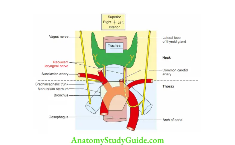
Recurrent Laryngeal Nerve
Recurrent Laryngeal Nerve Introduction: It is the nerve of 6th pharyngeal arch supplying muscles of larynx, pharynx and soft palate and mucous membrane of most posterior part of tongue.
Table of Contents
1. Recurrent Laryngeal Nerve Origin
- Right recurrent laryngeal nerve arises from right vagus nerve in the neck.
- Left recurrent laryngeal nerve arises from the left vagus nerve in the thorax.
2. Recurrent Laryngeal Nerve Course and relations: The relations are slightly different for the right and left recurrent laryngeal nerves.
Read And Learn More: Face Anatomy Notes And Important Questions
1. Right recurrent laryngeal nerve
- Winds around the right subclavian artery.
- Runs upwards and medially behind the subclavian and common carotid arteries and reaches the tracheo-oesophageal groove.
- In the upper part of groove, it is related to the inferior thyroid artery.
- The nerve passes deep to lower border of inferior constrictor. It enters the larynx behind cricothyroid joint.
2. Left recurrent laryngeal nerve
- It crosses the left side of arch of the aorta.
- It loops around the ligamentum arteriosum and reaches the tracheooesophageal groove.
- It is not have to pass behind the subclavian and carotid arteries.
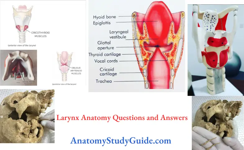
Larynx Anatomy
Recurrent Laryngeal Nerve Distribution
- It supplies all the intrinsic muscles of larynx except cricothyroid.
- It carries the sensations of the larynx below the vocal cords.
There are four cardiac branches from the right and left recurrent laryngeal nerves.
- These are
- Two superior, and
- Two inferior branches.
- Out of the four cardiac branches, the left inferior branch goes to superficial cardiac plexus.
- The other three cardiac branches join the deep cardiac plexus.
3. Branches to trachea and oesophagus
4. Branch to the inferior constrictor.
Larynx Anatomy
3. Recurrent Laryngeal Nerve Applied anatomy
Arytenoid Cartilage
- During thyroidectomy, the right recurrent laryngeal nerve is injured because of variable relation with inferior thyroid artery on right side.
- Left recurrent laryngeal nerve is damaged because of left atrial enlargement. The enlargement of left atrium compresses the left recurrent laryngeal nerve.
- Recurrent laryngeal nerves may be injured in thyroid surgery or compressed by a growing tumour, aortic aneurysm or from other causes.
- If only one recurrent laryngeal nerve is paralyzed, the affected vocal cord remains in the paramedian position and the vocal cord on the normal side compensates for phonation.
- If both recurrent laryngeal nerves are paralyzed, the vocal cords remain in the paramedian position (in between abduction and adduction). This results in
- Loss of phonation,
- Dyspnoea (difficulty in breathing) and respiratory stridor.
Larynx Anatomy
Stylopharyngeus
Stylopharyngeus Muscle Introduction: It is the longitudinal muscle of pharynx.
1. Stylopharyngeus Muscle Origin: It arises from medial side of base of styloid process.
2. Stylopharyngeus Muscle Insertion: Along with palatopharyngeus, it is inserted in the posterior border of lamina of thyroid cartilage.
3. Stylopharyngeus Muscle Action: It lifts the larynx during swallowing and phonation.
4. Stylopharyngeus Muscle Development: It is developed from 3rd pharyngeal arch.
5. Stylopharyngeus Muscle Features:
- Along with glossopharyngeal nerve (IX), it passes between two branches of common carotid arteries.
- It passes between two constrictors.
Give sensory nerve supply of larynx.
It is divided into two parts
- Above the glottis-internal laryngeal nerve, branch of external laryngeal nerve.
- Below the glottis-recurrent laryngeal nerve, branch of vagus nerve.
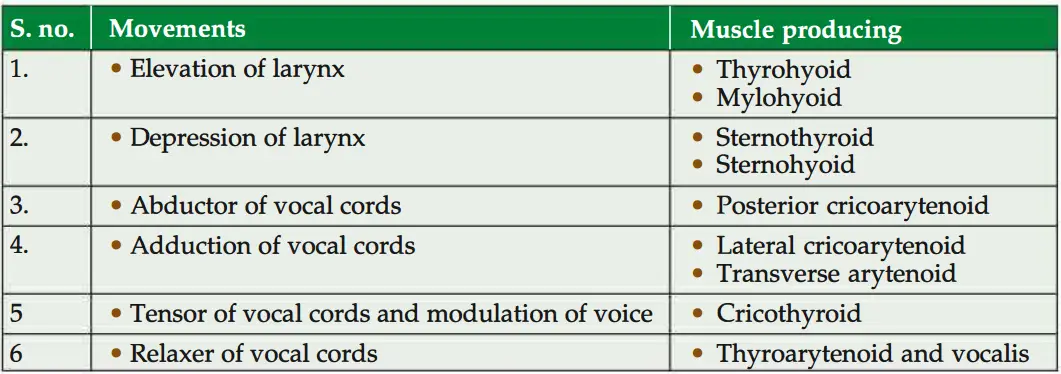
Question 1: Describe the movements of vocal cords. Name the muscles causing them.
Answer:
Vocal and vestibular folds
1. Inside the cavity of larynx, there are two folds of mucous membrane on each side.
1. The upper fold is the vestibular fold. The space between the right and left vestibular folds is the rima vestibule.
2. Lower fold is the vocal fold. The space between the vocal folds is the rima glottidis.
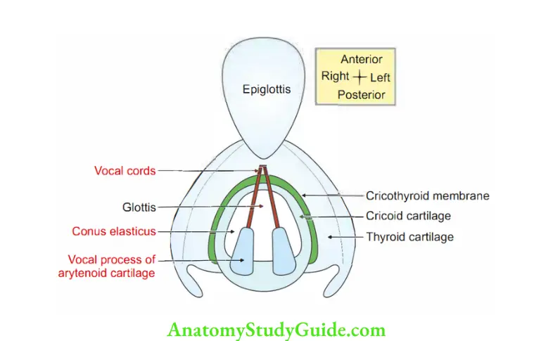
1. Vocal Cords Anatomy Attachments
- Anteriorly to the posterior aspect of middle of the angle of the thyroid cartilage.
- Posteriorly to the vocal process of the arytenoid cartilage.
Larynx Anatomy
2. Vocal Cords Anatomy Features
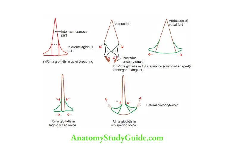
- The rima glottidis is limited posteriorly by interarytenoid fold of mucous membrane.
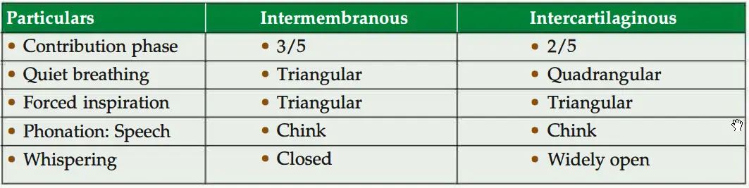
- The rima has an anterior intermembranous part (3/Sth) and a posterior intercartilaginous part (2/Sth).
- The rima is the narrowest part of the larynx.
- It is longer (23 mm) in males O ‘han in females S (17 mm).
2. The vestibular and vocal folds divide the cavity of the larynx into three parts.
- Supraglottic part: Part above the glottis.
- Sinus or ventricle of the larynx: Part between the vestibular and vocal folds.
- Infraglottic part: Part below the glottis.
Rima Glottidis
Rima Glottidis Introduction: It is the narrowest anteroposterior cleft, or space of laryngeal cavity.
It is lined by a stratified squamous non-keratinized epithelium.
It is without submucous coat.
Arytenoid Cartilage
1. Rima Glottidis Attachments:
- Anteriorly: The middle of angle of thyroid cartilage.
- Posteriorly: Vocal process of arytenoid cartilage.
Limited: Posteriorly by an interarytenoid fold of mucous membrane.
Shape and size of rima glottidis is changed by movements of vocal cords.
2. Rima Glottidis Nerve supply: All muscles of larynx are supplied by recurrent laryngeal nerve (vagus nerve) except cricothyroid, which is supplied by external laryngeal nerve (superior laryngeal, branch of vagus nerve).
Question 2: Describe Larynx under the following heads:
1. Larynx Formation ,
2. Cartilages of larynx,
3. Muscles of larynx,
4. Larynx Actions,
5. Larynx Nerve supply, and
6. Larynx Applied anatomy
Answer: 1. Formation: Larynx is formed by paired and unpaired cartilages. They are
- Unpaired: Thyroid, cricoid and epiglottis.
- Paired: Arytenoid, corniculate and cuneiform cartilages.
2. Cartilages of larynx: They are 11 in number. Four are paired and three are unpaired.
- Paired cartilages of larynx: They are 1 on each side.
- Unpaired
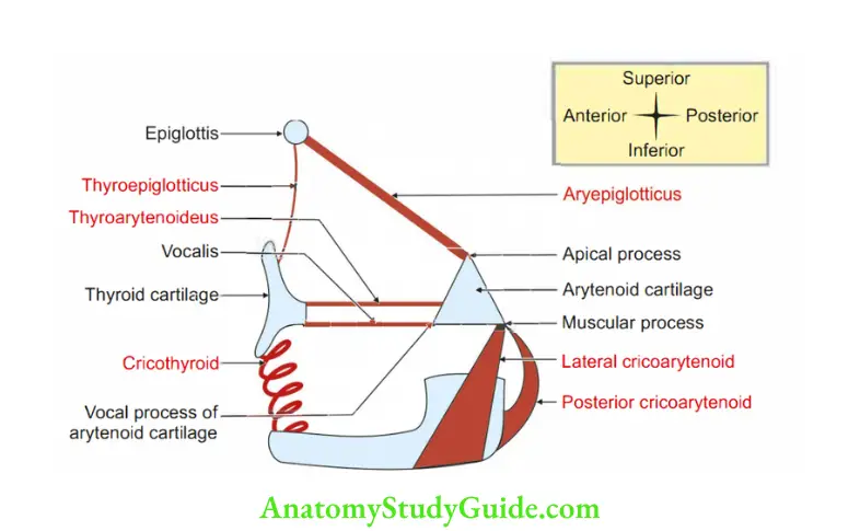
1. Paired: There are four paired cartilages
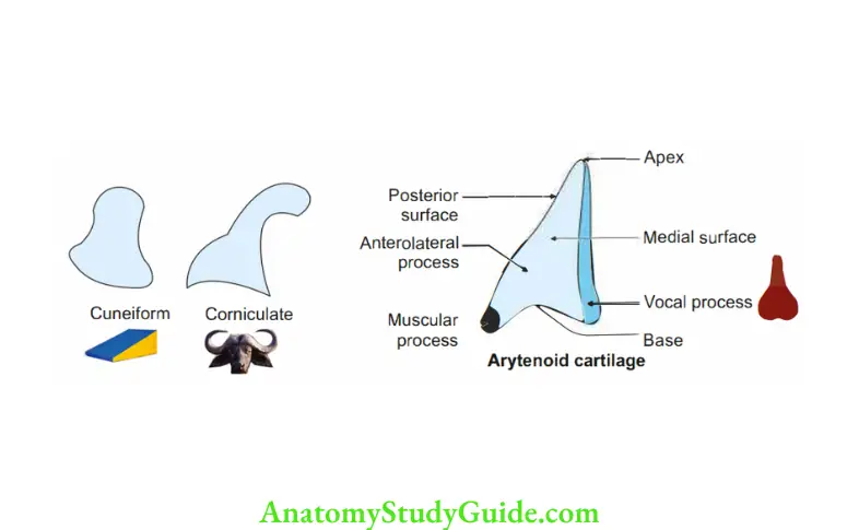
1. Aryenoid cartilage: It is a 3-sided pyramid with anterolateral, medial and posterior surfaces.
- Apex: It is directed upwards and articulates with corniculate cartilage. It gives attachments to oblique arytenoid muscle.
- Base: It is directed downwards and forms cricoarytenoid joint.
- Vocal process: It is pointed and projects horizontally forwards from the base.
It gives attachment to vocal ligament by its tip and vocalis ligament by its lateral aspect. - Muscular process: It gives attachment to
Posterior cricoarytenoid by the posterior aspect, and
Lateral cricoarytenoid by its anterior aspect. - Medial surface: It is flat and narrow and faces similar surface of the other arytenoid.
It forms the lateral boundary of the intercartilaginous part of rima glottidis.
6. Posterior surface: It is deep to oblique arytenoid muscle.
7. Anterolateral surface is convex and rough. It gives attachment to
- Vestibular ligament,
- Vocalis, and
- Lateral cricoarytenoid muscles.
- Thyroarytenoid muscle.
- Comiculate cartilage (of Santorini): It is conical nodule enclosed in the dorsal part of aryepiglottic fold.
- Cuneiform cartilage: It is present in the aryepiglottic fold and lies ventral and superior to the comiculate cartilage.
- Cartilago triticea: They are present at lateral part of thyrohyoid membrane.
Arytenoid Cartilage
2. Unpaired cartilages
- Cricoid cartilage: Refer SN-127
- Thyroid cartilage: Refer SN 128
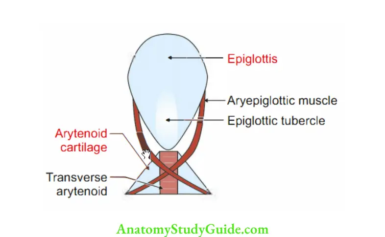
- Epiglottis:
- It is slightly curved, resembles a leaf. It is prolonged below into a slender process, the stalk of the leaf.
It is attached in the midline to the back of laryngeal prominence. - The posterior surface above the apex is pitted by mucous glands.
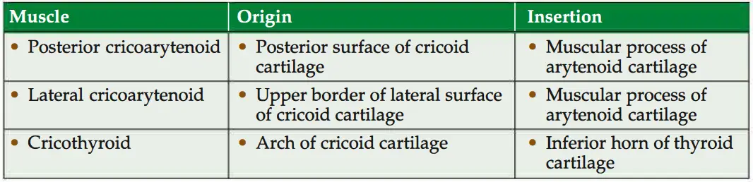
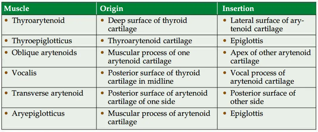
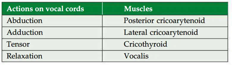
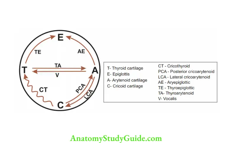
3. Muscles of larynx: Muscles of larynx are divided into extrinsic and intrinsic groups.
- Intrinsic muscles take origin and get inserted in the same organ. They are described as given in Table 16.3.
2. Extrinsic muscles take origin outside and get inserted inside the organ. They are
- Sternothyroid,
- Thyrohyoid,
- Omohyoid and
- Digastric.
4. Actions of intrinsic muscles
Nerve supply
1. Sensory
- Above vocal fold: Internal laryneal nerve, a branch ofsuperior laryngeal nerve, branch of vagus nerve.
- Below the vocal fold: Recurrent laryngeal nerve (branch of vagus nerve).
2. Motor (muscle): All muscles of larynx are supplied by recurrent laryngeal nerve, a branch of vagus except cricothyroid, which is supplied by external laryngeal nerve, a branch of superior laryngeal nerve, a branch of vagus nerve.
Arytenoid Cartilage
1. Embrylogical (developmental) correlation
- Cricothyroid muscle develops from 4th pharyngeal arch. Nerve of the 4th pharyngeal arch is superior laryngeal nerve, which gives external laryngeal nerve.
Hence, cricothyroid is supplied by external laryngeal branch. - Remaining all muscles of larynx are developed from 6th arch. The nerve of 6th pharyngeal arch is vagus nerve which gives a recurrent laryngeal nerve.
Hence, remaining all muscles are supplied by a recurrent laryngeal nerve.
2. Physiological (functional correlation): The cricothroid muscle acts as a tuning fork.
First, it receives impulses and starts vibrating.
The remaining muscles receive impulses few milli-second afterwards, which help in producing voice.
6. Larynx Applied anatomy.
- Examination of larynx is called laryngoscopy.
- Laryngitis is inflammation of larynx. It occurs in common cold.
- The swelling of vocal cords is rare in acute laryngitis because of following reasons:
- The vocal cords are lined by stratified squamous epithelium (rest of the larynx is lined by pseudostratified ciliated columnar epithelium).
- The mucous membrane is firmly attached to the underlying vocal ligaments.
- There is no submucous tissue and there are no glands over the vocal cords.
For the same reason, the vocal cords appear pearly white in colour. - Damage to internal laryngeal nerve produces anaesthesia (loss of sensation in supraglottic part of larynx).
- Foreign bodies can readily enter the larynx, if internal laryngeal nerve is damaged.
- Damage to the external laryngeal nerve causes paralysis of cricothyroid muscle.
It results in weakness of phonation. - When both recurrent laryngeal nerves are injured, vocal cords lie in cadaveric position.
- During swallowing, the larynx moves up and down by extrinsic laryngeal muscles (viz. palatopharyngeus, salphingopharyngeus and stylopharyngeus).
- Compression of larynx due to thyroid swelling may produce hoarseness of voice called dysphonia.
- Removal of foreign bodies from the piriform fossa may damage internal laryngeal nerve, leading to anaesthesia of the supraglottic portion of the larynx.
- Teacher’s nodules (or singer’s nodules): These nodules are seen in the vocal cords of the teachers and singers.
They are usually located at the junction of anterior and middle third of vocal cord.
Cricoid Cartilage
(Cricoid Gr. Krikos-a ring, -aid-resemblance)
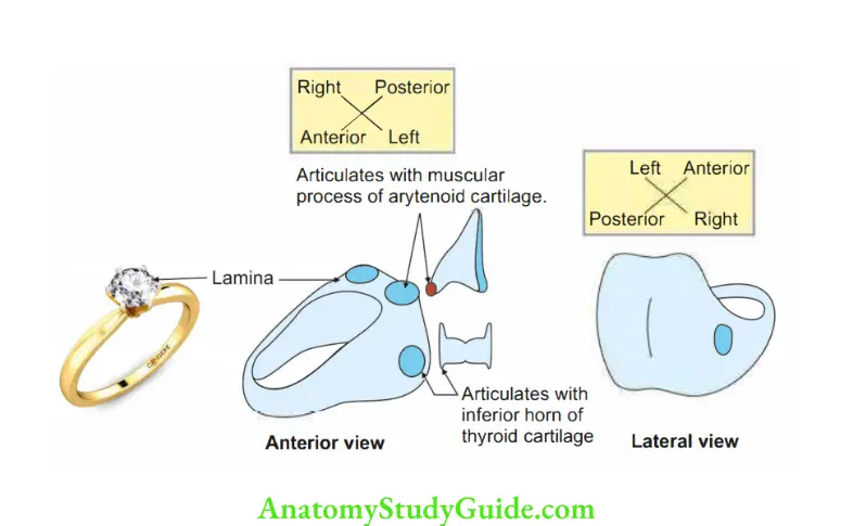
Cricoid Cartilage Introduction: It is a midline unpaired cartilage of larynx. It forms the foundation of larynx.
1. Cricoid Cartilage Morphology
- Shape: It is only complete ring O in all the structres of respiratory tract,
Situation: It encircles the larynx and is present at the level of 6th cervical vertebra.
Parts: It has
- Arch: The narrow anterior part is called arch. It has upper and lower borders and outer and inner surfaces.
- Lamina: Cricoid cartilage has broad, quadrilateral posterior part called lamina.
It has upper and lower borders, anterior and posterior surfaces.
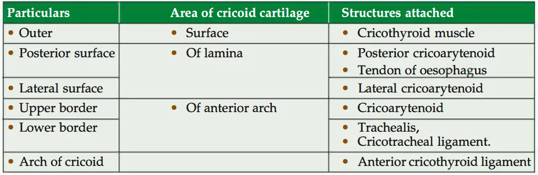
2. Cricoid Cartilage Attachments
3. Cricoid Cartilage Joints: It forms joint with thyroid and arytenoids. Both are synovial joints.
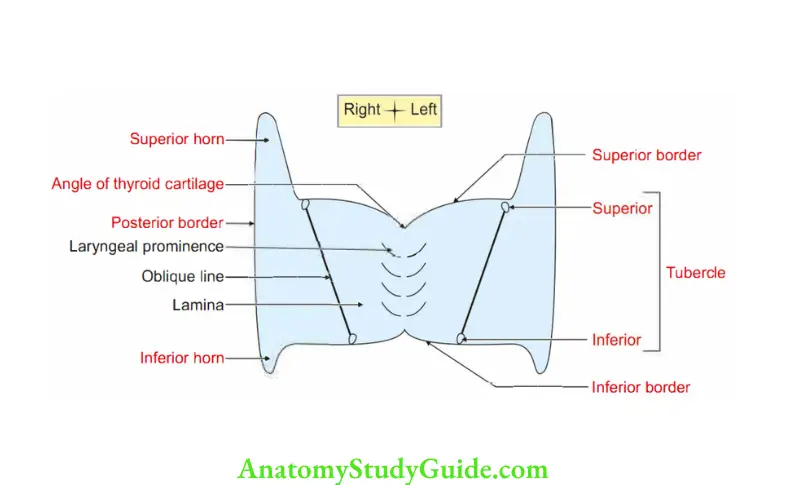
Thyroid Cartilage
(Gr. thyreos-a shield-aid-like)
Thyroid Cartilage Introduction: It is one of the unpaired cartilages of larynx.
1. Parts
- It has two conjoined laminae whose posterior borders are free.
They are projected upwards and downwards as superior and inferior comua. - The two laminae fuse in the midline and form the laryngeal prominence (Adam’sapple).
- Oblique line: It extends on the outer surface. It is bounded below and above by a tubercle.
2. Thyroid Cartilage Joints: Inferior horn articulates with the cricoid cartilage and forms cricothyroid joint.
3. Thyroid Cartilage Relations: The common carotid artery bifurcates at the tip of superior cornu into internal and external carotid arteries.
4. Attachments
1. Superior border gives attachment to thyrohyoid membrane.
2. Oblique line gives attachments to the following muscles.
- sternothyroid
- Inferior constrictor of pharynx
- Ihrohyoid
3. Thyroid Cartilage Inferior cornu and lower border of thyroid lamina give attachment to cricothyroid -om
4. Thyroid Cartilage Superior cornu gives attachment to thyroepiglottic ligaments.
5. Thyroid Cartilage Applied anatomy: The upward enlargement of the thyroid gland is prevented by the sternothyroid muscle attached to oblique line of thyroid cartilage.
Inlet of Larynx
Inlet of Larynx Introduction: It is the continuation of laryngopharynx.
1. Boundaries
- In front and above: Upper free margin of epiglottis.
- Below and behind: Interarytenoid fold of mucous membrane.
- Laterally: Aryepiglottic fold.
2. Inlet of Larynx Placement: Obliquely. The anterior wall is much longer than the posterior wall.
3. Inlet of Larynx Communication
- Above: Laryngopharynx
- Below: Trachea.
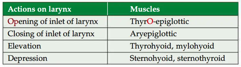
4. Inlet of Larynx Muscles acting on inlet of larynx
- Thyroepiglotticus: It helps in active opening of laryngeal inlet.
- Aryepiglotticus: It closes the inlet of larynx.
5. Inlet of Larynx Relations: On either side of inlet of larynx, there is a small recess termed the piriform fossa.
6. Inlet of Larynx Applied anatomy
- A malignancy tumour may grow in the piriform fossa without producing symptoms, until the patient presents with metastatic lymphadenopathy.
- The recesses are dangerous sites for perforation by an endoscope.
Thyrohyoid Membrane
Thyrohyoid Membrane Introduction: It is extrinsic membrane extending from
- Upper border of thyroid laminae, and
- Superior horns of thyroid cartilage to
- Greater comu of hyoid bone, and
- Upper border of body of hyoid bone.
1. Thyrohyoid Membrane Modifications
- Median thyrohyoid ligament: It is midline thickened part of thyrohyoid membrane.
- Lateral thyrohyoid ligament: It is thickening on the posterior free border.
2. Thyrohyoid Membrane Relations
- It forms the lateral boundary of piriform fossa.
- A bursa lies between the membrane and back of hyoid bone.
3. Thyrohyoid Membrane Structures piercing
- Internal laryngeal nerve.
- Superior laryngeal vessels.
Cricothyroid Muscle
Cricothyroid Muscle Introduction: It is the only intrinsic muscle present outside the larynx. It is
1. Cricothyroid Muscle Proximal attachment: It arises from the arch of cricoid cartilage.
2. Cricothyroid Muscle Distal attachment: Inferior horn and lower border of thyroid cartilage.
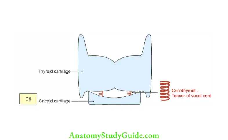
3. Cricothyroid Muscle Action: It brings the arch of cricoid cartilage near thyroid cartilage. Hence, tensor of the vocal cord.
4. Cricothyroid Muscle Development: It develops from the skeletal element of 4th pharyngeal arch.
5. Cricothyroid Muscle Nerve supply: External laryngeal nerve, branch of superior laryngeal nerve (branchof vagus nerve).
Posterior Cricoarytenoid
Posterior Cricoarytenoid Introduction: It is the intrinsic and most important muscle of larynx and probably in the body.
1. Posterior Cricoarytenoid Origin: It arises from the posterior surface of lamina of cricoid cartilage. It is also known as, “saf muscle of laryx”.
2. Posterior Cricoarytenoid Insertion: Posterior aspect of muscular process of arytenoid cartilage of the same side.
3. Posterior Cricoarytenoid Action: It abducts the vocal folds and opens the glottis.
4. Posterior Cricoarytenoid Nerve supply: Recurrent laryngeal nerve, branch of vagus nerve.
5. Posterior Cricoarytenoid Development: It develops from 6th pharyngeal arch.
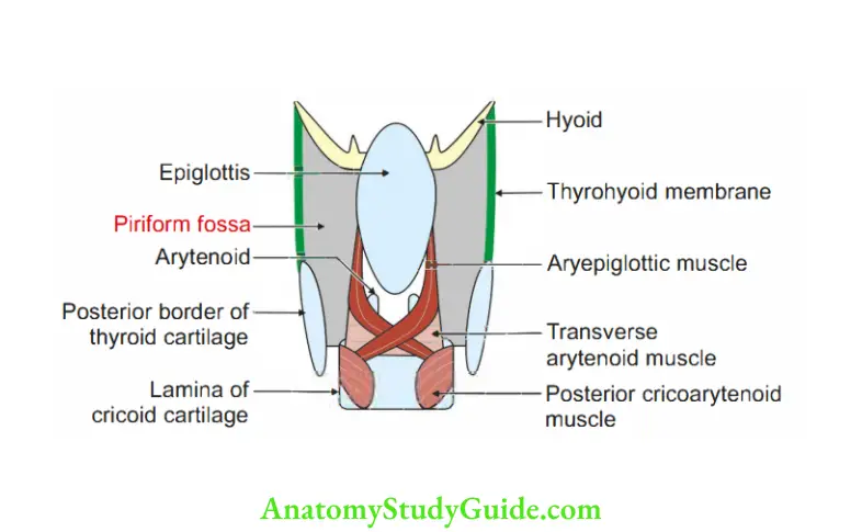
Piriform Fossa
Piriform Fossa Introduction: It is a space present on each side of inlet of larynx. It is broad above and narrow below.
1. Piriform Fossa Boundaries
- Medially: By the quadrate membrane below the aryepiglottic fold.
- Laterally: Mucosa covering the thyroid lamina and thyrohyoid membrane.
- Superiorly: Lateral glossoepiglottic fold. It separates pyriform fossa from vallecula of oropharynx.
2. Piriform Fossa Structures deep to piriformis
- Internal laryngeal nerve, and
- Superior laryngeal vessels.
3. Piriform Fossa Relations: It is traversed by internal laryngeal nerve.
4. Piriform Fossa Applied anatomy
- Foreign body in larynx: At times fish bones may get impacted in the vallecula or piriform fossa.
These foreign bodies scratch the mucosa.
The person feels discomfort and uneasiness due to a dull visceral pain. - The fossa is artificially deepened by smugglers to carry smuggled goods.
In old days, it was used to smuggle gold coins.
After introduction of metal detectors, it is used to carry precious stones, diamonds, etc.
Hence, it is called smuggler’s fossa. - Tumours in the piriform fossa cause dysphagia.
These also cause referred pain in the ear.
Pain of pharyngeal tumours may be referred to the ear, as vagus nerve supplies larynx, tympanic membrane and external acoustic meatus. - By laryngoscope, one can visualize piriform fossa.
- A malignancy may grow in the space without producing symptoms unless the patient presents with metastatic cervical lymphadenopathy.
- The recess is dangerous site for perforation by an endoscope.
- It acts as a site for lodgment of foreign body.
Leave a Reply